i
EXPRESSION OF FATTY ACID OXIDATION-RELATED GENES IN Acsl4 L -/- PRIMARY HEPATOCYTES
Dennis Lin
Senior Honors Thesis Department of Nutrition
University of North Carolina at Chapel Hill
April 2018
Approved by,
Rosalind Coleman
ii
Abstract
Long-chain acyl-CoA synthetases are a family of enzymes responsible for catalyzing the
reaction converting long chain fatty acids to fatty acyl-CoA. ACSL4, a specific member of this
family, plays a role in eicosanoid metabolism and has been found to be associated with liver
diseases including non-alcoholic fatty liver disease and hepatocellular carcinoma. In order to
study ACSL4 in the context of hepatic lipid metabolism, a murine liver-specific Acsl4 knockout
(Acsl4 L -/-) was developed and characterized in our lab. Based on a previous experiment in our lab, we hypothesized that Acsl4 L -/- would cause a defect in beta adrenergic signaling. In order to test this hypothesis, we conducted experiments to determine the effects of various treatments on
the expression of fatty acid oxidation-related genes in both knockoutand control hepatocytes.
Treatments with fatty acids caused increases in expression of several fatty acid oxidation-related
genes in both knockout and control hepatocytes. Hormone treatments did not cause anticipated
increases in expression of fatty acid oxidation-related genes in several of the control groups.
Because of this we were not able to make conclusions about the effects of the knockout. These
iii
Acknowledgements
First and foremost, I would like to thank Dr. Rosalind Coleman for allowing me to work
in her lab and shaping me into the scientist I am today. I appreciate all the time and patience she
invested in me at a point a when I had a lot less knowledge than I do now. I would especially like
to thank Dr. Pamela Young, for guiding me every step of the way through my projects and
teaching me almost everything I know about lipid research. She was instrumental in helping me
grow as a researcher and encouraged me during times of frustration and doubt. I would also like
to thank Dr. Eric Klett, Dr. Amanda Suchanek, Dr. Theresa D’Aquila, Dr. Shufen Chen, Liyang
Zhao, and everyone else in the Coleman lab for giving me advice and support for my
experiments. I have grown tremendously during my time in the Coleman lab and would not have
iv
Table of Contents
List of Tables ... v
List of Figures ... vi
Chapter I. Introduction ... 1
1.1 Liver and Lipid Metabolism ... 1
1.2 ACSL4 Gene and Protein ... 2
1.3 ACSL4 Abnormalities and Human Disease ... 3
1.4 ACSL4 Tissue and Substrate Preferences ... 3
1.5 Study Aims and Hypothesis:... 4
Chapter II. Methods and Materials ... 6
2.1 Primary hepatocyte isolation and preparation ... 6
2.2 Primary hepatocyte hormone treatments ... 6
2.3 Primary hepatocyte lipid treatments ... 6
2.4 Gene expression in ACSL4 L -/-and control primary hepatocytes ... 7
Chapter III. Results ... 8
3.1 Oleate and palmitate treatment effects on gene expression ... 8
3.2 Hormone treatment effects on gene expression ... 8
Chapter IV. Discussion ... 10
4.1 Adjustments to Methods and Materials ... 10
4.2 Hormone treatment effects on gene expression ... 10
4.3 Fatty acid treatment effects on gene expression ... 11
4.4 Future Directions ... 12
v
List of Tables
Table 1. Treatment and corresponding vehicle conditions ... 17
vi
List of Figures
Figure 1. Lipid Treatments Gene Expression (Experiment 1) ... 13
Figure 2. Lipid Treatments Gene Expression (Experiment 2) ... 13
Figure 3. Hormone Treatment Gene Expression (Experiment 1)... 14
Figure 4. Hormone Treatment Gene Expression (Experiment 2)... 15
1
Chapter I. Introduction
1.1 Liver and Lipid Metabolism
The liver is perhaps the most important organ when it comes to lipid metabolism, acting as the main site for fatty acid (FA) synthesis, fatty acid oxidation, complex lipid synthesis, and very low-density lipoprotein (VLDL) production. The liver acts as a mediator for fat metabolism to meet the energy requirements of the rest of the body. These metabolic pathways are
controlled by various enzyme interactions and regulated by the liver under different hormonal and physiological conditions.
Lipid metabolism starts with the digestion and absorption of dietary fats. In the mouth, lipids are first broken down by lingual lipases and further processed in the stomach and duodenum where dietary fats are emulsified with help from bile acids secreted from the gallbladder. Lipases secreted from the pancreas further digest lipids into free FA and
monoacylglycerol to be absorbed by enterocytes. Within the enterocyte, triacylglerol (TAG) molecules are resynthesized and repackaged along with cholesterol esters (CE), phospholipids (PL), lipoproteins, and fat-soluble vitamins into chylomicron lipoproteins that pass into lacteals that eventually enter circulation. Peripheral tissues hydrolyze TAG from chylomicrons via lipoprotein lipase to obtain FA while remnant chylomicrons and other lipoproteins are taken up by the liver. The liver incorporates the components of remnant lipoproteins particles into various metabolic pathways.
Long-chain FAs are activated by the long chain acyl-CoA synthetase (ACSL) enzyme family, which converts long chain FAs into long chain fatty acyl-CoAs and shuttles them into different metabolic pathways.1 Depending on energy status, activated FAs can enter synthetic pathways such as de novo lipogenesis, TAG synthesis, and phospholipid synthesis. TAG synthesis is initiated by ACSLs, with glycerol-3-phosphate acyltransferase (GPAT) catalyzing the formation of TAG by esterifying glycerol-3-phosphate with acyl-CoA. Acetyl-CoA
2
the liver also serves as the main distribution center. It exports lipids, TAG, CE, and
phospholipids by producing and secreting VLDL for peripheral utilization of lipids and FAs. Hepatic concentrations of lipid and apolipoprotein B100 directly affect VLDL production and secretion.
Beta-oxidation is the degradative pathway of FAs activated by ACSLs, which produces acetyl-CoA from fatty acyl-CoA to be used as energy production via the Krebs cycle and oxidative phosphorylation. The main regulated enzyme of beta-oxidation is carnitine palmitoyltransferase I (CPT-1), which catalyzes the entry of fatty acyl CoA into the
mitochondria via conversion to acyl-carnitine. CPT-1 is inhibited by malonyl CoA, which is produced by ACC during de novo lipogenesis. Beta-oxidation is also regulated by the
transcription factor peroxisome proliferator-activated receptor alpha (PPARa). PPARa ligand activation induces several genes controlling FA import including CPT-1 and several major enzymes within the beta-oxidation pathway including acyl-CoA dehydrogenases.2
1.2 ACSL4 Gene and Protein
While all enzymes in the ACSL family serve the same enzymatic function, the ACSL isoforms vary in substrate specificity, subcellular location, and potential impact on downstream metabolic functions. With the exception of ACSL4, which is localized to both the plasma membrane and cytosol, most of the ACSL isoforms are membrane associated. 3 Accurate methods for measuring enzymatic activity for individual ACSL isoforms have not been well established due to non-specificity of ACSL inhibitors. It is still unclear how each isoform’s distinct structure and subcellular location affect function and activity. This study aims to investigate the effects of a liver-specific ACSL4 knock out (ACSL4 L -/-) mouse model to study the role of ACSL4 in hepatic lipid metabolism.
3
1.3 ACSL4 Abnormalities and Human Disease
Sequence abnormalities and sequence deletions surrounding the Acsl4 gene have been associated with several conditions including elliptocytosis, mental retardation, and Alport syndrome. Alport syndrome is an X-linked condition that is characterized by a large deletion of the Xq22.3-23 (including the Acsl4 gene) region of the X-chromosome. Its symptoms include progressive renal failure, hearing loss, and vision loss. High Acsl4 expression in specific regions of the brain, including the cerebellum and hippocampus, and its deficiencies associated with mental retardation suggest that the enzyme may play an important role in brain development.6 Investigations of Acsl4 expression during mouse development showed high levels of expression in mice embryos and newborn brains.7
ACSL4 dysregulation has also been studied in the context of hepatic lipid dysfunction associated with non-alcoholic fatty liver disease (NAFLD) and hepatocellular carcinoma. Studies using CLOCK-deficient mouse models found that reduced Acsl4 and Fabp1 expression was associated with reduced steatosis on a high fat diet.8 This suggests that ACSL4 may play a role in the development of hepatosteatosis and circadian controlled lipid metabolism. In human studies, researchers identified polymorphisms in Acsl4 that were related to liver fat content. Higher hepatic lipid content of NAFLD patients, along with hyperinsulinemia and obesity, were
associated with the expression of a rare ACSL4 rs7887981 allele.9 ACSL4 was also found to be involved in hepatocellular carcinoma development.10 Several human hepatoma cell lines,
including Hep3B and HepG2, have exhibited high ACSL4 expression.11 In patients, ACSL4 has been found to be up-regulated (between 2.3 and 27.5-fold) in hepatocellular carcinoma compared to adjacent non-cancerous tissue.11 These findings suggest that ACSL4 expression may
contribute to dysregulation in hepatic lipid metabolism associated with both cancer and steatosis.
1.4 ACSL4 Tissue and Substrate Preferences
4
revealed the presence of Acsl4 transcripts in the adrenal cortex, brain, lung, ovaries, testes, and liver.6 Because of its preference for polyunsaturated FAs, ACSL4 may play an important role in eicosanoid metabolism. Arachidonate has potential to generate inflammatory products as a precursor to several clinically important eicosanoids and metabolites of eicosanoids.
Cyclooxygenases metabolize arachidonate to prostaglandins and thromboxanes that mediate inflammation and platelet aggregation. To prevent excessive eicosanoid synthesis, free arachidonate is converted to arachidonoyl-CoA by ACS enzymes and re-esterified into phospholipids.
1.5 Study Aims and Hypothesis:
Liver-specific ACSL4 knock out mice (ACSL4 L -/-) have been developed to further investigate the role of ACSL4 in hepatic lipid metabolism. Under fasting and ketogenic
conditions, ACSL4 L -/- mice and control mice do not exhibit differences in ACS activity, serum TAG, or serum glucose. Ongoing studies have suggested that ACSL4 L -/- may affect
transcriptional regulation of beta-oxidation by disrupting adrenergic signaling. Hepatic lipid metabolism is tightly controlled by hormone responses. Glucagon and catecholamines, such as norepinephrine, stimulate G-protein coupled receptors and initiate signaling cascades that regulate both glucose and lipid homeostasis via transcriptional regulation. Stimulation of
adrenergic and glucagon receptors increases production of cAMP and activates protein kinase A (PKA). PKA phosphorylates transcription factor CREB, which induces PGC-1α gene expression. Acting as a coactivator, PGC-1α is recruited by transcription factors such as PPARα that bind to the promoter regions of FA oxidation enzyme genes to increase expression.12
A previous study tested the effects of norepinephrine treatments on cultured primary hepatocytes from both control and ACSL4 L -/- mice. Results showed lowered gene expression of beta-oxidation genes among ACSL4 L -/- hepatocytes after treatment compared to control.
5
Therefore, our study aimed to examine the effects of Acsl4 deletion on the expression of FA oxidation genes after treatments with several hormones and metabolites that promote
6
Chapter II. Methods and Materials
2.1 Primary hepatocyte isolation and preparation
Primary hepatocytes were isolated from the livers of 3-month-old ACSL4 L -/- mice and floxed control littermates. Because Acsl4 is X-linked, we used male knockout mice for our experiments. Hepatocytes were plated at 4 X 10⁵ cells per well in 6-well plates with William’s E. media (5% serum). After two hours, William’s E. media was replaced with low glucose DMEM (1g/L glucose, 10% fetal bovine serum, penicillin-streptomycin). Before treatments, primary hepatocytes of both genotypes were serum starved in low glucose DMEM (5mM glucose, 1mM sodium pyruvate, 1mM L-Glutamine, serum free) media using 1 ml per well for 4 hours.
2.2 Primary hepatocyte hormone treatments
All treatments were prepared using the same glucose (5mM) DMEM media at 1 mL per well. Hormone solutions were added to three wells of each plate and the corresponding vehicle control solutions were added to the remaining three wells of each plate. The norepinephrine solution was prepared at 10µM from a 10mM norepinephrine stock (dissolved in water) diluted in media. The 8-Br-cAMP treatment solution was prepared at 1 mM from an 80mM stock (dissolved in water) diluted in media. The forskolin treatment solution was prepared at 40 µM from a 4mM stock (dissolved in DMSO) diluted in media. The glucagon treatment solution was prepared at 100mM from a 1mg/mL stock (dissolved in phosphate buffered saline) diluted in media. All vehicle control solutions were prepared by diluting the corresponding vehicle (Table 1) in media by the same dilution factor used for diluting the stock hormone solution. After incubating the hepatocytes for 1 hour, the cells were washed twice with chilled PBS and frozen at -80° C.
2.3 Primary hepatocyte lipid treatments
7
stock solutions were first diluted with fatty acid free bovine serum albumin (1:25 for oleate and 1:50 for palmitate) and allowed to complex at 37° C for 1 hour. Carnitine and serum free media was added to both solutions for final concentrations of 1mM carnitine and 500µM of FA. After treating the primary hepatocytes for 3.5 hours, the cells were washed twice with chilled PBS and stored at -80°C.
2.4 Gene expression in ACSL4 L -/- and control primary hepatocytes
Before testing expression of FA oxidation genes, the liver-specific knockout of Acsl4 was confirmed. RNA was extracted from both ACSL4 L -/- hepatocytes and littermate control
hepatocytes using phenol chloroform extraction (Invitrogen TRIzol reagent). RNA concentration was measured by spectrophotometry and cDNA was made by reverse transcription (iScript cDNA synthesis kit) using 1µg of RNA. The mRNA levels of Acsl4 were quantified with real time PCR using ACSL4 and β-actin primers (Table 1) to determine the relative expression of Acsl4 compared to Actin (Biorad iTaq universal SYBR green supermix). Acsl4 expression in
ACSL4 L -/- mice was less than 1% of control littermates, confirming the knockout. The same
8
Chapter III. Results
3.1 Oleate and palmitate treatment effects on gene expression
To determine whether ACSL4 L -/- disrupts oleate’s signaling effects on the expression of
beta-oxidation genes, ACSL4 L -/- and control hepatocytes were treated with oleate and palmitate.
Two experiments were conducted using a different set of hepatocytes. RT-PCR results from Experiment 1 showed increased Cpt1a, Acox1, Pgc1a, and Ppara expression (relative to Actin) following both oleate and palmitate treatments (Figure 1). These increases in gene expression occurred in both ACSL4 L -/- and control hepatocytes. Experiment 2 results, with palmitate
treatment, showed increased Cpt1a, Acox1, Ppara and Creb expression in control hepatocytes along with increased Acox1 and decrease Ppara expression in ACSL4 L -/- hepatocytes (Figure 2).
Experiment 2 results, with oleate treatment, showed increased Creb expression and decreased Pgc-1α and Pparα expression in control hepatocytes. In ACSL4 L -/- hepatocytes, oleate treatment
resulted in increased Creb and Acox1 expression and decreased Pgc-1α and Pparα expression.
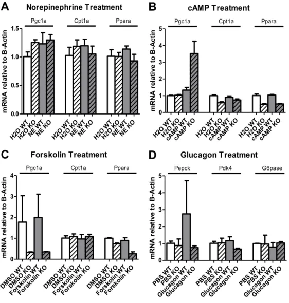
3.2 Hormone treatment effects on gene expression
To investigate whether ACSL4 L -/- disrupts adrenergic signaling effects on beta oxidation
gene expression, ACSL4 L -/- and control hepatocytes were treated with norepinephrine, forskolin,
glucagon, and 8-Br-cAMP. All of these hormone treatments increase expression of fatty acid oxidation-related genes by increasing intracellular cAMP (Figure 5). RT-PCR results from Experiment 1 showed increased Pgc1a, Cpt1a, and Ppara expression following norepinephrine treatment only in control hepatocytes (Figure 3A). With 8-Br-cAMP treatment, Pgc1a
expression increased only in ACSL4 L -/- hepatocytes. There was also lowered Cpt1a and Ppara
expression among all ACSL4 L -/-hepatocytes in the 8-Br-cAMP study compared to control
(Figure 3B). After forskolin treatment, Ppar expression decreased only in ACSL4 L -/- hepatocytes
(Figure 3C). After glucagon treatment, Pepck expression increased only in control hepatocytes (Figure 3C). There was also lowered Pdk4 expression in ACSL4 L -/- hepatocytes compared to
control following glucagon treatment.
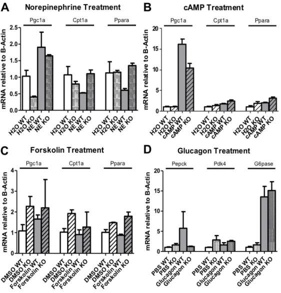
9
norepinephrine and 8-Br-cAMP treatments. Unlike in Experiment 1, norepinephrine treatment increased Pgc1a expression in both ACSL4 L -/- and control hepatocytes and increased Cpt1a
expression in only ACSL4 L -/- hepatocytes. Cpt1a and Ppara expression decreased among control
hepatocytes following norepinephrine treatment (Figure 4A). Treatment with 8-Br-cAMP only affected Pgc1a expression, with increased expression in both control and ACSL4 L -/- hepatocytes
(Figure 4B). Following forskolin treatment, only Pgc1a expression increased in control hepatocytes while only Cpt1a expression decreased in ACSL4 L -/- hepatocytes (Figure 4C).
Following glucagon treatment, both Pepck and G6pase expression increased in control
10
Chapter IV. Discussion
4.1 Adjustments to Methods and Materials
Experiment 1 produced gene expression data that were inconsistent with the expected treatment effects. The data also exhibited high variability between technical replicates within treatment groups. This was likely due to the poor quality of the extracted RNA and reagents used for treatments. Further analysis of A260/A230 ratios revealed poor RNA purity of Experiment 1 extracted RNA. Prior to performing Experiment 2 with new cells, several adjustments were made to improve experimental conditions. In order to improve the quality and purity of the extracted RNA, the extraction was performed with smaller batches of cells at a time to prevent
thawing/degradation that would disturb expression patterns. RNA was also eluted using spin columns to improve purity and yield for extraction. RNAase free micro-volume tubes were also used in place of standard micro-volume tubes during cDNA synthesis. New reagents were purchased for norepinephrine, 8-Br-cAMP, and glucagon treatments prior to the second experiment to replace expired reagents used for the first experiment.
4.2 Hormone treatment effects on gene expression
11
While all of these hormones have different mechanisms of action, they were expected to elicit similar increases in the expression of fatty acid oxidation genes in control hepatocytes. Not only did all of the treatments have inconsistent effects on increasing fatty acid oxidation gene expression, norepinephrine treatment actually caused a decrease in Cpt1a and Ppara expression in control hepatocytes. The Acsl4 L -/- hepatocytes did exhibit different expression patterns
compared to control, however results were inconclusive due to the inconsistent effects observed in the control samples. Overall, the hormone treatments did not produce the anticipated effects and did not validate the findings of our previous study. Because of this, we were unable to form conclusions about whether the liver specific ACSL4 knockout decreased beta adrenergic
signaling or other exogenous mechanisms of FA oxidation gene regulation.
4.3 Fatty acid treatment effects on gene expression
This study also explored the role of fatty acids as intracellular signaling molecules to modulate fatty acid oxidation. Oleate increases intracellular levels of cAMP and expression of fatty acid oxidation genes in C2C12 myotube cells.14 While the mechanism behind how oleate elicits this cellular response is unknown, this finding illustrates the potential for therapeutic manipulation of intracellular lipid signaling to alter fatty acid oxidation through transcriptional regulation. Similar to the results from hormone treatment experiments, the gene expression changes following the lipid treatments in our study were not consistent with the findings of the published study. Oleate treatments were expected to increase expression of fatty acid oxidation genes while palmitate was not expected to affect expression. In control hepatocytes palmitate treatment increased gene expression of all genes of interest except Pgc-1a while oleate treatment only increased Creb expression. Similar to what was observed in the hormone treatment
12
4.4 Future Directions
In an attempt to validate the findings of the previous study, most of the results from the hormone treatment experiment were inconclusive. Several steps can be taken to optimize treatment conditions to improve consistency of gene expression changes. Due to different
13
Figure 1. Lipid Treatments Gene Expression (Experiment 1)
Relative gene expression (Actin) of Cpt1a, Creb, Acox1, Pgc1a, and Ppara in ACSL4 L -/- and WT hepatocytes following 3.5-hour oleate or palmitate treatment.
Figure 2. Lipid Treatments Gene Expression (Experiment 2)
Relative gene expression (Actin) of Cpt1a, Creb, Acox1, Pgc1a, and Ppara in ACSL4 L -/- and
WT hepatocytes following 3.5-hour oleate or palmitate treatment.
Oleate and Palmitate Treatment
BSA WT BSA KO
OA WTOA K
O
BSA WT BSA KO
PA WTPA KOBSABSA WT KOOA WTOA K
O
BSA WT BSA KO
PA WTPA KOBSABSA WT KOOA WTOA K
O
BSA WT BSA KO
PA WTPA KOBSABSA WT KOOA WTOA K
O
BSA WT BSA KO
PA WTPA KOBSABSA WT KOOA WTOA K
O
BSA WT BSA KO
PA WTPA KO
0 2 4 6 8 Cpt1a Oleate
Creb Acox1 Pgc1a Ppara
Palmitate Oleate Palmitate Oleate Palmitate Oleate Palmitate Oleate Palmitate
Re la ti ve m R NA ( B-a ct in )
Oleate and Palmitate Treatment
BSA WT BSA KO OA WTOA KOBSA W
T BSA KO PA W
T PA K
O BSA
WT BSA KO OA WTOA KOBSA W
T BSA KO
PA W T PA K
O BSA
WT BSA KO OA WTOA KOBSA W
T BSA KO
PA W T PA K
O BSA
WT BSA KO OA WTOA KOBSA W
T BSA KO PA W
T PA K
O BSA
WT BSA KO OA WTOA KOBSA W
T BSA KO PA W
T PA K
O 0.0 0.5 1.0 1.5 2.0 2.5 Cpt1a Oleate Relative mRNA (B-actin)
Creb Acox1 Pgc1a Ppara
14
Figure 3. Hormone Treatment Gene Expression (Experiment 1)
Relative gene expression (Actin) of Pgc1a, Cpt1a, and Ppara following 1-hour (A)
15
Figure 4. Hormone Treatment Gene Expression (Experiment 2)
Relative gene expression (Actin) of Pgc1a, Cpt1a, and Ppara following 1-hour (A)
16
17
Treatment Vehicle Concentration Time (Hr)
Norepinephrine H2O 10µM 1
8-Br-cAMP H2O 1mM 1
Forskolin DMSO 40µM 1
Glucagon PBS 100mM 1
Oleate BSA 500µM 3.5
Palmitate BSA 500µM 3.5
18
Target Direction Sequence
Acsl4 Forward ACAGCCTTGGAACCCGGGAGA
Reverse CCCAATGCAGTGAGGCCACTTCC
β-Actin Forward GGCTCCTAGCACCATGAAGA
Reverse GAAAGGGTGTAAAACGCAGC
Pgc-1α Forward CAACATGCTCAAGCCAAACCAACA
Reverse CGCTCAATAGTCTTGTTCTCAAATGGG
Pparα Forward CCTCAGGGTACCACTACGGAGTT
Reverse TCGCCGAAAGAAGCCCTTA
Cpt1a Forward CAACTATTATGCCATGGATTTTGTGCTT
Reverse CGATACATGATCATGGCGTGAACG
Creb Forward TCAGCCGGGTACTACCATTC
Reverse TTCAGCAGGCTGTGTAGGAA
Acox1 Forward GGTGGACCTCTGTCTTGTTCA
Reverse AAACCTTCAGGCCCAAGTGAG
Pepck Forward TCAACACCGACCTCCCTTAC
Reverse CATTGTGCCGCTATCTCAAA
G6pase Forward TCTGTCCCGGATCTACCTTG
Reverse TGGCAAAGGGTGTAGTGTCA
Pdk4 Forward CCGCTGTCCATGAAGCA
Reverse GCAGAAAAGCAAAGGACGTT
19
References
1. Nguyen, P. et al. Liver lipid metabolism. J. Anim. Physiol. Anim. Nutr. (Berl). 92, 272– 283 (2008).
2. Rakhshandehroo, M., Knoch, B., Müller, M. & Kersten, S. Peroxisome proliferator-activated receptor alpha target genes. PPAR Res. 2010, (2010).
3. Küch, E. M. et al. Differentially localized acyl-CoA synthetase 4 isoenzymes mediate the metabolic channeling of fatty acids towards phosphatidylinositol. Biochim. Biophys. Acta
- Mol. Cell Biol. Lipids 1841, 227–239 (2014).
4. Piccini, M. et al. FACL4, a New Gene Encoding Long-Chain Acyl-CoA Synthetase 4, Is Deleted in a Family with Alport Syndrome, Elliptocytosis, and Mental Retardation.
Genomics 47, 350–358 (1998).
5. Piccini, M. et al. FACL4, a New Gene Encoding Long-Chain Acyl-CoA Synthetase 4, Is Deleted in a Family with Alport Syndrome, Elliptocytosis, and Mental Retardation.
Genomics 47, 350–358 (1998).
6. Kang, M. J. et al. A novel arachidonate-preferring acyl-CoA synthetase is present in steroidogenic cells of the rat adrenal, ovary, and testis. Proc. Natl. Acad. Sci. U. S. A. 94,
2880–2884 (1997).
7. Cao, Y., Murphy, K. J., McIntyre, T. M., Zimmerman, G. A. & Prescott, S. M. Expression of fatty acid-CoA ligase 4 during development and in brain. FEBS Lett. 467, 263–7
(2000).
8. Kudo, T., Tamagawa, T., Kawashima, M., Mito, N. & Shibata, S. Attenuating Effect of Clock Mutation on Triglyceride Contents in the ICR Mouse Liver under a High-Fat Diet.
J. Biol. Rhythms 22, 312–323 (2007).
9. Kotronen, A. et al. Genetic variation in the ADIPOR2 gene is associated with liver fat content and its surrogate markers in three independent cohorts. Eur. J. Endocrinol. 160,
593–602 (2009).
10. Sung, Y. K. et al. Fatty acid-CoA ligase 4 is overexpressed in human hepatocellular carcinoma. Cancer Sci. 94, 421–4 (2003).
20
Gastroenterol. 11, 2557–63 (2005).
12. Puigserver, P. & Spiegelman, B. M. Peroxisome Proliferator-Activated Receptor-γ
Coactivator 1α (PGC-1α): Transcriptional Coactivator and Metabolic Regulator. Endocr.
Rev. 24, 78–90 (2003).
13. Lim, J.-H. et al. Oleic acid stimulates complete oxidation of fatty acids through protein kinase A-dependent activation of SIRT1-PGC1α complex. J. Biol. Chem. 288, 7117–26 (2013).