Introduction
The mechanisms behind cellular motility are remarkably complex and are further obscured by
the wide range of migratory modes used by cells in different extracellular conditions.
Traditionally migratory phenotypes are classified as either amoeboid or mesenchymal,
descriptors that refer to the cell known to undergo this type of motion. In two dimensions cells
migrating with a blebby amoeboid phenotype are typically rounded with few protrusions, and
rely on strong cytosolic pressures generated by an oscillatory actin myosin contractile wave
causing periodic expansion and retraction driving net movement1,2. By contrast, two
dimensionally mesenchymal migrating cells are typically highly polarized3. Mesenchymal
migration is driven primarily by actin polymerization and generation of focal adhesions at the
leading edge of the cell, and concomitant degradation of focal adhesions at the trailing edge of
the cell3. Adding to the complexity of characterizing these systems it is established that the
behavior and mechanisms of migration in cells migrating on a flat continuous surface are quite
different from cells migrating within a three dimensional mesh of extracellular proteins like
collagen and fibronectin2,4. In three dimensions amoeboid migration is still driven by cytosolic
pressure increases, but instead manifests as expansion through the pores in a fibrous matrix
followed by contraction events driving net movement forward2. Mesenchymal migration in three
dimensions manifests with long psuedopodial protrusions containing focal adhesions used to
anchor the cell to the extracellular matrix5. The cell also contains metalloproteases known to
cleave extracellular fibers allowing the larger body of the cell to get through upon cortical
contraction and degradation of focal adhesions at the trailing edge of the cell6.
In two dimensions focal adhesions have long been known to play a role in mesenchymal
only recently gained mainstream acceptance5. In a recent Cell paper, Liu et al (2015) found that
reduction of cellular adhesion strength was capable of inducing the mesenchymal amoeboid
transition; these investigators demonstrated that knockdown of vinculin or reduction of proteins
the cell can adhere to was sufficient to drive a mesenchymal to amoeboid transition in cells
confined to a region 3 - 5 um in depth within a microfluidic chamber inducing a chemotactic
gradient within the small volume containing the cell7.
In addition to focal adhesion proteins there is strong evidence that well defined microtubule
structure plays a role on the establishment of migratory phenotypes. It is well established that
microtubules play a role in establishment of cell polarity in cells known to undergo mesenchymal
migratory phenotypes8. Furthermore microtubule polymerization has been shown to provide
structure and in some cases drive many of the pseudopod like protrusions seen in mesenchymal
migratory cells9. This dependence on microtubules for polarization and protrusions is not seen in
amoeboid movement as evidenced by a recent Cell paper by Ruprecht et al (2015) indicating that
polarity in some amoeboid phenotypes was based on a stochastic polarization of actin-myosin
cortical contractility, generated and maintained through a feedback loop in which contraction
stimulates recruitment of additional contraction components10.
In this paper we begin the investigation of underlying mechanisms of the mesenchymal
amoeboid transition in two and three dimensions using a comparative approach between the cell
line CHO (a Chinese Hamster Ovary cell line known to be blebby amoeboid migratory on/in
two/three dimensional collagen1) and the mesenchymally migratory cell lines NIH-3T3 (a mouse
embryo fibroblast) and DU-145 (a human prostate cancer cell line). We wish to probe how
microtubule polymerization and generation of focal adhesions influence this mechanism in vitro.
various cell treatments we provide support for the hypothesis that regulation of microtubule
structure and focal adhesion deposition are key aspects of the transition of mesenchymal cell
migration to a blebby amoeboid migratory phenotype.
Materials and Methods:
Cell Culture: CHO (Chinese Hamster Ovary) & NIH-3T3 cells were obtained from the
University of North Carolina Lineberger Comprehensive Cancer Center Tissue Facility. DU-145
cells were generously provided by Chenying Fu PhD (University of Oklahoma Health Services
Center). CHO was cultured in GIBCO DMEM : F12 (1:1) x 1 while DU and NIH was cultured
using GIBCO DMEM x1 without F12 additions. The following additions were made to each
medium 4 mM L-glutamine, 100 U/mL Penicillin/Streptomycin, and 10% (by volume) Gibco
Fetal Bovine Serum.
2D Collagen Dish Creation: Two dimensional collagen dishes were created by incubating serum
coated glass plates in 8uL (3.1 mg / mL) Bovine Collagen + 500uL 10mMol HCl (aq) for one
hour. Plates were then aspirated and washed in 1x DPBS before plating cells.
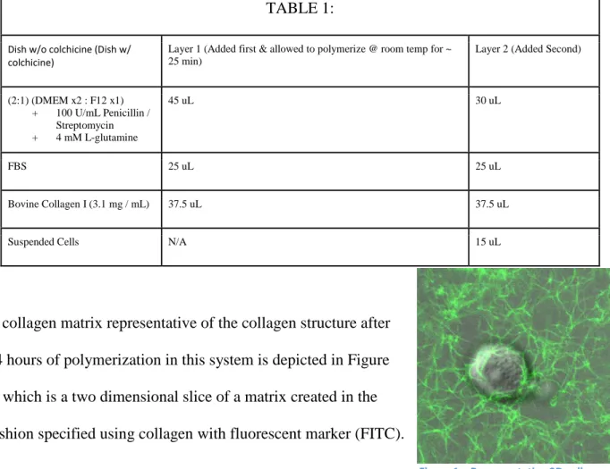
3D Collagen Matrix Creation: Three dimensional collagen matrices were produced in two layers
because when matrices were produced with only one layer cells fell to the bottom of the dish
before collagen fully polymerized. The first layer was allowed to polymerize for 20 – 30 minutes
at room temperature before the addition of the second layer. Plates were created either without
colchicine treatment or with colchicine treatment. The content of each layer is summarized in
Figure 1 – Representative 3D collagen matrix generated using a two layer polymerization technique labeled using FITC collagen
TABLE 1:
Dish w/o colchicine (Dish w/
colchicine) Layer 1 (Added first & allowed to polymerize @ room temp for ~ 25 min)
Layer 2 (Added Second)
(2:1) (DMEM x2 : F12 x1) + 100 U/mL Penicillin /
Streptomycin + 4 mM L-glutamine
45 uL 30 uL
FBS 25 uL 25 uL
Bovine Collagen I (3.1 mg / mL) 37.5 uL 37.5 uL
Suspended Cells N/A 15 uL
A collagen matrix representative of the collagen structure after
24 hours of polymerization in this system is depicted in Figure
1, which is a two dimensional slice of a matrix created in the
fashion specified using collagen with fluorescent marker (FITC).
Live Cell Imaging:
Image Capture: Fluorescent imaging was performed on
an Olympus Fluoview 1200 model laser scanning confocal microscope using an Olympus
40x silicon immersion objective lens.
Long term DIC imaging was performed using an Olympus VivaView Incubator
microscope provided through the UNC Olympus Imaging Center.
Visualization: Images were visualized using ImageJ Version 1.47n.
Image Processing: Cell migration speeds and persistence were determined using
https://github.com/NatPRoach/BIOLOGY692H) and publically available digitizing
scripts made available via the lab of Tyson Hedrick PhD in the UNC Biology Department
(available for download here: http://www.unc.edu/~thedrick/software1.html). Speeds
were determined by dividing total distance traveled in microns by the time taken in
minutes.
Three dimensional assays were also performed on CHO wild type and NIH wild type cells. The
matrices were created as specified in Table 1 above.
Cell Fixation:
Cells were fixed in 4% paraformaldehyde diluted in DPBS heated to 37° C in order to prevent
short term temperature effects on cell morphology as fixation occurs. Cells were then incubated
in blocking buffer solution (1x DPBS, 5% Goat Serum, .3% 100x Triton) for one hour, resulting
in permeabilized cells. Plates were then incubated for one hour with primary antibodies
suspended in 150uL Antibody Dilution Buffer (1x DPBS, 1% Bovine Serum Albumin (BSA),
.3% 100x Triton). Plates were again incubated for one hour, this time with secondary antibodies
suspended in 500 uL DPBS. Cells were washed three times in Dulbecco's Phosphate Buffer
Solution (DPBS) between each stage of fixation, and allowed a five minute period between each
DPBS aspiration. When not being imaged fixed dishes were sealed with Parafilm M® and stored
at 4o C with limited light exposure.
Results:
2D Migratory Assays Reveal Phenotypic Dependence On Microtubule Polymerization:
Two dimensional migratory assays were performed on NIH-3T3 cells on both serum-coated
presence and absence of microtubule depolymerizing agent colchicine and determine the effects
of microtubule perturbation on migratory phenotypes. When plating NIH-3T3 cells on a dish or
coverslip coated with a thin layer of collagen speed increased significantly relative to cells plated
on serum coated glass (p < 0.001). The phenotype of movement on collagen visually resembled
that of movement on serum coated glass.
In wild type conditions on serum-coated glass NIH-3T3 spread quickly rounding only to divide
after this point. Protrusions appear and disappear in nearly all cells, and movement occurs quite
slowly (0.71 + 0.20 um /min), and when it does the phenotype is clearly mesenchymal Figure 3.
The addition of microtubule depolymerizing agent colchicine on two dimensional serum coated
glass bottomed plates did not statistically significantly alter NIH -3T3 cell’s migration speed
(1.17 + 0.66 um /min), however the phenotype of migration was visually affected, as evidenced
by Figure 3, cells now exist primarily rounded, and protrude, seemingly independent of
lamellipodial growth. Colchicine treated cells also undergo a regular oscillatory pattern, not
visible in still frames, but evident in video (Supplemental Figures 2 & 4).
Plating NIH-3T3 cells on collagen also affects phenotype, increasing the speed of cells not
treated with colchicine to 1.74 + .19 um / min, and colchicine treated cells to 2.55 + .66 um /
min. Untreated cell phenotype was visually distinct from the serum coated glass counterpart as
well, with cells forming distinctive spines where adhesion points were being strained. Colchicine
treated cells however had a migratory phenotype similar in appearance to their counterparts on
serum coated glass, albeit with a statistically significant increase in speed (p < 0.155). Notably
on 2D collagen the addition of colchicine led to statistically significant increases in speed (p <
0.0067).
Upon plating cell lines in three dimensional collagen the phenotypes transitioned once again.
Wild type cells were observed to be highly polarized and contain long stable protrusions
characteristic of fibroblasts. The migration noted is clearly mesenchymal in nature, as cells
generate long pseudopod-like protrusions and move forward seemingly without any interference
from collagen fibers indicating metalloprotease driven remodeling of the collagen matrix
characteristic of mesenchymally migratory cells. Addition of colchicine to these plates led to a
shift in phenotype, resulting in the complete ablation of long pseudopod-like protrusions
characteristic of the untreated plates. Colchicine treated plates had significantly reduced net
migratory fidelity relative to untreated plates, though some cells retained migratory behavior.
Notably in colchicine treated plates a cell was seen undergoing a clearly blebby amoeboid
migratory process, moving extremely quickly with a large stable bleb localized in the direction
of movement, closely resembling a migratory behavior described by Liu et al and Ruprecht et al
in a pair of recent publications in Cell7,10. (Supplemental Figure 5)
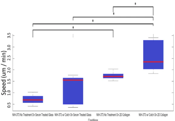
In addition to qualitative assessments speed was determined for different NIH 2D assays; the
results of this analysis are
summarized in Table 2 and the
accompanying figure.
TABLE 2:
Condition: Speed (um / min)
NIH-3T3 on serum treated glass no colchicine treatment n = 8
.71+ .20
NIH-3T3 on serum treated glass w/ colchicine treatment n = 5
1.17 + .66
NIH-3T3 on 2D collagen no colchicine treatment n = 5
1.74 + .19
NIH-3T3 on 2D collagen w/ colchicine treatment n = 6
Figure 2 – Distribution of cell speeds of NIH-3T3 cells under the conditions specified in each column. Statistical significance is indicated by the (*) symbol between columns, indicating a probability < 0.05 of the two sets having the same mean speed.
CHO cells underwent a clear blebby amoeboid form of migration in untreated assays, pushing
protrusions through pores in the collagen matrix, and driving motion forward by cortical
contraction and oscillation (Supplemental Image 6). CHO phenotype was not visually affected
by the addition of colchicine, still containing the characteristic pressure driven protrusion
followed by cortical contraction.
Fixed Cell Imaging Reveals Focal Adhesion Retention Is Strongly Correlated With Phenotype:
Cells were fixed 24 hours after plating. Focal adhesions (identified as areas with high vinculin
(green) concentration and low alpha tubulin concentration (red)) are clearly visible in both CHO
(Figure 4A) and NIH-3T3 (Figure 4B) cell lines when cells are spread on serum coated glass.
Note that cells spread on serum coating glass are attaching not to the glass itself but components
of the glass for which they have a binding integrin, in this case vitronectin. When embedded in a
3D collagen matrix, CHO cells completely lose any discrete vinculin localization (Figure 4c),
while NIH 3T3 cells retain visible focal adhesions (Figure 4d-g). Note that imaging resolution is
significantly reduced when cells are embedded in three dimensional collagen. The non-uniform
and fibrous nature of a three dimensional collagen matrix leads to a large amount of light
scattering, limiting the ability to resolve small scale structure11. Microtubule organization is also
well defined in both cell lines when cells were adhered to a glass bottom dish (Figure 4 a,b).
When placed in collagen CHO cells clearly have a localization of microtubules of some kind, the
fine structure of which is not resolvable (Figure 4c). Resolution of microtubule structure in NIH
was also reduced, however magnified portions of the image appear to contain fiber like peaks of
fluorescence (Figure 4h) which can be seen more clearly in the plot of average intensity values
Imaging Of Colchicine Treated Fibroblast Indicates Vinculin Localization Is Dependent On
Intact Microtubule Structure:
We imaged fixed cells of the fibroblastic cell line DU-145 in the presence and absence of
colchicine. Cells were stained with phalloidin (red) a fluorescent molecule known to localize to
filamentous actin, and vinculin (green). Focal adhesions (again identified as areas with high
vinculin signal and low actin signal) were seen in untreated controls (Figure 5, Row 1), but
localization was reduced by the addition of colchicine as evidenced by the elimination of points
with strong vinculin signal separate from any actin signal (Figure 5, Row 2).
Discussion:
The examination of 2D migratory phenotypes in NIH-3T3 revealed a notable increase in speed
when plating cells on collagen rather than glass. This change in speed is likely due to NIH-3T3
cells possessing a different binding affinity with collagen than the vitronectin and fibronectin
present in serum coated glass. The phenotypes of NIH on glass and on collagen however were
visually quite similar. This is expected given that NIH cells contain binding integrins for
collagen, fibronectin and vitronectin, as the cell should be able to adhere to both serum coated
glass and 2D collagen substrates 12,13. The addition of colchicine was capable of driving a
significant phenotypic shift in NIH-3T3 cells on both serum coated glass and on two dimensional
collagen, indicating that microtubule stability is somehow involved in the establishment of the
two dimensional mesenchymal migratory phenotype. However, addition of colchicine to cells on
two dimensional plates lined with collagen led to faster motion (p < .0289), in line with the
expectations of a transition from mesenchymal motion to amoeboid motion. This increase in
speed is well characterized in the conversion from mesenchymal motion to more amoeboid
motion, and is theorized to be one reason behind metastatic cancer cells undergoing a
mesenchymal to amoeboid transition in vivo4,7. In addition qualitative observations of migratory
phenotypes in the presence and absence of colchicine are quite different, with non-colchicine
treated plates having well defined polarity and stable protrusions, and colchicine treated plates
undergoing a notably oscillatory behavior (Figure 3).
NIH-3T3 cells in three dimensional collagen exhibit similar behaviors, with addition of
colchicine corresponding to a drastic reduction of long stable protrusions, and a visible effect to
migratory phenotype. NIH wild type phenotypes resembled physiological fibroblast movement,
with long stable polarization and protrusions preferentially occurring at the leading edge of the
cell. NIH cells treated with colchicine in a three dimensional collagen matrix had phenotypes
distinctly different, with complete ablation of long stable protrusions, yielding comparatively
rounded cells indicating that the principle of colchicine disrupting mesenchymal migratory
relationship. Given the role of microtubules in generating stable protrusions9 and development of
polarity8 their importance to the mesenchymal phenotype was expected. Whether colchicine
driven depolymerization of microtubules drives a full or partial mesenchymal to amoeboid
transition in NIH-3T3 cells remains to be seen, as it is uncertain if colchicine treated cells are
still migrating by a mesenchymal mechanism and only appear different due to depolymerizing
effects to cell morphology.
The indication that microtubule depolymerization was capable of driving phenotypic shift in 3D
mesenchymally migratory cells led us to question if intact microtubule structure was also
necessary for phenotype maintenance in blebby-amoeboid migration. We probed this question
using 3D collagen migratory assays of CHO in the presence and absence of colchicine and saw
no obvious alteration of migration phenotype in this blebby amoeboid model. An interesting
follow-up experiment would be treating CHO and NIH cells with Taxol, a microtubule stabilizer,
and examining the effect on its phenotype, as stable microtubules may actively inhibit blebby
amoeboid movement by reducing cell flexibility, a critical component of blebby amoeboid
migration.
The evidence that microtubule stability played a role in regulating or establishing migratory
phenotype in mesenchymal 3D motion led us to examine wild type microtubule structure of
CHO and NIH-3T3 cells plated on serum coated glass, and embedded in a 3D collagen matrix
using fluorescently labeled antibodies specific to the alpha-tubulin subunits of microtubules. The
presence of resolvable microtubule structure in untreated NIH cells embedded in 3D collagen is
expected, while CHO cells embedded in 3D collagen show some localization of microtubules
within the cell there is no identification of defined microtubule structure, which may explain the
microtubule structure in 3D collagen makes drawing conclusions from these data difficult,
however both the localization of microtubules in CHO and the polymerized structure of
microtubules in NIH-3T3 are interesting phenomena that require further study.
In addition to probing microtubule structure we examined the presence of focal adhesions in
NIH-3T3 and CHO cells, as focal adhesions are a structure known to be essential for
mesenchymal movement in both two and three dimensions.As predicted NIH-3T3 cells had
clearly defined focal adhesions when plated on glass (Figure 4B), and retained adhesions in 3D
collagen as well (Figure 4D). In contrast CHO cells have a network of focal adhesions when
spread on glass (Figure 4A), but completely lack any stable polarity or focal adhesion when
embedded in a three dimensional collagen matrix (Figure 4C).
The existence of defined microtubule structure and focal adhesion generation in mesenchymal
cells, and undefined microtubule structure or focal adhesion generation in amoeboid cell lines led
us to consider the possibility that these expression patterns were linked. We chose to examine the
dependence of focal adhesion generation on defined microtubule structure in the fibroblastic cell
line DU-145 (a human prostate cancer cell line). We found that colchicine addition led to
reduction of vinculin localization (indicative of focal adhesion presence) in these cells.
The dependence of vinculin localization on stable microtubules in DU-145 fixed
immunofluorescence imaging indicates that the mechanism by which microtubule
depolymerization affects phenotype may be linked to focal adhesion generation. It is possible
that the polarization established by microtubules plays a role in establishing the preferential
generation of focal adhesions at the leading edge of cells exhibiting a mesenchymal phenotype.
Comparison of focal adhesion generation of cells in the presence and absence of polymerized
Ultimately, the work summarized in this paper represents a small portion of the work that will
need to be done in order to substantiate the presented hypotheses in a formal publication. Before
publication transfection of an amoeboid migratory cell line with collagen binding integrin will
need to be performed, alongside a control mock transfection. If we see addition of a collagen
binding integrin is capable of converting an amoeboid migratory phenotype to a mesenchymal
phenotype as we would expect, this would provide further evidence for integrin as a keystone
protein regulating the transition. If however the addition of collagen integrin does not result in a
phenotype transition this would lend credence to the idea that well-structured microtubule
polarity is required in order to generate a mesenchymal phenotype.
Acknowledgements:
I’d like to thank Dr. Kenneth Jacobson, Dr. Maryna Kapustina, Dr. William Kier, and Dr. John
Bruno for their advice and guidance in this project.
I’d like to thank the UNC Olympus Imaging Center for providing access to confocal and
incubator microscopes.
Post Script:
Supplementary materials can be found at
https://drive.google.com/file/d/0B1E3oMs5NrVRajVBbUZTMUdkaFU/view?usp=sharing
References:
1. Kapustina M, Elston TC, Jacobson K. Compression and dilation of the membrane-cortex layer generates rapid changes in cell shape. J Cell Biol. 2013;200(1):95–108.
doi:10.1083/jcb.201204157.
3. Friedl P, Wolf K. Proteolytic interstitial cell migration: a five-step process. Cancer Metastasis Rev. 2009;28(1-2):129–35. doi:10.1007/s10555-008-9174-3.
4. Friedl P, Wolf K. Plasticity of cell migration: a multiscale tuning model. J Cell Biol. 2010;188(1):11– 9. doi:10.1083/jcb.200909003.
5. Cukierman E, Pankov R, Stevens DR, Yamada KM. Taking cell-matrix adhesions to the third dimension. Science. 2001;294(5547):1708–12. doi:10.1126/science.1064829.
6. Sabeh F, Shimizu-Hirota R, Weiss SJ. Protease-dependent versus -independent cancer cell
invasion programs: three-dimensional amoeboid movement revisited. J Cell Biol. 2009;185(1):11– 9. doi:10.1083/jcb.200807195.
7. Liu Y-J, Le Berre M, Lautenschlaeger F, et al. Confinement and Low Adhesion Induce Fast Amoeboid Migration of Slow Mesenchymal Cells. Cell. 2015;160(4):659–672.
doi:10.1016/j.cell.2015.01.007.
8. Magdalena J, Millard TH, Machesky LM. Microtubule involvement in NIH 3T3 Golgi and MTOC polarity establishment. J Cell Sci. 2003;116(Pt 4):743–56. Available at:
http://www.ncbi.nlm.nih.gov/pubmed/12538774. Accessed March 21, 2015.
9. Scaife RM, Job D, Langdon WY. Rapid microtubule-dependent induction of neurite-like extensions in NIH 3T3 fibroblasts by inhibition of ROCK and Cbl. Mol Biol Cell. 2003;14(11):4605–17.
doi:10.1091/mbc.E02-11-0739.
10. Ruprecht V, Wieser S, Callan-Jones A, et al. Cortical Contractility Triggers a Stochastic Switch to Fast Amoeboid Cell Motility. Cell. 2015;160(4):673–685. doi:10.1016/j.cell.2015.01.008. 11. Arifler D, Pavlova I, Gillenwater A, Richards-Kortum R. Light Scattering from Collagen Fiber
Networks: Micro-Optical Properties of Normal and Neoplastic Stroma. Biophys J. 2007;92(9):3260–3274. doi:10.1529/biophysj.106.089839.
12. Kern A, Marcantonio EE. Role of the I-domain in collagen binding specificity and activation of the integrins alpha1beta1 and alpha2beta1. J Cell Physiol. 1998;176(3):634–41.
doi:10.1002/(SICI)1097-4652(199809)176:3<634::AID-JCP20>3.0.CO;2-Y.
13. Regis S, Youssefian S, Jassal M, Phaneuf M, Rahbar N, Bhowmick S. Integrin α5β1-mediated attachment of NIH/3T3 fibroblasts to fibronectin adsorbed onto electrospun polymer scaffolds.