Published online 2016 February 3. Research Article
An Efficient DNA Extraction Method for
Lactobacillus casei
, a
Difficult-to-Lyse Bacterium
Mojtaba Alimolaei
1,2and Mehdi Golchin
1,*1Department of Pathobiology, Faculty of Veterinary Medicine, Shahid Bahonar University of Kerman, Kerman, IR Iran
2Department of Anaerobic Bacterial Vaccine Research and Production, Razi Vaccine and Serum Research Institute, Kerman Branch, Kerman, IR Iran
*Corresponding author: Mehdi Golchin, Department of Pathobiology, Faculty of Veterinary Medicine, Shahid Bahonar University of Kerman, Kerman, IR Iran. Tel/Fax: +98-343257447, E-mail: [email protected]
Received 2015 August 16; Revised 2015 September 6; Accepted 2015 September 22.
Abstract
Background: There are several protocols to extract DNA from Lactobacillus spp. In the case of L. casei it is harder because of its especial and thick cell wall.
Objectives: In this study, nine DNA extraction protocols (by lysozyme treatment) were evaluated and compared in two categories (traditional and kit-based protocols) and an improved method was presented.
Materials and Methods: DNA quantity and quality was determined by spectrophotometry, agarose gel electrophoresis and polymerase chain reaction (PCR).
Results: The results revealed that the yield of extracted DNA differed by each protocol (5.8 - 17.1 μg/100 μL), but provided appropriate DNA for PCR amplification. The modified protocol offered the best total DNA extraction method when both quality (DNA purity; 1.54 μg) and quantity (DNA yield; 17.1 μg) were considered.
Conclusions: We suggest this protocol for effective and inexpensive DNA isolation from L. casei for downstream biological processes such as PCR.
Keywords: DNA, Extraction, Modified Protocols, Lactobacillus casei
Copyright © 2016, Alborz University of Medical Sciences. This is an open-access article distributed under the terms of the Creative Commons Attribution-NonCom-mercial 4.0 International License (http://creativecommons.org/licenses/by-nc/4.0/) which permits copy and redistribute the material just in noncomAttribution-NonCom-mercial us-ages, provided the original work is properly cited.
1. Background
Cell wall of Gram-positive bacteria has several distinc-tive structures and protects the protoplast from me-chanical damage and osmotic rupture or lysis. The mu-rein layer (a thick peptidoglycan layer) is the ubiquitous component of the Gram-positive cell wall which provides shape, stability and viability. This layer contains almost equal amounts of polysaccharides and peptides (1, 2) and is composed of a polymer of disaccharide (glycan) chains of repeating N-acetylglucosamine and N-acetylmuramic acid residues (linked β1 → 4) and is cross-linked by short chains of amino acids (peptide) (1).
Undermining progress in Lactobacillus genetics has been the difficulty in achieving cell lysis with lysozyme (3, 4) and developing reliable procedures for DNA isola-tion (5). L. casei is a rod-shaped, Gram positive and highly lysis-resistant bacterium (6, 7). In this species, because of its special cell wall structure, genetic studies have several difficulties (8). Polysaccharide and peptidoglycan moi-eties are the major surface components of this bacterium (5, 7). The primary structure of its peptidoglycan has a common monomer GlcNAc–MurNAc–L-Ala–g-DGlu–L-Lys– D-Ala with an asparagine attached to the ω-amino group
of lysine (9). The peptide side chains are then cross-linked by a transpeptidase (1).
Abed evaluated five methods for the extraction and pu-rification of DNA from cultured Lactobacillus colonies isolated from dairy products. The results obtained in that study confirmed that wizard genomic DNA purification kit with modifications was superior to other methods because it produced a higher DNA yield with the high-est purity (10). Scornec et al. set up a rapid 96-well plate DNA extraction protocol for L. casei. They optimized the DNA extraction procedure based on silica membranes in 96-column format to obtain genomic DNA from a large number of mutants (8). De et al. presented a simple, inex-pensive and effective genomic DNA isolation procedure for Lactobacillus isolates. They verified the quality of the isolated genomic DNA by restriction digestion and poly-merase chain reaction (PCR) (11).
In recent years, various molecular techniques have been used for the lysis of L. casei by chemical and mechanical protocols (12). In chemical methods, the peptidoglycan can be lysed by cell wall hydrolase enzymes (e.g. lyso-zyme or mutanolysin). Mutanolysin is costly, not
gener-ally available, and thus unsuitable for routine use in labo-ratories. High resistance to lysozyme is also observed in several lactobacilli species (1).
2. Objectives
In the current study, we evaluated several different DNA extraction protocols using lysozyme treatment and com-pared them with a new modified method.
3. Materials and Methods
3.1. Bacterial Strains and Growth Conditions
For the analysis of both plasmid and genomic DNA, a re-combinant L. casei was constructed and used in this study. Briefly, beta toxin gene of Clostridium perfringens was syn-thesized by Generay biotechnology company (China) and cloned in NaeI and BamHI restriction sites of pT1NX vec-tor obtained from BCCM/LMBP plasmid collection of uni-versity of Ghent, Belgium (http://bccm.belspo.be/about/ lmbp.php). The modified vector was transformed to L. casei ATCC: 393 by a Gene PulserTM apparatus (Bio-Rad laboratories, Richmond, CA). This strain was grown stati-cally at 37°C for 24 hours in Lactobacillus de Man, Rogosa and Sharpe (MRS) broth (Himedia, India) supplemented with erythromycin (7.5 μg/mL), anaerobically. Wild-type L. casei was grown in MRS broth without erythromycin, too.
3.2. DNA Extraction
All the DNA manipulations were performed according to standard procedures (13). In this study, eight DNA ex-traction protocols in two categories (traditional and kit-based protocols) and an improved method were tested. After 24 hours of incubation at 37°C, cultures of L. casei (3 mL) in the exponential phase of growth (approximately 1.6 unit of OD600 nm) were centrifuged for three min-utes at 12000 rpm. These bacterial pellets were used for total DNA (genomic and plasmid) extraction.
3.3. Traditional Protocols
In this category, four protocols were used as follows.
3.3.1. P1 Protocol
This method, popularly known as boiling, was based on Abdulla (14) with no relevant modification. One milliliter of dH2O was added to the pellet. After vortexing, the sam-ple was boiled at 100°C for 15 minutes by placing in water bath. The suspension was cooled immediately to −20°C for 20 minutes and centrifuged at 13000 rpm for five minutes and the supernatant was kept frozen until used (14).
3.3.2. P2 Protocol
In this protocol, the cell pellet was suspended in 750 μL of 50 mM EDTA. Then, a volume of 100 μL of lysozyme solu-tion (50 mg/mL) was added to the cell suspension and
in-cubated overnight at 37°C. Subsequently 50 μL of protein-ase K (20 mg/mL) was added and the tube was incubated for 30 minutes at 55°C. The suspension was centrifuged at 12000 rpm at room temperature for five minutes and the pellet was gently resuspended in 950 μL of lysis solution (10 mM Tris-HCl, PH 8.0, 1 mM EDTA, 0.1% (w/v) sodium do-decyl (SDS)). Then, 15 μL of RNase A (20 mg/mL) was added to the lysate and incubated for 45 minutes at 37°C with gentle inversion. For protein precipitation, 300 μL of pro-tein precipitation solution (6 mL of 5 M potassium acetate, 1.15 mL of glacial acetic acid and 2.85 mL of distilled water) was added to the lysate mixture and vortexed at medium speed for 20 seconds. The lysate was centrifuged at 12000 rpm for 20 minutes and the supernatant was transferred to a clean 1.5 mL microtube. One additional centrifugation step at 12000 rpm for 10 minutes was performed to remove any residual protein. To precipitate DNA, 600 μL of cold isopropanol was added and the sample was centrifuged at 12000 rpm for 20 minutes; then, the pellet was washed with 70% ethanol before air drying for 15 minutes. Finally, the pellet was resuspended in 100 μL of Tris-EDTA buffer (10 mM Tris Hcl, 1 mM EDTA, pH = 8.0) and kept at 65°C for 15 minutes and stored at −20°C till further analysis (14).
3.3.3. P3 Protocol
In the third protocol, the pellet was washed thrice with 2 mL of NaCl-EDTA (30 mM NaCl, 2 mM EDTA, pH = 8.0) and resuspended in 100 μL of this buffer and then 100 μL of freshly prepared lysozyme solution (10 mg/mL in NaCl-EDTA) was added and mixed. This mixture was incubated at 37°C for one hour with periodic shaking. To remove RNA, 1 μL of RNase A solution (20 mg/mL) was also added to the mixture before incubation. The volume of the mix-ture was then made up to 500 μL with additional NaCl-EDTA, 50 μL of a 10% SDS solution and 10 μL of proteinase K solution (20 mg/mL). The contents were mixed thor-oughly and incubated at 55°C for one hour. After incuba-tion, 200 μL protein precipitation solution (same as the P2 protocol) was added and vortexed at medium speed for 20 seconds and kept on ice for five minutes. The lysate was centrifuged at 12000 rpm for three minutes and the supernatant was transferred to a clean 1.5 mL tube. DNA in the supernatant was precipitated with 600 μL of cold isopropanol and pelleted by centrifugation at 12000 rpm at room temperature for three minutes. The supernatant was discarded and the DNA pellet was washed once with freshly prepared 70% ethanol and air-dried. The final pel-let obtained was dissolved in 100 μL TE buffer (10 mM Tris HCl, 1 mM EDTA, pH = 8.0) and kept at 65°C for 15 minutes and stored frozen at −20°C till further analysis (11).
3.3.4. P4 Protocol
In this protocol, firstly, the bacterial pellet was resus-pended in 480 μL of 50 mM EDTA and gently vortexed. Then, a volume of 120 μL lysozyme (20 mg/mL) was added to the cell suspension and incubated at 37°C for two hours
with periodic mixing. The suspension was centrifuged at 12000 rpm for three minutes in room temperature and the supernatant was removed. The pellet was gen-tly resuspended in 600 μL genomic lysis buffer (10 mM Tris-HCl, pH = 8.0, 1 mM EDTA, 0.1% (w/v) SDS) containing 6 μL proteinase K solution (20 mg/mL). The sample was incubated at 60°C for one hour and after the incubation, 200 μL protein precipitation solution (same as P2 proto-col) was added and kept on ice for five minutes. After this step, the protocol continued as same as P3 protocol.
3.4. Kit-Based Protocols
In this category, five kit-based protocols were performed as described below.
3.4.1. P5 Protocol
This protocol was performed exactly based on DNA ex-traction kit (DN8115C, SinaClon, Iran) according to the manufacturer’s instruction.
3.4.2. P6 and P7 Protocols
In these protocols, the pellets were resuspended in 180 μL of lysis buffer composed of TE (Tris-HCl 20 mM, EDTA 2 mM, pH = 8.0), lysozyme (20 mg/mL) and triton X-100 (1% v/v). After this step, in P6 protocol we used kit and performed the protocol according to the manufacturer’s instruction, but in P7 protocol we incubated the mixture for two hours at 37°C and after the incubation, we fol-lowed the kit protocol from the lysis step.
3.4.3. P8 Protocol
In this procedure, the pellet was washed once with 500 μL NaCl-EDTA (30 mM NaCl, 2 mM EDTA, pH = 8.0) and re-suspended in 100 μL NaCl-EDTA buffer and 100 μL of freshly prepared lysozyme solution (25 mg/mL). To remove RNA, 1 μL of RNase A solution (20 mg/mL) was also added and the mixture was incubated at 37°C for two hours with periodic shaking. The lysate was centrifuged at 13000 rpm for two minutes and the supernatant was poured off. After this step, we used kit and performed the protocol from the lysis step according to the manufacturer’s protocol.
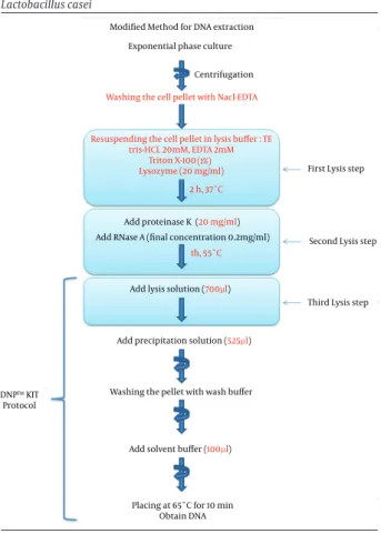
3.4.4. P9 Protocol (Modified Method)
In this new modified protocol, several modifications were made. Three lysis steps were used in this protocol (Figure 1). The pellet was washed once with 500 μL NaCl-EDTA (30 mM NaCl, 2 mM NaCl-EDTA, pH = 8.0) and resuspend-ed in 180 μL of lysis buffer composresuspend-ed of TE buffer (Tris-HCl 20 mM, EDTA 2 mM, pH = 8.0), lysozyme (20 mg/mL) and triton X-100 (1% v/v). The mixture was incubated for two hours at 37°C. Then, proteinase K (20 mg/mL) and RNase A (final concentration: 0.2 mg/mL) were added to the mix-ture and incubated for one hour at 55°C. After the incuba-tion, we continued according to the kit protocol, except for the buffer volumes which are indicated in Figure 1.
Figure 1. Schematic Representation of Modified DNA Extraction from
Lactobacillus casei
Modified Method for DNA extraction Exponential phase culture
Centrifugation
Washing the cell pellet with Nacl-EDTA
Resuspending the cell pellet in lysis buffer : TE tris-HCL 20mM, EDTA 2mM
Triton X-100 Lysozyme (20 mg/ml)
(1%) 2 h, 37˚C
First Lysis step
Add proteinase K
Add RNase A (final concentration 0.2mg/ml) (20 mg/ml)
1h, 55˚C
Second Lysis step
Third Lysis step Add lysis solution (700µl)
Add precipitation solution (525µl)
Washing the pellet with wash buffer
Add solvent buffer (100µl) DNP™ KIT
Protocol
Placing at 65˚C for 10 min Obtain DNA
The main protocol modifications are in red letters.
3.5. Evaluation of Quantity and Quality of Extracted
DNA
The presence and integrity of the extracted DNA from dif-ferent protocols were evaluated by agarose gel (0.7%) elec-trophoresis using horizontal elecelec-trophoresis system (Bio-Rad). The type of band pattern indicated the quality of DNA. The extracted DNA from each protocol was also quanti-fied by spectrophotometry using a BioPhotometer Plus (Eppendorf, Germany) at 260 nm and 280 nm. The qual-ity of DNA was determined by A260/A280 ratio value. The system software provides the DNA concentration (ng/ μL) and automatically calculates the absorption ratio of 260/280 (A260/A280). The total DNA yield (in 100 μL of samples) was calculated as described by Sambrook (13):
DNA yield (μg) = DNA concentration (ng/μL) × total sam-ple volume (μL)
3.6. Polymerase Chain Reaction Using Extracted
DNA
To check the efficiency and applicability of the extracted genomic and plasmid DNA, each DNA was tested by PCR. The extracted total DNA samples were used as template for selective amplification of DNA from the 16S rRNA gene of L. casei and cloned beta toxin gene (cpb) of C. perfringens. The primers used for different PCRs are listed in Table 1.
PCR reaction was performed using 5 μL of the extract-ed DNA with 25 μL of ready-to-use PCR master mix 2x (PR901638, SinaClon, Iran), 2.5 μL (20 pmol/μL) of each primer and dH2O till 50 μL volume was reached. Ampli-fication of DNA from the 16S rRNA gene of L. casei was performed as described previously (15). Amplicons of cpb were obtained with 35 cycles following an initial denatur-ation step at 95°C for 10 minutes. Each cycle involved de-naturation at 94°C for one minute, annealing at 52°C for one minute, synthesis at 72°C for one minute, and a final extension step at 72°C for 10 minutes. The PCR products were then examined for clarity and intensity. The ampli-fied products were electrophoresed in 1.7% agarose gel and observed with gel documentation system.
4. Results
4.1. Quantity and Quality of Extracted DNA
This study evaluated different DNA extraction methods of L. casei. In the nine protocols described in this work, total DNA was isolated from L. casei by the lysozyme treat-ment method and was estimated
spectrophotometrical-ly. The results revealed that DNA extraction with modified protocol produced acceptable DNA purity (1.54) and high-est DNA yield (17.1 μg) when compared with other proto-cols (Table 2). The DNA yield varied significantly depend-ing on the category of DNA extraction used (traditional and kit-based protocols). The DNA yields were lower with traditional protocols when compared to the kit-based protocols. In almost all DNA extraction protocols it was possible to visualize the DNA. Agarose gel electrophoresis showed better results for kit-based protocols (Figure 2).
4.2. Polymerase Chain Reaction Amplification of
DNA
All nine protocols provided effective DNA for PCR ampli-fication with the pairs of primers used. In all the samples, a single band of 196 bp of target cloned beta toxin gene was amplified and visualized on agarose gel (Figure 3). In addition, in all the protocols, the 16S rRNA gene was am-plified (Figure 4). These results indicated that there was no difference for PCR amplification of the target genes between different protocols.
Table 1. Primers to Detect 16S rRNA and Clone Beta Toxin Genes
Gene Primer Primer Sequence (5' – 3') Amplicon Size, bp Reference
16SrRNA 290 (15)
Casei-F CCCACTGCTGCCTCCCGTAGGAGT Casei-R TGCACTGAGATTCGACTTAA
cpb 196 (16)
CPB-F GCGAATATGCTGAATCATCTA CPB-R GCAGGAACATTAGTATATCTTC
Table 2. Yield and Quality of DNA Extracted From Lactobacillus casei by Different Protocols
Protocols Traditional Protocols Kit-Based Protocols
P1 P2 P3 P4 P5 P6 P7 P8 P9
DNA Concentration, ng/μL 62 58 112 108 120 158 90 145 171
Total Yield of DNA, μg/100 μL 6.2 5.8 11.2 10.8 12.0 15.8 9.0 14.5 17.1
Quality of DNA (A260/A280 ratio) 1.23 1.81 1.92 1.56 1.50 1.48 1.44 1.45 1.54
Figure 2. Agarose Gel Electrophoresis Pattern of Extracted Total DNA From Lactobacillus casei
Ten microliters of DNA samples were run in each lane of a 0.7% agarose gel. Lanes P1 - P9, Nine DNA extraction protocols performed in this study. Lane M: 1 kb DNA marker (Fermentas).
Figure 3. Amplified Polymerase Chain Reaction Products From
Lactobacillus casei With the Primer Set of Beta Toxin Gene
Lanes 1 - 9, Amplified PCR products (196 bp) from nine extracted DNA protocols, respectively; Lanes M, 50 bp DNA markers (Fermentas); Lane C, negative control (wild-type L. casei).
Figure 4. Results of 16s rRNA Polymerase Chain Reaction Amplification for the Identification of Lactobacillus casei
Lanes 1 - 9, PCR products amplicons (290 bp) from nine extracted DNA protocols, respectively; Lanes M: 100 bp DNA markers (Fermentas); Lane C+, Positive control (wild-type L. casei); Lane C−, Negative control (dH2O).
5. Discussion
The use of reproducible and efficient strategies for DNA extraction is essential for most protocols in molecular bi-ology analyses (10, 17). In this study, we tried to evaluate different methods to find the most efficient, economic and performable way associated with the acceptable pu-rity of the extracted DNA from L. casei. Our findings indi-cated that the use of the modified protocol for the extrac-tion of genomic and plasmid DNA from recombinant L. casei resulted in superior performance when compared to the other methods applied under similar conditions.
Previous studies have been performed to evaluate differ-ent DNA extraction methods in L. casei, but an efficient, suitable and economic method for L. casei extraction is still required (8, 10, 11, 14). In our study, we sequentially tested several traditional and kit-based methods to ex-tract DNA from L. casei and an improved method was de-signed for this purpose. In the traditional category, only four protocols were evaluated, as described. We did not perform other old protocols such as phenol-chloroform DNA extraction. The disadvantages of this method are the toxicity of phenol/chloroform, troubles of leftovers with enzymes (PCR digestion, etc.) and being time-consum-ing. In kit-based protocols, DNA is extracted much faster, cheaper and easier than traditional methods.
Several methods are used to isolate DNA from bacteria, but they often involve multiple time-consuming steps (10). These methods can vary due to the efficiency of physical and chemical characteristics of samples (17). In the current work, we used one type of bacterium in the same cultivating condition and time. With this policy, the effect of physical and chemical characteristics of culture medium was eliminated.
The failure of complete lysis of L. casei is due to the in-herent nature and specific cell wall which contains a high concentration of peptidoglycan (11). Cleavage of the covalent cross-links in the peptidoglycan by enzymes can help to disrupt the cell wall. Various enzymes such as ly-sozyme, mutanolysin and labiase have been discovered over the years and utilized with varying success rates by different researchers (11). Mutanolysin and labiase are
costly, not generally available and thus unsuitable for routine use in laboratories. Lysozyme is the best known among hydrolases as binds the bacterial surface and at-tacks peptidoglycans (18). For this reason, this suitable and economical hydrolase was used in our protocols. The use of lysozyme alone is insufficient for the lysis of L. ca-sei and results in lower yield of DNA (11). For this reason, we tested and analyzed lysozyme treatment at various time, temperature and chemical conditions to obtain a higher yield of pure DNA. Lysozyme is especially effective in disrupting bacterial cells when used in combination with EDTA (10, 19), which was also confirmed in our ex-perience. As our results showed, the complete lysis of L. casei was achieved in concurrent use of lysozyme, EDTA and Triton X-100.
The yield of DNA was significantly higher in the kit-based protocols (ranged: 90 - 171 ng/μL) in comparison to traditional procedures (ranged: 58 - 112 ng/μL). The high-est yield of DNA was extracted from the modified proto-col (17.1 μg). This was due to concurrent use of lysozyme, EDTA, Triton X-100 and proteinase K in multiple lysis steps of the protocol.
Another key issue in the sensitivity and usefulness of biological analyses such as PCR is the quality of extracted DNA from bacterial isolates (10). In the present study, the purity of extracted DNA varied between 1.23 - 1.92 in dif-ferent protocols. In the boiling method (P1 protocol), the lowest quality product was obtained (A260/A280 = 1.23). This ratio was due to the high protein contamination and can lead to an overestimation of the real concentration of DNA (20). P2 and P3 protocols had high-purity products (A260/A280 1.81 and 1.92, respectively). This may be due to the use of high concentration of lysozyme (50 mg/mL) for P2 protocol and the additional step of protein pre-cipitation and RNase (20 mg/mL) for P3 protocol, which may have resulted in the removal of contaminants and increased the purity, similar to the previously described investigations (10, 21). The purity of DNA in all kit-based protocols was ~1.50 which was lower than that of the tra-ditional protocols. This may be due to using a single tube during protein precipitation and purification steps.
The time taken for the isolation of DNA by the modified protocol was slightly longer than the other protocols, due to the incubation times required for multiple lysis steps. However, considering the yield, purity and econo-my of the presented method, it made it ideal. Hence, this method can be an economical and efficient method for the isolation of DNA from the difficult-to-lyse bacteria: Lactobacillus (11).
In conclusion, the comparison of nine DNA extraction protocols from recombinant L. casei showed that the modified protocol can be the best method for total DNA extraction from this difficult-to-lyse bacterial cell. There-fore, we offer it for many purposes such as screening of L. casei colonies after transformation. Overall, this univer-sal protocol is an inexpensive, safe and effective DNA iso-lation procedure with acceptable quality and quantity.
Footnotes
Authors’ Contribution:Mehdi Golchin designed the project and supervised the entire experiments and man-uscript writing. Mojtaba Alimolaei performed the experi-ments and data analysis and wrote the manuscript.
Funding/Support:This work was supported by a grant from Iran national science foundation (INSF) under grant number 920238680.
References
1. Piuri M, Sanchez-Rivas C, Ruzal SM. Cell wall modifications during osmotic stress in Lactobacillus casei. J Appl Microbiol.
2005;98(1):84–95. doi: 10.1111/j.1365-2672.2004.02428.x. [PubMed: 15610420]
2. Delcour J, Ferain T, Deghorain M, Palumbo E, Hols P. The biosyn-thesis and functionality of the cell-wall of lactic acid bacteria.
Antonie Van Leeuwenhoek. 1999;76(1-4):159–84. [PubMed: 10532377] 3. Chassy BM, Giuffrida A. Method for the lysis of Gram-positive,
asporogenous bacteria with lysozyme. Appl Environ Microbiol.
1980;39(1):153–8. [PubMed: 6986847]
4. Neujahr HY, Borstad B, Logardt IM. Factors affecting the re-sistance of Lactobacillus fermenti to lysozyme. J Bacteriol.
1973;116(2):694–8. [PubMed: 4745431]
5. Klaenhammer T. A general method for plasmid isolation in lacto-bacilli. Current Microbiol. 1984;10(1):23–8. doi: 10.1007/BF01576043. 6. Mac Faddin JF. Biochemical tests for identification of medical
bacte-ria. 3rd ed. USA: Lippincott Williams and Wilkins; 1976. 7. Nagaoka M, Muto M, Nomoto K, Matuzaki T, Watanabe T,
Yo-kokura T. Structure of polysaccharide-peptidoglycan complex from the cell wall of Lactobacillus casei YIT9018. J Biochem.
1990;108(4):568–71. [PubMed: 2292584]
8. Scornec H, Tichit M, Bouchier C, Pedron T, Cavin JF, Sansonetti PJ, et al. Rapid 96-well plates DNA extraction and sequencing pro-cedures to identify genome-wide transposon insertion sites in a difficult to lyse bacterium: Lactobacillus casei. J Microbiol Meth-ods. 2014;106:78–82. doi: 10.1016/j.mimet.2014.08.001. [PubMed: 25135488]
9. de Ambrosini VM, Gonzalez S, Perdigon G, de Ruiz Holgado AP, Oli-ver G. Chemical composition of the cell wall of lactic acid bacteria and related species. Chem Pharm Bull (Tokyo). 1996;44(12):2263–7. [PubMed: 8996856]
10. Abed TA. Evaluation of methods for the extraction and purifica-tion of DNA of cultured Lactobacillus colony isolated from dairy products. Int J Appl Microbiol Biotechnol Res. 2013;1:20–5. 11. De S, Kaur G, Roy A, Dogra G, Kaushik R, Yadav P, et al. A Simple
method for the efficient isolation of genomic DNA from lactoba-cilli isolated from traditional indian fermented milk (dahi). In-dian J Microbiol. 2010;50(4):412–8. doi: 10.1007/s12088-011-0079-4. [PubMed: 22282608]
12. Keer JT, Birch L. Molecular methods for the assessment of bac-terial viability. J Microbiol Methods. 2003;53(2):175–83. [PubMed: 12654489]
13. Sambrook J, Fritsch EF, Maniatis T. Molecular cloning. New York: Cold spring harbor laboratory press; 1989.
14. Abdulla AA. Optimization of DNA extraction of lactobacillus spp for Identification by tuf B gene–based polymerase chain reac-tion. J Biol Agric Healthcare. 2014;4(8):122–6.
15. Ward LJ, Timmins MJ. Differentiation of Lactobacillus casei, Lactobacillus paracasei and Lactobacillus rhamnosus by poly-merase chain reaction. Lett Appl Microbiol. 1999;29(2):90–2. [PubMed: 10499296]
16. van Asten AJ, van der Wiel CW, Nikolaou G, Houwers DJ, Grone A. A multiplex PCR for toxin typing of Clostridium perfringens isolates. Vet Microbiol. 2009;136(3-4):411–2. doi: 10.1016/j.vet-mic.2008.11.024. [PubMed: 19157726]
17. Leite DC, Balieiro FC, Pires CA, Madari BE, Rosado AS, Coutinho HL, et al. Comparison of DNA extraction protocols for microbial communities from soil treated with biochar. Braz J Microbiol.
2014;45(1):175–83. [PubMed: 24948928]
18. Touch V, Hayakawa S, Fukada K, Aratani Y, Sun Y. Preparation of antimicrobial reduced lysozyme compatible in food ap-plications. J Agric Food Chem. 2003;51(17):5154–61. doi: 10.1021/ jf021005d. [PubMed: 12903984]
19. Moore E, Arnscheidt A, KrUger A, StrOmpl C, Mau M. Simplified protocols for the preparation of genomic DNA from bacterial cultures. Molecular microbial ecology manual. 2nd ed. Netherland: Kluwer Academic Publishers; 2004. pp. 3–18.
20. Oliveira CF, Paim TG, Reiter KC, Rieger A, D'Azevedo PA. Evalua-tion of four different DNA extracEvalua-tion methods in coagulase-negative staphylococci clinical isolates. Rev Inst Med Trop Sao Paulo. 2014;56(1):29–33. doi: 10.1590/S0036-46652014000100004. [PubMed: 24553605]
21. Rantakokko-Jalava K, Jalava J. Optimal DNA isolation method for detection of bacteria in clinical specimens by broad-range PCR. J Clin Microbiol. 2002;40(11):4211–7. [PubMed: 12409400]