To Forbear or not to Forbear? A Behavioral Perspective of Multimarket Competition
257
0
0
Full text
(2) with significant selectivity over duplex sequences. A series of biophysical studies were conducted to test the groove-binding mode of DB832, along with the selectivity for diverse quadruplex forming sequences. To gain better understanding of quadruplex grooverecognition by DB832, a series of structurally similar heterocyclic diamidines were also evaluated. The unique binding mode of DB832 as well as its selectivity for its DNA target may allow it to serve as a paradigm for the design of new class of highly selective quadruplex groove-binding molecules.. Beyond the alternative secondary structures, it is also becoming increasingly apparent that the structure and dynamics of the canonical Watson–Crick DNA double helix play pivotal roles in diverse biological functions. In recent years, compounds preferentially binding to duplex GC sequences have attracted the scientific community to further understand the DNA recognition rules. DB1878, a phenyl-furan-indole diamidine, was shown to recognize a mixed GC/AT motif as a stacked antiparallel dimer. Here, we have conducted detailed NMR structural studies of the complex of DB1878 with a GC/AT sequence. The structure reported here is completely different from the traditional DNA recognition exhibited by lexitropsins, and represents an entirely new motif for DNA minor groove recognition.. INDEX WORDS:. DNA, Quadruplex, Duplex, Telomeres, Oncogenes, Telomerase, Diamidines, Groove binding, Selectivity, Specificity, Inhibition, Circular dichroism, Nuclear magnetic resonance, Surface plasmon resonance, Isothermal calorimetry, Thermal melting, Dimer, GC recognition.
(3) SEQUENCE-SPECIFIC AND CONFORMATION-SPECIFIC TARGETING OF DUPLEX AND QUADRUPLEX DNA GROOVES WITH SMALL MOLECULES. by. RUPESH KUMAR NANJUNDA. A Dissertation Submitted in Partial Fulfillment of the Requirements for the Degree of Doctor of Philosophy in the College of Arts and Sciences Georgia State University 2010.
(4) Copyright by Rupesh Kumar Nanjunda 2010.
(5) SEQUENCE-SPECIFIC AND CONFORMATION-SPECIFIC TARGETING OF DUPLEX AND QUADRUPLEX DNA GROOVES WITH SMALL MOLECULES. by. RUPESH KUMAR NANJUNDA. Committee Chair:. Dr. W. David Wilson. Committee: Dr. Markus Germann Dr. David W. Boykin. Electronic Version Approved:. Office of Graduate Studies College of Arts and Sciences Georgia State University December 2010.
(6) iv. It cannot be stolen by thieves, nor can it be taken away by kings. It cannot be divided among brothers; it does not cause a load on your shoulders. If spent... it indeed always keeps growing. The wealth of Knowledge… is the most superior wealth of all! -Bhamini (from Sabhataranginii).
(7) v. …. Dedicated to my dearest Maa and Paa The greatest parents one can ever have!!!.
(8) vi. ACKNOWLEDGEMENTS It is absolutely impossible to list all the people who have influenced me throughout my doctoral career, nevertheless, first and foremost, I would like to express my deep sense of gratitude in acknowledging Dr. David Wilson for the excellent guidance, encouragement and affection he has showered on me during the course of my work. I have been highly inspired by his keen interest and ever-increasing enthusiasm in research. I consider myself lucky to have such a long time association with him both as a scientific advisor and as a caring friend, and, most importantly, for making me feel left out while other students complained about their advisors. Along with science, he has also taught me how to be a great individual; his passion for science is something every student should imbibe. He is the greatest mentor one can ever have!. I am deeply indebted to my committee members Dr. David Boykin and Dr. Markus Germann for their invaluable input in my research project, for their constructive criticism of this manuscript and, most importantly, for considering me worthy enough to receive a PhD degree! Special thanks to Dr. Germann for letting me use his well maintained NMR facility. My current knowledge of NMR is a result of his inspiring teaching techniques and due to the enlightening discussions I had with him. Special thanks to Dr. Boykin for his excellent compounds, without which none of this research would have ever been possible. Also, my deep gratitude for Dr. Sekar Chandrasekaran for helping me to get started on the Varian NMR instruments and for his constant advice on technical issues.. My thanks go out to all the members of Dr. Wilson‟s group, both past and present, especially to: Dr. Elizabeth White and Dr. Lei Wang, whose doctoral studies with small molecules to target quadruplexes and duplexes, respectively, formed the starting point of my.
(9) vii. studies. Special thanks to Dr. Binh Nguyen (“The Technical Guru”), Farial Tanious, Dr. Manoj Munde and Dr. Yang Liu for their constant help with all the instruments in the lab and for useful discussions. I would like to thank Dr. Prashanth Athri for being such a great friend and his exceptional advice on all matters. Special thanks to Caterina Musetti – the prettiest Italian I. ever met!! – for all her help with quadruplex research and showing me that there is life outside lab. Ti voglio bene!. The amount of work involved in a doctoral research is impossible without collaboration with other high-quality research groups. Many thanks for Dr. Nick Hud from Georgia Tech and Dr. Stephen Neidle from University of London for providing excellent compounds for quadruplex research.. Life outside the lab has been wonderful all throughout my graduate years and I thank each one of my friends: Ajay, Vijay, Stan, Sooka, Googs, Rishi, Ved, Vamsi, Sai, Ritesh, Jigar and many more for making it a memorable experience. Thank you Fools!. In order to complete a PhD thesis one needs plenty of endurance and patience. While these characteristics do come in handy for the one actually working on the thesis, they are even more important for the loved ones. On the one hand, this means the family, and I am deeply grateful to them for enduring my unwarranted snappy responses during seemingly endless times of stress. I thank them for their love and support as well as their confidence. I thank my brother who has been a great moral support and for taking care of all the affairs at home. My little nieces – Aishu, Aditi, Divya and Teja – the angels who always made me laugh and forget the rest of the world at the end of the day with their cute smiles, hugs and constant fights!.
(10) viii. No word is big enough to thank my Maa and Paa for all their support and the hardship they faced to fulfill all my ambitions. They put my education as a first priority in their lives, and raised me to set high goals and standards for myself. They taught me to value honesty, courage, and humility above all other virtues. I dedicate this work to them, to honor their love, patience, and support.. On the other hand – even more critical – the partner, who has to bear all moods firsthand, ranging from just plain bad through to foul, all the way to hostile. If there were ever degrees for such achievements and for putting-up with me then Hetal certainly deserves a dozen of them. At the end of the day, it was she who had to handle all the piled-up bad moods and stress situations and, at the same time, keep her sanity. I thank her for her true love, her selfless support, and from-bottom-of-the-heart understanding! My deepest love, gratitude, and the biggest Thank you for her–the love of my life!.
(11) ix. TABLE OF CONTENTS ACKNOWLEDGEMENTS ..................................................................................................................... vi LIST OF TABLES................................................................................................................................... xv LIST OF FIGURES.............................................................................................................................. xvii LIST OF ABBREVIATIONS.............................................................................................................. xxvii. 1. INTRODUCTION .........................................................................................................................1 1.1 PERSPECTIVE ......................................................................................................................1 1.2 THE CENTRAL DOGMA ....................................................................................................2 1.3 BEYOND THE DOUBLE HELIX .........................................................................................3 1.4 G-QUADRUPLEXES............................................................................................................4 1.4.1 Biological Role of G-Quadruplexes....................................................................5 1.4.2 Telomeres .............................................................................................................6 1.4.3 End-Replication Problem and Telomerase ........................................................7 1.4.4 Oncogenes ......................................................................................................... 10 1.4.5 Other G-Quadruplex Families ........................................................................ 13 1.5 TARGETING G-QUADRUPLEXES WITH SMALL MOLECULES ................................... 14 1.6 RESEARCH GOALS .......................................................................................................... 15 1.7 REFERENCES .................................................................................................................... 21.
(12) x. 2. EVALUATION OF DB832 AS A POTENTIAL GROOVE BINDER FOR HUMAN TELOMERIC QUADRUPLEX DNA AS A STACKED SPECIES ........................................................................ 27 2.1 INTRODUCTION............................................................................................................. 27 2.2 MATERIALS AND METHODS......................................................................................... 31 2.2.1 Oligonucleotides, compounds and buffer ...................................................... 31 2.2.2 Circular Dichroism Studies ............................................................................. 32 2.2.3 Thermal Melting Studies .................................................................................. 32 2.2.4 Nuclear Magnetic Resonance Studies ............................................................. 33 2.2.5 Quadruplex-Fluorescence Intercalator Displacement Assay (G4-FID)....... 33 2.2.6 Surface Plasmon Resonance Studies................................................................ 34 2.2.7 Isothermal Calorimetry .................................................................................... 35 2.3 RESULTS AND DISCUSSION .......................................................................................... 36 2.3.1 DB832 Binds to the Human Telomeric DNA as a Stacked Species in Multiple Grooves .............................................................................................................. 36 2.3.2 DB832 is the First Reported Quadruplex Groove Binder for Biologically Relevant Telomeric Quadruplex Sequences ................................................... 40 2.3.3 DB832 Induces the Formation of Hybrid Conformation in Telomeric Quadruplex DNA .............................................................................................. 42 2.3.4 DB832 Exhibits High Selectivity for Human Telomeric Quadruplex DNA over Duplex DNA ............................................................................................. 44 2.3.5 Multiple Binding Modes Observed in NMR ................................................... 44.
(13) xi. 2.3.6 DB832 Binds to the Human Telomere Cooperatively with High Stoichiometry .................................................................................................... 48 2.4 CONCLUSION ................................................................................................................. 51 2.5 REFERENCES .................................................................................................................... 65. 3. SELECTIVE RECOGNITION OF MIXED PARALLEL/ANTIPARALLEL HYBRID QUADRUPLEX DNA BY DB832 AS A STACKED SPECIES ............................................................................... 69 3.1 INTRODUCTION............................................................................................................. 69 3.2 MATERIALS AND METHODS......................................................................................... 73 3.2.1 Sample Preparation .......................................................................................... 73 3.2.2 Thermal Denaturation Studies ........................................................................ 73 3.2.3 Circular Dichroism (CD) Studies .................................................................... 74 3.2.4 Nuclear Magnetic Resonance Studies ............................................................. 74 3.3 RESULTS AND DISCUSSION .......................................................................................... 75 3.3.1 DB832 is Highly Selective for Quadruplex DNA over Duplex DNA ........... 75 3.3.2 Effect of Salt on DB832 Binding to Human Telomeric Quadruplex DNA .. 76 3.3.3 DB832 Binds Selectively as Stacked Species to the Hybrid Conformation of the Human Telomere ........................................................................................ 77 3.3.4 Selectivity of DB832 to Diverse Quadruplex-Forming Sequences .............. 79 3.3.5 DB832 Induces the Formation of a Single Quadruplex Structure ............... 84 3.4 CONCLUSION ................................................................................................................. 86 3.5 REFERENCES .................................................................................................................... 98.
(14) xii. 4. HETEROCYCLIC DIAMIDINES AS POTENTIAL G-QUADRUPLEX TARGETING AGENTS ....... ................................................................................................................................................. 102 4.1 INTRODUCTION........................................................................................................... 102 4.2 MATERIALS AND METHODS....................................................................................... 107 4.2.1 Sample Preparation ........................................................................................ 107 4.2.2 Circular Dichroism Experiments .................................................................. 108 4.2.3 Thermal Melting Studies ................................................................................ 108 4.2.4 Surface Plasmon Resonance Studies.............................................................. 108 4.2.5 Nuclear Magnetic Resonance Studies ........................................................... 110 4.3 RESULTS AND DISCUSSION ........................................................................................ 111 4.3.1 Compounds are Highly Selective for Quadruplex-DNA over Duplex-DNA ... .......................................................................................................................... 111 4.3.2 DB Compounds Bind to the Human Telomeric DNA with Multiple Binding Modes............................................................................................................... 114 4.3.3 Multiple Binding Modes Observed in NMR ................................................. 117 4.3.4 Surface Plasmon Resonance Study Reveals Multiple Binding Events......... 119 4.4 CONCLUSION ............................................................................................................... 121 4.5 REFERENCES .................................................................................................................. 134. 5. EVALUATION OF AZACYANINES, NAPHTHALENE DIIMIDES AND DIARYL UREAS AS NEW. SCAFFOLDS. FOR. G-QUADRUPLEX. RECOGNITION:. SURFACE. PLASMON. RESONANCE STUDIES ........................................................................................................... 138.
(15) xiii. 5.1 INTRODUCTION........................................................................................................... 138 5.2 MATERIALS AND METHODS....................................................................................... 144 5.2.1 Sample Preparation ........................................................................................ 144 5.2.2 Immobilization of DNA and Biosensor SPR Experiments ............................ 145 5.3 RESULTS AND DISCUSSION ........................................................................................ 147 5.3.1 Evaluation of Azacyanines ............................................................................. 147 5.3.2 Evaluation of Naphthalene Diimides ............................................................ 150 5.3.3 Evaluation of Diaryl Ureas ............................................................................. 155 5.4 CONCLUSION ............................................................................................................... 156 5.5 REFERENCES .................................................................................................................. 182. 6. NMR STRUCTURAL CHARACTERIZATION OF A NOVEL HETEROCYCLIC DIAMIDINE THAT RECOGNIZES A UNIQUE GC/AT MOTIF AS AN ANTIPARALLEL STACKED DIMER ................................................................................................................................................. 187 6.1 INTRODUCTION........................................................................................................... 187 6.2 MATERIALS AND METHODS....................................................................................... 191 6.2.1 Surface Plasmon Resonance (SPR) Studies ................................................... 191 6.2.2 Nuclear Magnetic Resonance (NMR) Studies .............................................. 192 6.3 RESULTS AND DISCUSSION ........................................................................................ 193 6.3.1 Proton Assignments of Oligo2-1 ................................................................... 193 6.3.2 Proton Assignments of DB1878 .................................................................... 195 6.3.3 DB1878 Binds as a Highly Cooperative Dimer ........................................... 195.
(16) xiv. 6.3.4 Characterization of the 2:1 Complex ........................................................... 196 6.3.5 Detailed Assignment of DB1878:Oligo2-1 Complex .................................. 198 6.3.6 Extreme Ring Current Effects on H4‟ Protons .............................................. 200 6.3.7 Major Ligand-DNA and Ligand-Ligand Interactions .................................. 201 6.3.8 Importance of the Central G•C Basepair ...................................................... 203 6.3.9 Docking Studies .............................................................................................. 204 6.4 CONCLUSION ............................................................................................................... 204 6.5 REFERENCES .................................................................................................................. 225.
(17) xv. LIST OF TABLES Table 1.1: Quadruplex forming sequences most commonly found in the genome. .................. 20. Table 3.1: DNA sequences selected for CD studies with DB832. ................................................ 97. Table 4.1: Thermal melting resultsa at different ratios (listed in bracket) for Tel22b and two duplex sequences (Dickerson and GC20) with 5-5-6 and related ring systems at 100 mM K+. .............................................................................................................. 132 Table 4.2: Equilibrium binding constantsa determined by SPR for representative DB compounds with Tel22. ................................................................................................................ 133. Table 5.1: Equilibrium binding constants determined by SPR for azacyanines with Tel26 and Tel24. ........................................................................................................................ 177 Table 5.2: Equilibrium binding constantsa determined by SPR for Naphthalene Diimidies with several quadruplex-forming motifs. ........................................................................ 178 Table 5.3: FRET melting studiesa,b of selective intramolecular quadruplex-forming sequencesc and a duplex sequence with naphthalene diimides. ............................................... 179 Table 5.4: Equilibrium binding constantsa determined by SPR for Diaryl Ureas with several quadruplex-forming motifs. .................................................................................... 180 Table 5.5: FRET melting studiesa,b of selective intramolecular quadruplex-forming sequencesc and a duplex sequence with diaryl ureas. ............................................................... 181. Table 6.1: Exchangeable and non-exchangeable proton assignment of free oligo2-1 at 5 °C. ..... ................................................................................................................................... 220 Table 6.2: Assignments of the free drug and the bound drugs in the complex. ....................... 221.
(18) xvi. Table 6.3: Exchangeable and non-exchangeable proton assignment of the 2:1 complex of DB1878 with oligo2-1 at 5 °C. ................................................................................ 222 Table 6.4: Major intermolecular crosspeaks between two DB1878 molecules........................ 223 Table 6.5: Major intermolecular crosspeaks between the two ligands and DNA protons. ...... 224.
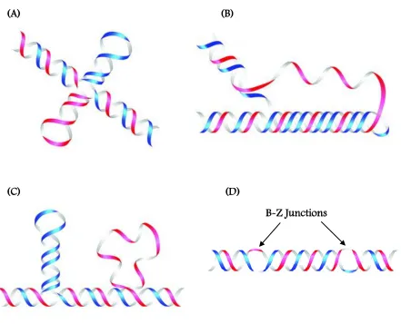
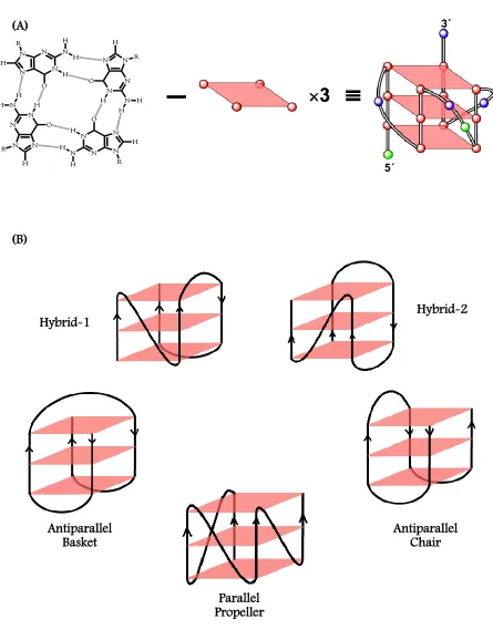
(19) xvii. LIST OF FIGURES Chapter 1 Figure 1.1: Some non-B DNA conformations commonly adopted by DNA. (A) cruciforms, (B) triplexes, (C) slipped structures, and (D) Left-handed Z-DNA. .............................. 18 Figure 1.2: Organization of a G-Quadruplex (A), and some commonly observed intramolecular quadruplex folds (B). ................................................................................................ 19.
(20) xviii. Chapter 2 Figure 2.1: Chemical structures of RHPS4 (A), DODC (B), DB832 (C), Distamycin (D) and F1190 (E). .................................................................................................................. 53 Figure 2.2: Folding patterns of different quadruplex-forming motifs: Tel24. [32]. (A), c-myc. [40]. (B), ODN9 [34] (C), and ODN4 [12a] (D) used in this study. The bulky bromine atoms are represented as green ball and stick. ................................................................... 54 Figure 2.3: (A) Induced CD spectra of DODC with intermolecular quadruplex forming sequences and a duplex sequence as reported by Shafer et al. (B) CD spectra of DODC with a dimeric quadruplex sequence, U6U7 in K+, and (C) CD spectra of Tel22 in K+ with RHPS4, a well-known human telomeric quadruplex end-stacking agent. ......................................................................................................................... 55 Figure 2.4: UV melting profiles of the hairpin duplex, d(CGCGAATTCGTCTCCGAATTCGCG), monitored at 260 nm (A) modified human telomere sequences, Tel24 (B), and Tel26. (C) monitored at 295 nm in the absence and presence of DB832 in. phosphate buffer containing 100 mM K+. ............................................................... 56 Figure 2.5: CD spectra of DB832 titrated into (A) Tel22 (B) Tel24, (C) ODN9, and (D) ODN4 quadruplex forming sequences. ............................................................................... 57 Figure 2.6: CD spectra of Distamycin with Tel22 (A), F1190 with Tel22 (B), and DB832 with. c-myc (C) in K+. ........................................................................................................ 58 Figure 2.7: Fluorescence Displacement assay (A) and competition CD experiment (B) performed with Tel22 sequence using TO as the fluorescent probe. (C) Close-up of the DNA absorbance region highlighted in blue in (B). .......................................... 59 Figure 2.8: Imino proton spectra of Tel24 (hybrid-1) with DB832 at 25 °C in 10 mM K2HPO4/80 mM KCl, pH 7.0. ................................................................................... 60 Figure 2.9: Aromatic proton spectra of Tel24 with DB832 at 25 °C in 10 mM K 2HPO4/80 mM KCl, pH 7.0. ............................................................................................................... 61.
(21) xix. Figure 2.10: Plot of the ICD signal at wavelength corresponding to the maximum absorbance of the bound DB832 (434 nm) as a function of DB832 added molar ratio with different telomeric quadruplex sequences. ........................................................... 62 Figure 2.11: SPR sensorgrams for binding of DB832 analogs with human telomeric quadruplex sequences. ............................................................................................................... 63 Figure 2.12: Isothermal titration calorimetry plot of DB832 (400 µM) titrated into a 10 µM (A) and 20 µM (B) Tel24 quadruplex sequence in TRIS buffer containing 100 mM K+. ........................................................................................................................... 64.
(22) xx. Chapter 3 Figure 3.1: (A) Chemical structure of DB832. (B-H) Major folding topologies of quadruplex forming sequences used in this study. ...................................................................... 88 Figure 3.2: UV melting profiles of the hairpin duplex, d(CGAGATCAAAAGATCTCG), monitored at 260 nm (A) and the human telomere sequence, Tel22, monitored at 295 nm (B) in the absence and presence of DB832 in phosphate buffer containing 100 mM K+. (C) CD spectra of DB832 titrated into d[(GC)7] in HEPES buffer containing 50 mM KCl. Compound:DNA ratios ranged from 1:1 to 5:1. (D) CD spectra of DB832 titrated into d(GCGAATTCGC) in HEPES buffer containing 50 mM KCl. .......................................................................................... 89 Figure 3.3: CD spectra of DB832 titrated into Tel22 in HEPES buffer containing 50 mM KCl (A), NaCl (B), or LiCl (C). (D-F) Close-up of the wavelength region of DNA absorbance for the spectra shown in A-C respectively. ........................................... 90 Figure 3.4: CD spectra of DB832 titrated into (A) Tel22, (B) Tel26, (C) wtTel26, and (D) Tel22 (with PEG 400).......................................................................................................... 91 Figure 3.5: CD spectra of DB832 titrated into (A) Tetrahymena telomeric sequence, d(T 2G4)4, (B). Oxytricha,. d[G4(T4G4)3],. and. (C). bcl-2,. d(G3CGCG3AG2A2T2G3CG3). quadruplex sequences. ............................................................................................. 92 Figure 3.6: CD spectra of DB832 titrated into (A) TBA, d(G 2T2G2TGTG2T2G2), (B) d(G2T4)3G2, (C) TG4T, and (D) c-myc27, d(TG4AG3TG4AG3TG4AAG2) quadruplex sequences. .... ................................................................................................................................... 93 Figure 3.7: Imino proton spectra of DB832 with Tel22 (top), Tel26 (middle), and wtTel26 (bottom) at 25 °C in 10 mM K2HPO4/80 mM KCl, pH 7.0..................................... 94 Figure 3.8: NMR imino proton titrations of (A) bcl-2 promoter sequence and (B) TG4T with DB832. ...................................................................................................................... 95.
(23) xxi. Figure 3.9: NMR imino proton titration of U6U7, d(TAGGGUUAGGGT) dimeric hairpin quadruplex with DB832 (A). (B) TOCSY spectra of uracil H5-H6 cross-peaks for 0.5 mM U6U7 at 0:1 (top), and 2:1 (bottom) DB832 molar ratios at 308K. ......... 96.
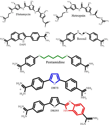
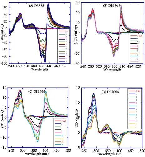
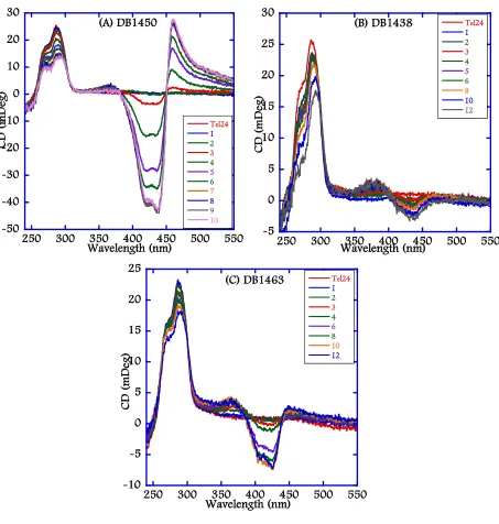
(24) xxii. Chapter 4 Figure 4.1: Chemical structures of representative Duplex-DNA minor groove binders. (A) Distamycin, (B) Netropsin, (C) DAPI, (D) Berenil, (E) Pentamidine, (F) DB75, and (G) DB293. .............................................................................................................. 122 Figure 4.2: Chemical structures of 5-5-6 and related ring systems used in this study............ 123 Figure 4.3: CD spectra of DB compounds (A) DB832, (B) DB1949, (C) DB1999, and (D) DB1093 with Tel24 quadruplex sequence. ........................................................... 124 Figure 4.4: CD spectra of thiophene containing DB compounds (A) DB1450, (B) DB1438, and (C) DB1463 with Tel24 quadruplex sequence. ..................................................... 125 Figure 4.5: CD spectra of DB compounds (A) DB1972, (B) DB2037, (C) DB934, (D) DB1693, and (E) DB1694 with Tel24 sequence. .................................................................. 126 Figure 4.6: Plot of mole ratio versus the ICD signal at wavelength corresponding to the maximum absorbance of the bound ligand with Tel24. ....................................... 127 Figure 4.7: Imino proton spectra of Tel24 with (A) DB1450 and (B) DB1463 at 25 °C in 10 mM K2HPO4/80 mM KCl, pH 7.0. ......................................................................... 128 Figure 4.8: Imino proton spectra of Tel24 with (A) DB1438 and (B) DB2037 at 25 °C in 10 mM K2HPO4/80 mM KCl, pH 7.0. ......................................................................... 129 Figure 4.9: SPR sensorgrams for binding of representative thiophene analogs with Tel22 quadruplex sequence. DB1450 (top left), DB1463 (top middle) and DB1438 (top right) to Tel22 in 10 mM TRIS buffer containing 100 mM K+ at 25 °C. .............. 130 Figure 4.10: SPR sensorgrams for binding of DB832 analogs with Tel22 quadruplex sequence. DB934 (top left), DB1693 (top middle) and DB1694 (top right) to Tel22 in 10 mM TRIS buffer containing 100 mM K+ at 25 °C. .............................................. 131.
(25) xxiii. Chapter 5 Figure 5.1: Structures of (A) TMPyP4, (B) Telomestatin, (C) RHPS4, (D) Pt-MPQ, (E) BRACO19, and (F) G-quartet with bound cation shown as a blue circle. ........................ 158 Figure 5.2: Folding patterns of different quadruplex-forming motifs used in SPR-Biosensor studies. ..................................................................................................................... 159 Figure 5.3: Chemical Structures of Azacyanines. ...................................................................... 160 Figure 5.4: Chemical Structures of Naphthalene Diimides. ...................................................... 161 Figure 5.5: Chemical Structures of Diaryl Ureas. ...................................................................... 162 Figure 5.6: Interaction of Azacyanine-3 with Tel24 quadruplex motif. .................................. 163 Figure 5.7: Molecular models of ligands complexed with a parallel topology of human telomeric quadruplex conformation. (A) 4ND08 (B) WD313 (C) WD308 (D) WD263. ................................................................................................................... 164 Figure 5.8: SPR sensorgrams for binding of Aza3 to the immobilized G-quadruplexes formed by Tel26 (top left), Tel24 (top middle) and GC(20) duplex (top right) in HEPES buffer containing 80 mM KCl at 25 °C. ................................................................. 165 Figure 5.9: SPR sensorgrams for binding of Aza4 to the immobilized G-quadruplexes formed by Tel26 (top left), Tel24 (top middle) and GC(20) duplex (top right) in HEPES buffer containing 80 mM KCl at 25 °C. ................................................................. 166 Figure 5.10: SPR sensorgrams for binding of Aza5 to the immobilized G-quadruplexes formed by Tel26 (top left), Tel24 (top middle) and GC(20) duplex (top right) in HEPES buffer containing 80 mM KCl at 25 °C. .............................................................. 167 Figure 5.11: SPR sensorgrams for binding of 4ND01 to the immobilized G-quadruplexes formed by Tel22 (top left), ckit-1 (top middle) and ckit-2 (top right) in HEPES buffer containing 80 mM KCl at 25 °C. .............................................................. 168.
(26) xxiv. Figure 5.12: SPR sensorgrams for binding of 4ND02 to the immobilized G-quadruplexes formed by Tel22 (top left), ckit-1 (top middle) and ckit-2 (top right) in HEPES buffer containing 80 mM KCl at 25 °C. .............................................................. 169 Figure 5.13: SPR sensorgrams for binding of 4ND03 to the immobilized G-quadruplexes formed by Tel22 (top left), ckit-1 (top middle) and ckit-2 (top right) in HEPES buffer containing 80 mM KCl at 25 °C. .............................................................. 170 Figure 5.14: SPR sensorgrams for binding of 4ND08 to the immobilized G-quadruplexes formed by Tel22 (top left), ckit-1 (top middle) and ckit-2 (top right) in HEPES buffer containing 80 mM KCl at 25 °C. .............................................................. 171 Figure 5.15: SPR sensorgrams for binding of 4ND09 to the immobilized G-quadruplexes formed by Tel22 (top left), ckit-1 (top middle) and ckit-2 (top right) in HEPES buffer containing 80 mM KCl at 25 °C. .............................................................. 172 Figure 5.16: SPR sensorgrams for binding of 3ND03 to the immobilized G-quadruplexes formed by Tel22 (top left), ckit-1 (top middle) and ckit-2 (top right) in HEPES buffer containing 80 mM KCl at 25 °C. .............................................................. 173 Figure 5.17: SPR sensorgrams for binding of WD263 to the immobilized G-quadruplexes formed by c-myc (top left), Tel22 (top middle) and Dickerson (top right) in HEPES buffer containing 80 mM KCl at 25 °C. ................................................... 174 Figure 5.18: SPR sensorgrams for binding of WD308 to the immobilized G-quadruplexes formed by c-myc (top left), Tel22 (top middle) and Dickerson (top right) in HEPES buffer containing 80 mM KCl at 25 °C. ................................................... 175 Figure 5.19: SPR sensorgrams for binding of WD313 to the immobilized G-quadruplexes formed by c-myc (top left), Tel22 (top middle) and Dickerson (top right) in HEPES buffer containing 80 mM KCl at 25 °C. ................................................... 176.
(27) xxv. Chapter 6 Figure 6.1: Chemical structure of DB1878 with the atom naming and coloring schemes used in this study and the hairpin duplex, Oligo2-1. The possible binding site of DB1878 is highlighted in green................................................................................................ 206 Figure 6.2: (A) Aromatic (H6/H8) to H1‟ backbone connectivity region of oligo2-1 at 5 °C. (B) Schematic H6/H8-H1‟ connectivity observed. ...................................................... 207 Figure 6.3: Expanded region of the observed exchangeable proton 2D NOESY spectrum of free oligo2-1 at 5 °C (A), and imino proton spectra of free oligo2-1 as a function of temperature (B). ...................................................................................................... 208 Figure 6.4: The expanded aromatic region of COSY spectrum of DB1878 in D 2O at 25 °C (A), and the corresponding 1D proton spectra (B). ...................................................... 209 Figure 6.5: SPR sensorgrams of DB1878 binding to oligo2-1 with the corresponding Scatchard plot (A), and the 2D COSY spectra of the T: CH3-H6 region for DB1878-oligo2-1 interaction at 5 °C with listed ratios (B). ................................................................ 210 Figure 6.6: Aromatic proton spectra of DB1878 with oligo2-1 at 5 °C (A), and the aromatic region of the COSY spectrum of 2:1 complex at 5°C (B). ..................................... 211 Figure 6.7: Expanded region of the 1H-13C-HSQC spectrum of oligo2-1 with 2:1 ratio of DB1878 at 5 °C without decoupling. ..................................................................... 212 Figure 6.8: The aromatic to H1‟ backbone connectivity region of 2:1 complex of DB1878 with oligo2-1 at 5 °C. ...................................................................................................... 213 Figure 6.9: Plot of ligand induced changes for H1‟ (A), and H6/H8 (B) in the 1H-NMR chemical shifts for the 2:1 complex of DB1878 with oligo2-1. The loop residues are not included. ..................................................................................................... 214 Figure 6.10: Expanded NOESY spectrum of the upfield region (A), and. 31P-1H. correlation. spectrum (B) of 2:1 complex of DB1878 with oligo2-1 at 5 °C. ........................ 215.
(28) xxvi. Figure 6.11: Intermolecular crosspeaks between the two DB1878 molecules and between DB1878 and DNA protons in the aromatic region (A), H1‟ region (B) of the NOESY spectrum of the 2:1 complex at 5 °C. The 2D stacked plot illustrating the strong interactions between the B1 protons and adenine residues are in (C). ......... ............................................................................................................................... 216 Figure 6.12: Some of the major NOE interactions observed between the two drug protons and the DNA. ............................................................................................................... 217 Figure 6.13: Expanded NOESY spectrum of the imino proton region of 2:1 complex of DB1878 with oligo2-1 at 5 °C. ........................................................................................... 218 Figure 6.14: Preliminary docking studies of DB1878 complexed with oligo2-1 based on NMR data. (A) View into the groove of oligo2-1 with the two DB1878 molecules (B) View along the groove of the helix (C) interaction between M2B1 and M2NH with A15H2 and (D) possible H-bond between G5NH2 and furan oxygen. ..... 219.
(29) xxvii. LIST OF ABBREVIATIONS 1D. –. One-dimensional. 2D. –. Two-dimensional. Å. –. Angstroms. A. –. Adenine. Aza. –. Azacyanines. C. –. Cytosine. COSY. –. Correlated Spectroscopy. CD. –. Circular Dichroism. d1. –. Relaxation delay. dA. –. Deoxyadenosine. dT. –. Deoxythymidine. DNA. –. Deoxyribonucleic Acid. DQF. –. Double Quantum Filtered. DSS. –. 4,4-dimethyl-4-silapentane-1-sulfonic acid. EDTA. –. Ethylenediaminetetraethanoic acid. G. –. Guanine. G4-FID. –. Quadruplex-Fluorescence Intercalator Displacement. HEPES. –. 4-(2-hydroxyethyl)-1-piperazineethanesulfonic Acid. HETCOR. –. Heteronuclear Correlation Spectroscopy. HSQC. –. Heteronuclear Single Quantum Correlation. HTel. –. Human Telomeric DNA. IC50. –. 50% Inhibitory Concentration. ITC. –. Isothermal Calorimetry. mdeg. –. millidegrees.
(30) xxviii. MES. –. 2-(N-morpholino)ethanesulfonic Acid. ms. –. milliseconds. ND. –. Naphthalene Diimides. nm. –. nanometers. NMR. –. Nuclear Magnetic Resonance. NOE. –. Nuclear Overhauser Effect. NOESY. –. NOE Spectroscopy. ODN. –. Oligodeoxyribonucleotide. Oligo. –. Oligonucleotide. ppm. –. parts per million. RNA. –. Ribonucleic acid. RU. –. Response Units. SPR. –. Surface Plasmon Resonance. ss. –. single stranded. T. –. Thymine. TBA. –. Thrombin Binding Aptamer. TetTel. –. Tetrahymena Telomere. Tm. –. Thermal melting temperature. TO. –. Thiazole Orange. TOCSY. –. Total Correlation Spectroscopy. TRIS. –. Tris Hydroxymethylaminoethane. UV. –. Ultraviolet.
(31) 1. 1 1.1. INTRODUCTION PERSPECTIVE A great mystery of life is how a single-celled entity, beginning from the time of. conception to birth to development, dividing and subdividing in an orderly manner, becomes in time a complex, individual organism. Science has, to a great extent, helped us in our neverending quest for understanding the complex process of development by probing down to the level of atoms and molecules, to decode their fundamental properties how they are related to, contribute, and control diverse biochemical processes within the cells. The last fifty years have completely changed the way biological and medical researchers study and understand life, susceptibility to infectious and inherited diseases, and the molecular mechanisms of metabolic processes. One reason that brought about this understanding lies in the ability to access the information contained in biological macromolecules. Breakthroughs, combined with unanticipated discoveries, at various stages over decades have led to the creation of what is termed as modern-day medicine that has significantly altered the perception of human body. The reductionist's approach – the study of chemistry and physics of life – created an enormous wealth of biochemical and genetic data available for the rational design of drugs and the manipulation of the genome. The scientific advancements over the years have considerably increased our potential to understand and defend against lethal diseases. Science has gone to a great extent to solve the mystery of life, but it still is at an early stage. With the current knowledge, we can at least make an assertion that many of the underlying processes that guide the complex biochemical processes occurring in our body can be explained through science. Nevertheless, the fact remains that the more we decipher the secrets of the human body through science, the more complex it seems to be..
(32) 2. 1.2. THE CENTRAL DOGMA Within the countless number of cells in the human body, there are infinite number of. biochemical processes undergoing, at any given time in a well-orchestrated manner, almost all of which involve the basic biomolecules: carbohydrates, lipids, proteins and nucleic acids. The five bases and twenty amino acids, which make the repertoire of nucleic acids and proteins respectively, are very well conserved throughout nature and, essentially, control and guide most of the biochemical processes occurring in the body. The central dogma of molecular biology, which formulates the sequential information from DNA to protein to be deterministic, was first articulated by Francis Crick in 1958[1]. This model summarizes the transfer of the most complex hereditary information in a very simplistic, but elegant way. Nucleic acids in all organisms carry the genetic information in the form of DNA, although some viruses use RNA as their genetic material. By the process of transcription, a stretch of DNA for a gene dictates the sequence for its correlative mRNA, and then the same mRNA serves as a template for the translation of amino acids into a sequence of polypeptides, which form a protein. Thus, the central dogma reveals a predictable chain of information from DNA to protein, whereupon sequential information for any one component offers determinative sequence information for the other components. The transfer of this information in unidirectional (i.e., DNA RNA PROTEINS), and each step is tightly regulated to maintain a balance between the individual components, a hallmark of normal cells. Cancer cells and viruses, however, exhibit deregulated signaling of all or any of the components of the central dogma, resulting in their unbridled proliferation. Also, the flow of information in cancer cells is not in accord with the simplest principles set forth by central dogma, reverse-transcriptase[2] and prion proteins[3] being prime examples.. Complex human diseases encompass a spectrum of genetic and environmental attributes that together affect the normal functioning of several molecular and cellular.
(33) 3. pathways. They occur commonly in the population and are a major source of disability and death worldwide. The genes that contribute to complex diseases are notoriously difficult to identify, because they typically exert small effects on disease risk; in addition, the magnitude of their effects is likely to be modified by other unrelated genes as well as environmental factors[4]. One of the greatest challenges facing researchers today is to sort out how these contributing factors interact in a way that translates into effective strategies for disease diagnosis, prevention, and therapy. While major inroads have been made in identifying the genetic causes of disorders, little progress has been made in the discovery of common gene variations that predispose to complex diseases. On an exciting note, the genome sequencing projects have provided a step toward a broader and more complete understanding of challenges and opportunities in addressing some of the underlying questions for a successful therapeutic intervention. Further extensive investigation will help discover hitherto unknown biological functions and provide clues to the occurrence and control of many diseases.. 1.3. BEYOND THE DOUBLE HELIX Ever since the discovery of double stranded DNA, various higher order DNA structures. have been discovered. [5].. The remarkable ability of single strand oligonucleotides to form a. multitude of structures is attributed to an excellent array of hydrogen bond donors and acceptors that nature has precisely incorporated among the nucleobases, and also various external factors such as salt, ion concentration, pH and the extent of hydration. The network of hydrogen bond donors and acceptors among the bases enable them to fold into structures that deviate from the classical Watson-Crick base pairing. Various triplexes[6], cruciforms[7], and imotif[8] architectures have been reported in designed DNA sequences that have shown to exhibit interesting hydrogen-bonding patterns between the nucleotides (Figure 1.1). Multistranded DNA structures have assumed great importance in recent years with the realization.
(34) 4. that they play important roles in various biological processes such as DNA recombination, replication, translation, gene expression and disease control, to name a few[9]. There is mounting evidence that DNA structural properties beyond the double helix significantly affect its interactions with proteins and play an important role in a number of biological processes[10].. 1.4. G-QUADRUPLEXES Guanosine molecules show a remarkable ability to self-assemble into highly complex. patterns [11]. The self-association of guanine-rich DNA motifs under physiologically feasible ion concentrations led to the hypothesis that these structures might have significant biological importance. Guanine-rich DNA sequences, under certain physiological conditions, can form unique structures known as G-quadruplexes that have structural properties very different from the canonical DNA [5a, 12]. Stacking of G-tetrads leads to the formation of G-quadruplexes; complex and highly ordered helical structures. The G-tetrad motif is composed of four guanines arrayed in a square planar configuration and can self-assemble in the presence of cations (Figure 1.2, A). Adjacent guanine bases in a tetrad are hydrogen-bonded on their Watson-Crick and Hoogsteen edges, with their carbonyl groups oriented towards the centre of the quadruplex. Within a tetrad, the N1 imino proton and the C2 amino proton of a guanine residue are hydrogen bonded respectively to the C6 carbonyl oxygen and the N7 of the neighboring guanine residue, and a similar hydrogen bonding pattern continues until all the four guanines in a tetrad are cyclically connected by hydrogen bonds. Overall, eight hydrogen bonds are observed in a tetrad adding significant stability to the tetrads. The quadruplex is further stabilized through π-π stacking interactions of the stacked tetrads as well as by coordination with cations located between or within the tetrads. These structures exhibit extensive structural diversity and polymorphism relative to duplex DNA. In general, structural.
(35) 5. polymorphism arises mostly from the nature of the loop, such as variations of strand stoichiometry, strand polarity, glycosidic torsion angle, and the location of the loops that link the guanine strands. [5a, 12].. Meanwhile, the solution environment, such as proteins, ligands, or. molecular crowding conditions, may also influence the topology of quadruplex. [13].. G-. quadruplexes can be folded from a single G-rich sequence intramolecularly or by the intermolecular association of dimeric or tetrameric strands. Other self-assembling motifs, like G-ribbons, were also identified in lipophilic guanosine derivatives. [14].. Switching between G-. ribbon and G-quadruplex structures was observed as a function of the solvent and cations in the solution [15].. 1.4.1 Biological Role of G-Quadruplexes Despite the fact that these unique structures were discovered and have been known for more than half a century, it is only during the past decade or so these G-quadruplex structures have come into the limelight, due to an overwhelming rise in the evidences supporting various hypotheses about the relevant role of G-quadruplexes in wide range of biological processes [16]. Potential quadruplex forming guanine rich sequences are found in biologically important regions of the genome including telomeres. [12, 17],. transcriptional regulatory regions of genes. (such as insulin, VEGF, fragile X mental retardation gene). [8a, 18],. immunoglobulin switch. regions [19], promoter regions of oncogenes (such as c-myc, c-kit, bcl-2, ret, k-ras) [20] thereby raising exciting possibilities of moderating gene expression with the aid of G-quadruplexes. Because of their apparent role in controlling gene expression and their putative involvement in the inhibition of telomerase activity, G-quadruplexes are potential targets in cancer research [21].. Recent bioinformatics studies have shown that human genome contain as many as. 376,000 potential quadruplex-forming sequences and an astounding 40% of these gene promoter regions have at least one quadruplex forming motif [22]..
(36) 6. The discovery of a number of proteins in various organisms, such as yeast and oxytricha that can bind to telomeric DNA suggests that these quadruplex structures may also exist in vivo. [23]. and, interestingly, recent studies from different labs have provided evidence. that these quadruplex structures do form in vivo. Autoradiography studies of metaphase chromosomes of normal and cancer cell lines have shown that a quadruplex binding ligand, 3H-360A,. accumulates in the nuclei of these cells and preferentially binds to the terminal. regions of metaphase chromosomes of both normal and cancer cells[24]. Biochemical assays have shown that the cationic porphyrin, TmPyP4, binds to and stabilizes a particular conformation of G-quadruplex structure in the NHE III1 of c-myc promoter region thereby directly repressing c-myc transcriptional activation[25]. The strongest evidence to date for Gquadruplex existence in vivo synthetic single chain antibody fragments specifically targeted against a parallel intermolecular G-quadruplex assembled from the ciliated protozoan. Stylonichia lemnae telomeric sequence[26]. The presence of a strong signal in the macronucleus by one of the antibody fragments confirmed the presence of quadruplex structures in vivo. These exciting results demonstrate that quadruplex structures can also form in vivo and ligands can be invariably employed to exploit these G-quadruplex structures as “druggable targets”[27].. 1.4.2 Telomeres Telomeres are specialized non-coding structural units capping the ends of chromosomes in eukaryotic cells of parasites and higher organisms. [17d, 28].. They are found to. play a vital role in maintaining the stability of chromosomes by preventing end-to-end fusion, recombination, nuclear degradation and cellular senescence. [17d, 28a, 29].. Telomere length is. regarded as a potential biomarker of aging[30]; there is a growing body of evidence indicating that shorter telomeres are associated with various diseases, including cancer, infectious.
(37) 7. diseases, psychological stress, and cardiovascular disease[28a,. 31].. Telomere dysfunction limits. the proliferative capacity of human cells by activation of DNA damage responses, inducing senescence or apoptosis[30]. In humans, telomere shortening occurs in the vast majority of tissues during aging, and telomere shortening is accelerated in chronic diseases that increase the rate of cell turnover[30]. The leading hypothesis for telomere attrition is due to inflammation, exposure to infectious agents, and other types of oxidative stress, which damage telomeres and impair their repair mechanisms[32]. Several lines of evidence support this hypothesis, including observational findings that people exposed to infectious diseases have shorter telomeres.. 1.4.3 End-Replication Problem and Telomerase The semi-conservative nature of DNA replication prohibits DNA polymerase from replicating the 3‟-end of the lagging strand of the DNA (termed as the “end replication problem”) due to the absence of a RNA primer with a free 3‟-OH group. Therefore, after each round of cell division and DNA replication, the extreme 3‟-end of the telomeres shorten by about 50-150 base pairs and after about 60-70 cycles of cell division, the telomeres reach a critical length when the cell enters the senescence stage eventually leading to apoptosis and finally results in cell death[17d, 31d]. However, the telomere length is maintained at a constant level in tumor cell lines as well as in germ-line cells. [29].. This is due to the activation of an. enzyme called telomerase, which is absent for the normal function of most somatic cells that usually have longer telomeres, whereas widely expressed in immortal cells. In fact, telomeres are highly maintained in length in 80-85% of human tumor cells, which divide indefinitely by the action of telomerase[21d]. Telomerase is a ribonucleoprotein reverse-transcriptase enzyme consisting of an eleven bases long RNA template and a catalytic subunit (hTERT) with reverse transcriptase activity. With the aid of accessory proteins, the enzyme extends the 3‟ ends of the.
(38) 8. DNA by consecutively adding the TTAGGG hexanucleotide repeats using the RNA template as a primer[21d]. However, in order to continue the elongation process, it is highly imperative that the primer maintain the single stranded conformation. Formation of any secondary structures, such as G-quadruplexes, impedes the hybridization of template RNA subunit of telomerase onto the primer during the elongation process and consequently resulting in the inhibition of telomerase activity. Therefore, inducing or stabilizing the telomeric quadruplex conformation might be one of the important methods of controlling and inhibiting the activity of telomerase. Moreover, the telomerase holoenzyme itself has potential recognition sites that can be exploited for the development of inhibitors. Antisense oligodeoxynucleotides[33], hammerhead ribozymes[34], peptide nucleic acids[35], chimeric RNA modules[36], reverse transcriptase inhibitors[37] and immunotherapy agents[38] are some of the agents that are shown to potentially recognize different structural and functional units of telomerase holoenzyme, and are actively studied as telomerase inhibitors. Since telomerase is necessary for the immortality of many cancer types, it is thought to be a potentially highly selective and attractive drug target for several anti-tumour strategies. Its action is detected in most primary human tumor specimens and tumour-derived cell lines, such as those of the prostate, breast, colon, lung and liver. [21c-e, 31c, 39].. Hence, inactivation of telomerase may play an important role in cancer. therapy. As aforementioned, telomerase activity can also be inhibited by stabilization of quadruplex conformation of telomeres and, therefore, small molecules that stabilize these quadruplex structures could act as telomerase inhibitors and can be employed as potential therapeutic agents.. Human telomeric sequences are one of the most extensively studied motifs due to their critical role in maintaining chromosomal integrity. Human telomeres consists of highly conserved tandem repeats of guanine rich hexanucleotide d[TTAGGG] ranging from 5-15 kb, while the extreme 3‟ terminus of this telomeric sequence is single stranded and composed of.
(39) 9. only about 100-200 bases that folds into a “t-loop” structure[40]. The hexanucleotide repeats of d[TTAGGG] can fold into an array of quadruplex topologies in vitro under different physiological conditions[5a]. Structural information of telomeric quadruplex DNA under in vivo conditions is essential from drug design stand point of view. The first structure of the human telomeric DNA sequence, AG3(T2AG3)3 by NMR in Na+ has shown that this sequence folds into an intramolecular quadruplex termed as an antiparallel basket structure with a mixture of diagonal and lateral TTA loops[41]. In the presence of K+ in a crystalline state, an intramolecular parallel, propeller-type G-quadruplex conformation has been reported. Propeller-type Gquadruplexes have the loops running diagonally between the G-strands with the G-strands in a parallel arrangement[42]. Because the structure in K+ solution is considered to be biologically more relevant, due to the high intracellular K+ concentration, several attempts have been made to elucidate the folding topology of human telomeric quadruplex in K+, and several structures were found that are inconsistent with the crystal structure. The equilibrium of G-quadruplex species in K+ solution can be altered by several additional factors. For example, platinum-based cross-linking studies have shown that the basket-type structure coexists with other quadruplexes in both Na+ and K+ solutions[43]. A subsequent. 125I-radioprobing. study has. revealed that a chair-type conformation is the major species in K+ solution[44]. Recently, sedimentation and fluorescence studies have revealed that the crystal structure of telomeric DNA is unlikely to be the major species in K+ solution, and various forms are energetically similar [45]. A mixture of chair-type and parallel/antiparallel hybrid structures may coexist for telomeric DNA in K+ solution. Circular dichroism studies of several modified human telomeric sequences with bromo-guanine substitutions revealed the formation of highly stable hybridtype conformations, and were later further confirmed by high resolution NMR studies. [46].. Recent structural studies by several groups also showed that the human telomeric quadruplex folds into a mixture of “hybrid-type” mixed parallel/antiparallel quadruplex conformations [47].. However, the NMR structures all have flanking sequences that confer additional.
(40) 10. stabilization, so they cannot be directly compared with the crystal structure. Recently, it was reported that human telomeric DNA forms parallel-stranded intramolecular G-quadruplexes in K+ solution under molecular crowding conditions. [48].. Moreover, various labs have. suggested a compact stacking structure for multimers of hybrid-type and parallel-type Gquadruplexes in human telomeric DNA. The “hybrid-type” structures of telomeric quadruplexes under physiological K+ conditions maybe the predominant conformation in vivo.. 1.4.4 Oncogenes The c-myc gene belongs to the Myc gene family and functions as a gene-specific transcription factor through its protein product, c-Myc, for a wide range of human cancers. The c-Myc protein regulates almost 20% of all cellular genes and is also involved in cell cycle regulation, apoptosis, metabolism, cellular differentiation, and cell adhesion[49]. As a result, the aberrant over expression of c-myc is associated with a variety of malignant tumors including those of breast, colon, cervix, and myeloid leukemia[50]. In particular, c-Myc has been identified as one of the main activating factors for the human telomerase reverse transcriptase (hTERT) catalytic domain of the telomerase enzyme[51]. The nuclear hypersensitivity element III1 (NHE III1) upstream of the P1 promoter of c-myc is a G-rich strand containing a 27-base-pair sequence (Pu27, Table 1.1), which has the propensity to adopt a G-quadruplex structure. The presence of a quadruplex within this promoter region was initially proposed based on chemical probe studies, gel mobility measurements, and fluorescence resonance energy transfer (FRET) spectroscopy[52]. In later studies, the topological structures of several c-. myc quadruplex sequences were determined by circular dichroism (CD), NMR, and mutational experiments. [16a, 53].. The G-rich region of c-myc contains more than four. consecutive G-strands, resulting in the formation of a dynamic mixture of four parallel Gquadruplex loop isomers in the native Pu27 region. Furthermore, two different sequences.
(41) 11. derived from the Pu27 region have been analyzed by NMR[20d], revealed that both myc-2345 (Table 1.1) and myc-1245 (Table 1.1) fold into intramolecular propeller-type G-quadruplexes in K+ solution. In this case, the core of three G-tetrads is formed by four G-stretches oriented in the same direction, with all the guanines in an anti glycosidic conformation and the three loops adopting double-chain-reversal structures, very similar to the crystalline state of telomeric G-quadruplex conformation in K+. Similar structures have also been found in the. Pu24I (Table 1.1) and myc22-G14T/G23T (Table 1.1) sequences. [54].. A interesting structural. feature of Pu24I revealed by NMR studies show that a guanine base (G24) at the 3 end plugs back into the G-tetrad core by participating in G-tetrad formation and displacing another guanine (G10) of a continuous guanine tract in a loop. This configuration is maintained by a stable diagonal loop, which contains a G•(A-G) triad stacking on and capping the G-tetrad core. These new folding features result from the presence of five guanine tracts in the sequence that are different from the four guanine tracts in the c-myc sequences studied previously.. bcl-2 is a proto-oncogene, and its oncogenic property arises from decreasing the rate of cell death[55]. Its protein product, Bcl-2, is a mitochondrial membrane protein, which is present in delicate balance with related proteins and is involved in the control of programmed cell death, functioning as an apoptosis inhibitor. [56].. Over-expression of bcl-2 has been found in a. wide range of human cancers, including B-cell and T-cell lymphomas, breast, cervical, prostate, and colorectal. In addition, it also functions in chemotherapy-induced apoptosis[55], which indicates its potential role in drug resistance. The human bcl-2 gene contains a GC-rich region upstream of the P1 promoter, which is critical for the regulation of bcl-2 gene expression. It can form a mixture of three distinct intramolecular G-quadruplexes (5 G4, MidG4, and 3 G4) resulting from the six runs of guanines, including one run of five guanines, two runs of four guanines each, and three runs of three guanines each (Table 1.1). With more than four consecutive G-tracts in the sequence, the G-quadruplex in bcl-2 has the ability to.
(42) 12. form either three or six different loop isomers. [57].. The central G-quadruplex (MidG4, Table. 1.1), which is the most stable of the major species formed in the bcl-2 promoter region, is likely to form a mixed parallel/antiparallel structure consisting of three tetrads connected by loops and to give rise to three possible loop isomers. An NMR study of the shorter and mutated. bcl-2 quadruplex bcl2MidG4Pu23-G15T/G16T (Table 1.1) has shown that one of the topologies for this mixed parallel/antiparallel intramolecular quadruplex has two lateral loops and one propeller loop, similar to one of the Tel22 telomeric quadruplex topologies [20c, 58]. The G-rich strand located in the bcl-2 P1 promoter plays a significant role in the regulation of bcl-. 2 transcription. [59].. Although the effects of G-quadruplex ligands on bcl-2 expression remain. to be deciphered, some studies have shown that some G-quadruplex ligands can induce apoptosis [60]. In particular, the ligand 12459 has been found to induce apoptosis characterized by dysfunction of Bcl-2 [61]. These findings suggest the possible role of G-quadruplex formation in the bcl-2 promoter during apoptosis.. Recently, two G-rich sequences (c-kit native and c-kit21, Table 1.1) in the promoter region of the human c-kit gene have been identified, and biophysical studies have shown that these sequences can form G-quadruplexes. [20e, 62].. In the c-kit native sequence, 87 base pairs. upstream of the transcription start site of the human c-kit gene, a single G-quadruplex structure forms in K+ solution. An NMR study has shown that the c-kit87up (Table 1.1) sequence forms a new intramolecular G-quadruplex[63]. Most strikingly, an isolated guanine (G10) is involved in G-tetrad core formation, despite the presence of four three-guanine tracts. There are four distinctive loops, including two single-residue and double-chain-reversal loops (A5, C9), a two-residue loop (C11, T12), and a five-residue stem-loop (A16, G17, G18, A19, G20). In view of the importance of predicting G-quadruplex topologies from sequence information, these new folds in which G residues in non-G-tract regions can participate in structural core formation are particularly worthy of attention. In the case of the c-kit21.
(43) 13. sequence, a variety of quadruplex conformations have been identified. This sequence needs to be mutated in order to form a single quadruplex species, probably with a parallel fold.. 1.4.5 Other G-Quadruplex Families Guanine-rich sequences of human vascular endothelial growth factor (VEGF) and the. neuroblastoma (Rb) oncogene promoter have also been shown to form G-quadruplexes by chemical footprinting and CD studies [16c, 64]. The G-rich sequences in these regions are known to exist in duplex form and ligands such as TMPyP4 and Telomestatin have been shown to induce parallel quadruplex conformations. Putative quadruplex forming sequences have also been discovered in hypoxia-inducible factor 1α (HIF-1α) promoter region, k-ras and ret oncogenes. [65].. Other types of G-quadruplexes have also been found in the RNA region[66],. peptide nucleic acids (PNA)[67] and locked nucleic acids (LNA)[68].. There are now reasons to believe that G-quadruplex structures are not merely an in. vitro artifact. Both in vitro and in vivo data strongly support the physiological relevance of this nucleic acid structure at the telomere, one of the signature guanine-rich regions of the genome. While the precise role of this structure at chromosomal ends remains a matter of conjecture, a number of scenarios have been put forward, taking into account the diverse conformations of the G- quadruplex. The inability of telomerase to utilize the G-quadruplex fold of telomere has led to the emergence of a novel avenue for cancer therapy, through Gquadruplex stabilizing agents. These agents can trap DNA in a quadruplex conformation and, in this manner, may either inhibit telomere extension by telomerase or perturb telomerecapping mechanisms. This approach may be complicated by the structural heterogeneity of Gquadruplexes that arise from human telomeric DNA. In addition, questions remain regarding the in vivo mechanism of action of existing G-quadruplex stabilizers. Nevertheless, given the.
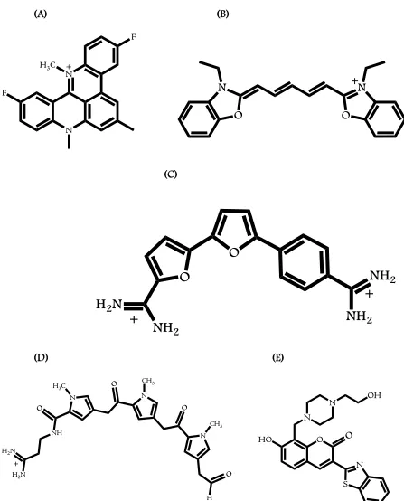
(44) 14. clinical significance of the G-quadruplex, research activities on telomeric and genomic quadruplexes will continue to grow. With a better understanding of the biological functions and structural properties of G-quadruplexes, it is expected that a wealth of new drugs that are less cytotoxic and that have higher selectivity will emerge in the near future.. 1.5. TARGETING G-QUADRUPLEXES WITH SMALL MOLECULES Studying small molecule interactions with quadruplex forming structures is a rapidly. developing area in the field of drug design and synthesis driven by the need for understanding at the molecular level for the development of potential candidates into effective chemotherapeutic agents. The basic principles that aid in the design and synthesis of small molecules for duplex DNA recognition can also be employed for quadruplex recognition. Compounds that intercalate between the base pairs of duplex DNA can be used as lead candidates to develop small molecules that can interact with quadruplexes by either intercalation or end-stacking. Several compounds to-date have been shown to bind and stabilize the human telomeric quadruplex conformations. The quadruplex-small molecule interactions, in most cases, occur via the external stacking of the ligand on the G-quartets of either one or both ends of the quadruplex. These compounds are traditionally planar and have aromatic moieties that can efficiently stack on the large aromatic surface area of the G-quartet via π-π interactions. Some of the compounds designed by exploiting the structural properties of G-quartets and have shown to inhibit telomerase activity are di- and tri-substituted acridines such as BSU6039[69] and BRACO-19[60d, 70], cationic porphyrins such as TmPyP4[53, 71] and Se2SAP[72], macrocycles such as PIPER[73], BOQ1[74] and MMQ3[75], carbazoles[76], metal mediated compounds such as Pt-MPQ[77], Ni(II)-salphen[78] and Cu-ttpy[79], and neutral molecules such as telomestatin[16d, 60b, 71b] and HXDV[80]. These compounds have been studied in great detail, and in all cases end stacking is the observed primary mode of binding with.
(45) 15. telomeric quadruplex structure, whereas, some compounds such as TmPyP4 exhibit nonspecific binding such as external binding to the loops or the backbone of the quadruplex structure. Quadruplex grooves offer an attractive recognition site for interaction of small molecules, but there has been very little success in designing molecules that target quadruplex grooves. Small molecules that have been discovered to sequence-specifically interact with the grooves of duplex DNA can be used as paradigms to develop compounds to target the grooves of the quadruplex.. Spectroscopic studies have shown that DODC, a carbocyanine dye,. interacts with the grooves of a dimeric hairpin quadruplex DNA[81]. Recent NMR studies have shown that distamycin, a classic duplex-DNA minor groove binder, also binds to an intermolecular quadruplex conformation as an antiparallel dimer in the opposite grooves making specific hydrogen bonding with the donor and acceptors along the grooves of the quadruplex[82]. 1H-NMR studies have shown that diaryl amides can potentially recognize grooves of a parallel quadruplex conformation formed in the promoter region of c-kit2 oncogene with high degree of selectivity over duplex DNA[83]. However, small molecules that can target grooves of biologically relevant telomeric quadruplex conformation have not been reported so far. Nonetheless, these results point out that quadruplex groove recognition is indeed a viable strategy for drug design and needs much further attention.. 1.6. RESEARCH GOALS DB832, a bifuryl-phenyl diamidine, was recently reported to selectively recognizing. human telomeric G-quadruplex conformation with significant selectivity over duplex sequences. Circular dichroism analysis revealed that DB832 can potentially recognize multiple grooves of the telomeric quadruplex as a stacked agent. The first goal of this research described.
(46) 16. in Chapter 2 involved a multifaceted approach providing complementary lines of evidence to comprehensively characterize the binding mode of DB832. An array of established spectroscopic techniques, circular dichroism, NMR, thermal melting, fluorescence intercalator displacement (G4-FID) assay, surface plasmon resonance, and isothermal calorimetry techniques have been used to test this. DB832 serves as a paradigm to show that the grooves of the human telomere can indeed be selectively targeted and can serve as a starting point for the design of new molecules that may have therapeutic use as anti-cancer or anti-trypanosomal agents. With the abundance of various quadruplex forming structures in the human genome, selectivity within different quadruplex conformations becomes an important task. Binding to non-targeted quadruplex sequences results in compound loss and may have unintentional effects on regulation of non-targeted genes. The second goal of this research was to evaluate the selectivity of DB832 to various quadruplex forming sequences in the genome and is presented in Chapter 3. CD studies show that DB832 is highly selective for different human telomeric quadruplex conformations and potentially interacts as a stacked species in the grooves. DB832 binding to non-telomeric sequences such as c-myc, bcl-2, TBA and intermolecular quadruplex sequences are non-specific in nature, and most likely interact as monomers by stacking at their terminal tetrads. NMR studies with the wild-type and modified telomeric sequences further suggest that DB832 can bind to a selective quadruplex conformation from a mixture of structures. With the goal of gaining a better understanding of all the structural elements of DB832 contributing towards quadruplex recognition, a series of structurally similar aromatic diamidines, designed and developed by the laboratory of Dr. David Boykin, with DB832 as the prototype, were evaluated. Particular emphasis was directed towards compounds that.
(47) 17. maintained the core structure of DB832: the 5-5-6 ring systems. Systematic atom-wise and group-wise modifications were undertaken on the 5-5-6 scaffold of DB832 that were hypothesized to be important for the compound binding. Chapter 4 describes the characterization of these ligands with telomeric quadruplex-DNA using an array of biophysical techniques. These ligands form a novel class of quadruplex-interactive agents with significant selectivity over duplex-DNA, and can be used as lead candidates to design compounds with improved selectivity and affinity. The goal of the work described in Chapter 5 was the characterization of different classes of quadruplex-interactive agents with telomeric and oncogenic quadruplex forming sequences using surface plasmon resonance. All the ligands exhibited significant selectivity for quadruplex structures over duplex sequences. The synthetic ease of these ligands can facilitate in carrying out more systematic substitutions, coupled with the opportunities to modulate the side-chains, for better quadruplex recognition. The goal of understanding groove-binding was also probed with a new duplex target compound, DB1878, with a unique GC-containing duplex sequence, and is described in Chapter 6. Detailed NMR studies show that DB1878 interacts as a cooperative antiparallel dimer in the minor groove of –ATGA– containing sequences. However, the stacking of the ligand in the minor groove is unique from all the ligands that have been reported as dimers so far. The presence of guanine is of utmost importance for the dimer formation. Structural details of the dimer complex could potentially aid in design and development of small molecules that can better interact with the grooves of the guanine-rich quadruplex-DNA..
(48) 18. (A). (C). (B). (D) B-Z Junctions. Figure 1.1: Some non-B DNA conformations commonly adopted by DNA. (A) cruciforms, (B) triplexes, (C) slipped structures, and (D) Left-handed Z-DNA.. Cartoon representations directly obtained from Bacolla et al[84], without further permission..
Figure
![Figure 2.2: Folding patterns of different quadruplex-forming motifs: Tel24 [32] (A), c-myc [40]](https://thumb-us.123doks.com/thumbv2/123dok_us/9130360.987674/84.612.90.523.67.498/figure-folding-patterns-different-quadruplex-forming-motifs-tel.webp)
+7


Outline
End-Replication Problem and Telomerase
INTRODUCTION
MATERIALS AND METHODS
DB832 Binds to the Human Telomeric DNA as a Stacked Species in Multiple
Multiple Binding Modes Observed in NMR
Circular Dichroism Experiments
Compounds are Highly Selective for Quadruplex-DNA over Duplex-DNA
DB Compounds Bind to the Human Telomeric DNA with Multiple Binding
Evaluation of Azacyanines
Evaluation of Naphthalene Diimides
Related documents
Pemupukan susulan menghasilkan viabilitas benih yang lebih tinggi pascasimpan empat bulan daripada tanpa pemupukan susulan, hasil ini didukung oleh persentase perkecambahan,
Pada penelitian ini, pemberian kitosan diharapkan dapat menghambat proses tersebut dan mengurangi kerusakan hati yang ditimbulkan oleh plumbum. Dalam hal ini, kitosan
2 Kebebasan memperdagangkan sediaan farmasi dan kosmetik tanpa Izin edar tidak dapat ditindak secara hukum baik oleh Kepolisian dan lembaga pemerintah yang
Berdasarkan dari uraian diatas, peneliti tertarik untuk melakukan penelitian mengenai analisis faktor-faktor resiko (paparan debu, usia, masa kerja, status gizi dan
penelitian ini diberi judul: “Implikasi Struktur Kepemilikan Perusahaan terhadap Nilai Perusahaan (Studi pada Perusahaan Sektor Pertambangan yang Terdaftar di BEI Tahun
Berdasarkan penelitian-penelitian yang telah dilakukan sebelumnya, dimana perbedaan penelitian ini dengan penelitian sebelumnya yaitu struktur kepemilikan perusahaan
kepemilikan saham oleh salah satu pihak dalam perusahaan, diukur oleh proporsi saham yang dimiliki oleh salah satu pihak yang besarnya lebih dari 50%.. >
Selain untuk mengendalikan tingkat perubahan parameter tersebut, hasil dari penelitian ini khususnya tentang faktor ekologis yang berpengaruh terhadap terjadinya
