R E S E A R C H A R T I C L E
Open Access
Effects of transcutaneous electrical nerve
stimulation in the Management of
Post-Injection Sciatic Pain in a non-randomized
controlled clinical trial in Nnewi, Nigeria
Uchenna Prosper Okonkwo
1*, Sam Chidi Ibeneme
2, Ebere Yvonne Ihegihu
1, Afamefuna Victor Egwuonwu
3,
Ikechukwu Charles Ezema
2, Adesina Fatai Maruf
3, Emmanuel Chiebuka Okoye
3, Olanrewaju Peter Ibikunle
3and Antoninus Obinna Ezeukwu
2Abstract
Background:Many studies on transcutaneous electrical nerve stimulation (TENS) had been undertaken to explore
its pain relieving efficiency on several medicals/surgical conditions but none, specifically, had been carried out to determine the effect it has on post-injection sciatic pain (PISP) which comes about from wrong administration of intramuscular pain. This study aims to assess the effects of TENS in the management of PISP.
Methods:A total of 72 PISP subjects comprising 40 test subjects and 32 control subjects participated in a non-randomized controlled clinical trial in the current study. Participants were recruited from Department of Physiotherapy, Nnamdi Azikiwe University Teaching Hospital, Nnewi and Landmark Physiotherapy Services, Nnewi. The participants were however blinded to the intervention method they will receive before being allotted conveniently to test/experimental group (TG) or control group (CG). A written informed consent was obtained from participants before enrollments in the study. TENS and sham TENS (STENS) was applied to 40 test and 32 subjects respectively, 3 times a week, and 1 hour per session for the 10 weeks the study lasted. The Visual Analogue Scale was used to collect baseline data as well as those of 2nd, 4th, 6th, 8th and 10th weeks after TENS and STENS interventions. The data analysis was performed with the Descriptive statistic of Mean ± SD, mean comparison test, repeated analysis of variance and paired wise t-test. Statistical level of significance was set atP< 0.05.
Result:Results of repeated measure ANOVA showed that the pain level among participants in the treatment group at the end (after 10 weeks) of the intervention was significantly lower than that of their counterparts in the control group (F = 16.26;p= 0.01); with the intervention accounting for the 19% of the variance. The effect size (partial eta squared) = 0.19. Conclusion:The outcome of this research has proved the effectiveness of TENS in the management of PISP and is being recommended in the management of PISP.
Trial registration:Pan Africa Clinical Trial Registry (PACTR201805003408271). The study was registered retrospectively on the 29th May, 2018.
Keywords:Sciatic nerve, Post-injection sciatic pain, Transcutaneous electrical nerve stimulation, Sham TENS
* Correspondence:[email protected];[email protected]
1Department of Physiotherapy, Nnamdi Azikiwe University Teaching Hospital,
Nnewi, Anambra State PMB 5025, Nigeria
Full list of author information is available at the end of the article
Background
Nerve injection injury (NII) is a common complication following intramuscular injection; the sciatic nerve is the most frequently affected nerve [1, 2]. Sciatic nerves in-jection injury (SNII) has been recognized for many years: ‘sciatic neuritis due to injection’ was first reported in 1882 [3], sciatic nerve injuries were reported after quin-ine injections as early as 1920 [4]. However, SNII re-mains a persistent global problem that affects patients in both wealthy and poorer healthcare systems [5]. The World Health Organization has estimated that of the 12 billion injections administered globally every year, 50% of them are unsafely administered and 75% are unneces-sarily administered [6]. Post-injection sciatic pain is a particular type of pain that stems from an injury to the sciatic nerve and its clinical presentations mimic that of sciatica only that its pain routes from the injection site downward. Due to its sensitive anatomical location and its supply of most of the muscles of the lower limbs the sciatic nerve is often times directly or indirectly trauma-tized during the process of administering an intramuscu-lar injection or direct pressure on scar formation. The sciatic nerve can also be irritated by some other medical problems such as a herniating disc. The consequence of these on the body system is the generation of painful sensation that traverses partially or completely the route of the sciatic nerve and is known as sciatica. PISP has an intriguing nature and could present with the symptoms of pain, weakness, numbness and other discomforts along the sciatic nerve. It can afflict adults and non-adult from time to time and subsequently continues to interfere with the activities of daily living (ADL). There are varied causes/manifestations of pain; as such differ-ent medical options aimed at alleviating it may include surgical and non-surgical methods. The results of surgi-cal approach or intervention in most cases are very dis-appointing. The non-surgical management involves administration of medicines, acupuncture, chiropractic, and physical therapy. One of the physical therapy modal-ities used in this regard is transcutaneous electrical nerve stimulation (TENS).
Significantly, when giving gluteal injections, it is safe to use the upper outer quadrant. The choice of site for injection must be based on good clinical judgment, using the best evidence available and individualized client as-sessment. There is wide agreement on the literature that the ventrogluteal site is preferable [7]. Review of the lit-erature on relevant injection procedure found that injury to the sciatic nerve was associated with the use of the dorsogluteal site for injection. Sciatic pain affects one side of the lower limb; presenting with dull, sharp, or ac-companied by intermittent shocks of shooting pain be-ginning at the buttock, travelling downward into the back or side of the thigh and / or leg. Sciatic pain then
extends over the knees and may be felt in the feet. Sometimes symptoms may also include tingling sensa-tion, sitting and trying to stand up is painful and diffi-cult. Coughing and sneezing can intensify the pain [8]. Some medical disorders that can cause sciatica include: herniating discs, degenerative diseases of the lumbosa-cral spine, lumbar spinal stenosis, spondylolisthesis, spinal tumors, infections and Intramuscular injection [9]. The management of PISP can pose great difficulty to physicians and other medical professionals, as it some-times does not arise immediately after an intramuscular injection. Authors experience in many years of clinical practice shows many clients have even forgotten about the injection experience. The modalities available for pain relief in physiotherapy practice include but not lim-ited to infra-red radiation, manipulative therapy, inter-ferential therapy, and Transcutaneous Electrical Nerve Stimulations (TENS), amongst others. Most times these options are used in combination in order to achieve maximal benefit [10].
For years, clinicians have been using TENS in an at-tempt to manage pain. It has been widely used in the treatment of various types of pain. It has also been shown that TENS is highly effective alleviating pain and reducing analgesic use following cesarean section, ortho-pedic and thoracic operations as well as mixed surgical procedures [11]. TENS is defined by the American Phys-ical Therapy Association as the application of the elec-trical stimulation to the skin for pain relief [12]. Usually, the frequency, intensity, and pulse duration of the stimu-lation can be varied [10]. Conventional TENS is the most common mode used clinically and applies high fre-quency (> 50 Hz) and low intensity (below motor con-traction, sensory only) stimulation parameters. Another common mode of stimulation uses low frequency (< 50 Hz) and high intensity (motor contraction) stimulation parameters [13]. Furthermore, increasing stimulation in-tensity to produce a painful noxious response is usually given at low frequency, and is called acupuncture-like TENS and is the least common [13].
Pain is a subjective sensation and therefore difficult to quantify. It is, however, important to quantify it for sev-eral reasons; one of the most compelling reasons is that assigning a measurement of pain gives patients a sense of control over their condition and has positive effects on their cop abilities. Pain measurements also provide a means of assessing the efficacy of response to treatment and prognosis. The Visual Analogue Scale (VAS) is a well-studied method of measuring both acute and chronic pain; its usefulness has been validated by several investigators [14–16].
effects being that many of the patients with PISP that were later referred for physiotherapy are in chronic stages of the problem. This study, therefore, examines the possibility of the use of TENS to bridge this gap. There were no previous empirical studies on the effect of TENS in managing PISP. The nearest were several case reports and a small number of controlled trials which reported improvements in pain symptoms in people with peripheral neuropathy or nerve damage [17,18]. How-ever, these studies suffer deficits of poor design or reporting hence additional researches are needed before a firm con-clusion can be drawn about effectiveness. Consequently, there was not enough reliable evidence to draw a firm con-clusion of this area [19,20]. This lack of precedence over this research had created the problem of readymade stand-ard protocol for a research of this nature. However; the em-pirical studies on the effect of TENS in managing other medical /surgical pains would be strongly relied upon. The working hypothesis is that there will be no significant differ-ence between the test group and the control after 10 weeks of TENS and STENS application.
Methods
The current study was a non-randomized controlled clinical trial involving seventy-two subjects−40 test and 32 control participants. Participants were recruited from patients referred to Department of Physiotherapy, Nnamdi Azikiwe University Teaching Hospital, Nnewi and Landmark Physiotherapy Services, Nnewi. The pur-posive sampling technique was applied; all the subjects were required to meet certain selection criteria before participation in the study. Participants were blinded to the intervention they would receive by the investigator; two plain sheet papers had inscriptions T or C and folded. As participants came they were asked to pick ei-ther of the papers. Those that pick T will go to the TG while those that pick C will go to the CG. Ethical ap-proval was sought from Nnamdi Azikiwe University Teaching Hospital Ethics Committee (NAUTHEC), Nnewi. A written informed consent was obtained from the participants in the study. The Visual Analogue Scale (VAS) was presented and described to participating sub-jects who were instructed to describe their level of pain by signifying a number on the VAS scale; 10 cm is the highest level of pain and 0 cm shows no pain. The base-line VAS scores were recorded for all the participants; it will constitute the basis of comparison of subsequent VAS scores. By this procedure, the mean pre and post VAS scores were obtained for the TG and CG at 2nd, 4th, 6th, 8th, and 10th weeks. The data analysis was per-formed with student t-test and independent t-test. Statis-tical level of significance was set atP< 0.05. The current study adheres to CONSORT guidelines.
Sample size
The sample size determination was based on the 17% prevalence of injection palsy yearly as reported by Fatunde and Famulusi in Nigeria [21] and [22].
Sample size nð Þ ¼Z 2p 1ð −pÞ
d2 p¼prevalence¼17%¼0:17
Z¼Z statistic for 95%level of confidence¼1:96 d¼precision¼0:05
it is recommended by various authors that a precision of 5% is appropriate for prevalence rates between 10 and 90% [22–24].
n¼1:96
20:17 1ð −0:17Þ
0:05
ð Þ2
n¼3:84160:1411 0:0025
n¼0:54197921 0:0025
n¼217
Calculated sample size¼217
Inclusion criteria
the age range of 20 to 50 years
post injection sciatic pain of not more than 1 year
participants that stopped the medication for 2 weeks before intervention
participants without foot drop
participants without significant wasting of the muscles
Exclusion criteria
Spondylosis
osteoarthritis of the knee
metallic implant
mentally unbalanced participants
participants that refused to stop the medication
very elderly people
Intervention procedures
Test group
The Electrodes Placement
The adhesive electrodes were four in number in the dual channel type of TENS. They were securely placed along the route or course of the presenting sciatic pain (Figs. 1, 2 and 3) as maximum pain relief is obtained when the electrodes are placed on the painful area [25]. These electrode placement methods were applied throughout the period of study for the two groups. Pa-tients’education on the workings of TENS and skin toi-leting preceded the electrode application.
TENS Mode
TENS which uses lower frequency stimulation (2-5 Hz) and a wider (longer) pulse width of (200-250us) with an intensity greater than that of the traditional TENS was chosen because of its “carryover effect”. With all settings on zero, the TENS machine was switched on and the output increased until the pa-tient perceives a fairly strong buzzing or pulsating sensation. The parameters of pulse frequency, pulse width, and pulse amplitude were varied from mini-mum to maximini-mum rates of the chosen TENS mode to demonstrate the range to the subjects. The rate was varied (because each patient/subject felt and ex-perienced each of these parameters differently) until the level that was most comfortable to the subject and which did not produce motor contraction was found. When the subject ceased to feel the stimulus after a few minutes probably because of nervous ac-commodation the output intensity was turned up until some strong sensation was felt again.
Duration of TENS Application
Each subject received a total of 1 h of TENS per treat-ment session comprising 20 min per each 3 electrode placement methods. This added up to 3 h of TENS treat-ments per week which amounted to 30 h for the 10 weeks the study lasted.
Control group (sham TENS)
The 32 subjects that fell into this group were available as a control group. The sham TENS application followed the same procedure of application except that the sham TENS was not switched on throughout the period the treatment lasted. To ensure that the participants were
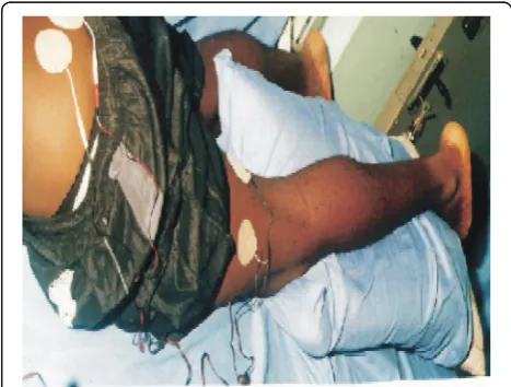
Fig. 1Showing the first tens electrodes placement on one of the subjects. The electrode was placed in such a way to cover the area of pain perception. From the sciatic nerve root origin down the route of sciatic nerve during a treatment session, in this position treatment lasted 20 min and electrodes changed to reflect the position in Fig.2
Fig. 2Showing the second tens electrode placement of on the same subject. The electrode was placed in such a way to cover the area of pain perception. Treatment lasted for 20 min and electrode placement was changed to reflect what is obtained in Fig.3
[image:4.595.305.538.86.271.2] [image:4.595.57.289.493.673.2] [image:4.595.304.538.514.691.2]not biased they were not told the TENS modality on them were just a mere sham. The VAS scores before and after the application of the sham TENS were taking from the patients and appropriately recorded. The subjects positioning and other procedural formalities were the same as described for the test group above.
Materials/equipment of study
A dual channel TENS (EZ 105 Model) with a variable pulse frequency of 2-250 Hz, the variable pulse width of 50–250 microseconds and variable pulse intensity (amplitude) of 0-80 mA produced by Avionix Medical Devices, Texas, USA (Appendix 4) was used for both the test group and the control group. But that of the control was not activated to deliver electrical impulses to the control subjects.
Stethoscope - Littman’s model
Mercury Sphygmomanometer (Accusson model)
Cotton and pin for skin sensation test.
Measuring tape for muscle bulk measurement.
Toilet soap, distilled water, and hand towel for skin cleansing.
Visual analog scale by Price et al. [26]. Used for pain assessment pre and post-treatment.
A Seca model weighing scale calibrated in a kilogram.
A Seca model stadiometer calibrated in centimeter and inches.
Statistical analysis
The SPSS software package version 23 was applied for data analysis. Descriptive statistic of Mean ± SD and mean com-parison test was used to analyze baseline characteristics of subjects. Repeated analysis of variance was (ANOVA) was used to compare mean VAS scores between the TG and CG. A paired t-test was used for pair-wise comparison of pain level in each group across 10 weeks the Statistical level of significance was set atP< 0.05.
Results
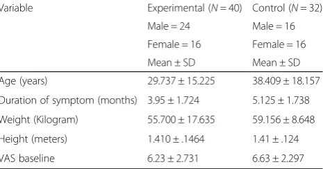
In Table1 baseline characteristics shows 72 patients that participated in the study 40 were males while 32 were fe-males. In the test group 40 patients participated, 24 were males while 16 were females. Their mean age was 29.737 ± 15.225 years and the mean duration of symptom was 3.95 ± 1.724. The mean body weight, height, and VAS were 55.700 ± 17.635 kg, 1.410 ± .1464 m and 6.23 ± 2.731 respectively. For the control group, of the 32 participants, 16 were males while 16 were females. The mean age for the control group was 38.409 ± 18.157 years and mean duration of symptom was 5.125 ± 1.738. The mean body weight, height, and VAS were 59.156 ± 8.648 kg, 1.41 ± .124 m and VAS 6.63 ± 2.297 respectively.
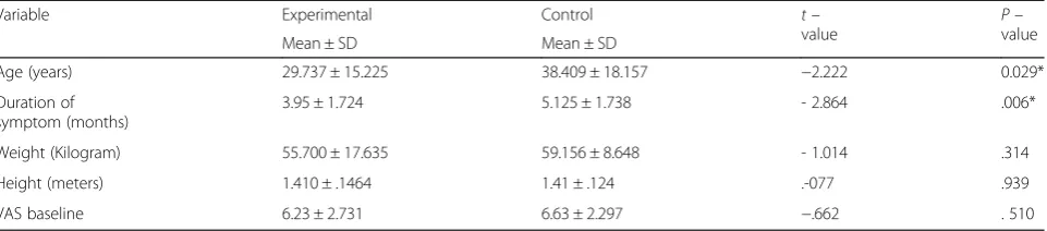
In Table 2there was no significant difference between the test and control group for weight, height, and VAS at baseline. In contrast, there was a significant difference between test and control groups for age and duration of the symptom.
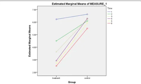
Table3shows the mean pain levels (visual analog scale scores) of the participants in both experimental and con-trol groups across 10 weeks. Unlike in the concon-trol group, there was a continuous decrease in pain levels in the ex-perimental group across the duration of the study. Re-sults of repeated measure ANOVA showed that the pain level among participants in the treatment group at the end (after 10 weeks) of the intervention was significantly lower than that of their counterparts in the control group (F = 16.26; p= 0.01); with the intervention ac-counting for the 19% of the variance (Table4).
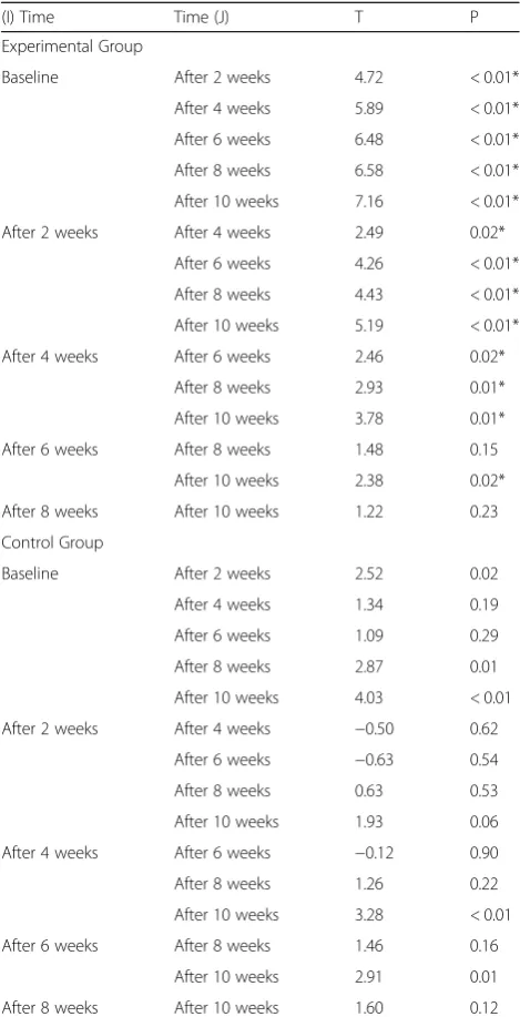
The paired comparison revealed that each of the pain level scores at the end of second, fourth, sixth, eightieth and tenth weeks was significantly lower than the base-line pain level score among the participants in the ex-perimental group. In the exex-perimental group, there was a significant difference in the pain levels in each pair of baseline, second, fourth, sixth, eightieth and tenth week (p< 0.05) except between pain levels at sixth and eight-ieth, and between those at eightieth and tenth weeks. In the control group, the baseline pain level was signifi-cantly lower than that at second, eightieth and tenth weeks (p < 0.05) (Table5).
Discussions
[image:5.595.306.539.111.233.2]This study evaluated the effect of Transcutaneous Elec-trical Nerve Stimulation (TENS) in the management of sciatic pain following intramuscular injection. In carrying out the current study the authors noted that no similar studies had been done in the past regarding the effect of TENS in the management of PISP; however, the effect of TENS in managements other medical and surgical condi-tions were well documented. Interestingly, these studies used TENS device as adjunctive therapy, but most of the outcomes had not equivocally established combination Table 1Baseline characteristics of the groups evaluated at initial assessment
Variable Experimental (N= 40) Control (N= 32)
Male = 24 Male = 16
Female = 16 Female = 16
Mean ± SD Mean ± SD
Age (years) 29.737 ± 15.225 38.409 ± 18.157
Duration of symptom (months) 3.95 ± 1.724 5.125 ± 1.738
Weight (Kilogram) 55.700 ± 17.635 59.156 ± 8.648
Height (meters) 1.410 ± .1464 1.41 ± .124
therapy as producing lasting pain relief on the patients, This notwithstanding, the use of TENS in combination with other therapies were suggested by most previous studies in contrast to using it in monotherapy form as ap-plied in this current study [18, 27]. The authors have noted that previous studies provided promising prelimin-ary evidence about TENS but did not include clear de-scriptions of research design or results. This lack of detailed design has led to most of the published studies on TENS producing conflicting outcomes about the actual ef-ficacy of TENS application. The authors had identified several factors which could contribute to these conflicting reports such as unspecified stimulation parameters, stimu-lation variables not controlled during the research process, different outcome measures, different electrodes place-ments, lack of placebo control, patients presenting at dif-ferent stages in disease process, and failure to monitor or document patient’s compliance [18,28–31]. The outcome of this current study has brought to limelight the import-ance of one of the physical therapy modalities in managing PISP.
The result in Table3shows a trend in mean value var-iations between the two groups, unlike in the control group, there was a continuous decrease in pain levels in the experimental group across the duration of the study. Figure 4 shows the pictorial comparison of the mean pain levels between the two groups at baseline, second, fourth, sixth, eightieth and tenth weeks. This is an indi-cation that TENS, a non-invasive modality, commonly
used in physiotherapy is able to reduce PISP in the treat-ment group, unlike the placebo group. This study agreed with previous studies on the efficacy of TENS in pain management [32–34]. Specifically, the study by White et al. showed that TENS effectively decreased pains in 64 adults with disc herniation related sciatic pain by 23%, while the oral drugs intake was reduced by 15% [35]. This current study was done on the assumption that since TENS had been widely reported to be useful in managing various kinds of pain from dental proce-dures; osteoarthritis of the knee; angina pectoris; low back pain and chronic pain of all sorts; peripheral neur-opathy to rheumatoid arthritis, that it could also be beneficial in managing PISP [17,18,36–42].
[image:6.595.59.540.99.205.2]Results of repeated measure ANOVA showed that the pain level among participants in the treatment group at the end (after 10 weeks) of the intervention was signifi-cantly lower than that of their counterparts in the con-trol group (F = 16.26; p= 0.01); with the intervention accounting for the 19% of the variance. The effect size (partial eta squared) was 0.19 (large) (Table4). The clin-ical implication is that those in the test group responded better to TENS application than those who received STENS. Though there was some improvement in the control group as shown in Table 5 (pair-wise compari-son of pain level), the authors were of the opinion that the said improvement, which might have resulted from the placebo effect and the possibility of subjects taking pain-relieving drugs, did not equal the improvement in the test group. The finding from the current study has rejected the working null hypothesis that there will be no significant difference (p> 0.05) between the TG and the CG after 10 weeks of TENS and STENS applications. It has to be emphasized that TENS achieves its pain relief: pain gate mechanism or the endogenous opioid Table 2Baseline mean comparison of age, duration of symptom, weight, height and visual analogue scale
Variable Experimental Control t–
value Pvalue–
Mean ± SD Mean ± SD
Age (years) 29.737 ± 15.225 38.409 ± 18.157 −2.222 0.029*
Duration of symptom (months)
3.95 ± 1.724 5.125 ± 1.738 - 2.864 .006*
Weight (Kilogram) 55.700 ± 17.635 59.156 ± 8.648 - 1.014 .314
Height (meters) 1.410 ± .1464 1.41 ± .124 .-077 .939
VAS baseline 6.23 ± 2.731 6.63 ± 2.297 −.662 . 510
[image:6.595.56.291.621.731.2]*significant atp< 0.05
Table 3Mean visual analogue scale scores of the participants in each group across 10 weeks
Time Mean ± Standard deviation
Treatment Control
Baseline Scores 6.23 ± 2.73 6.63 ± 2.30
After 2 Weeks 4.50 ± 3.07 6.09 ± 2.28
After 4 Weeks 3.63 ± 3.44 6.25 ± 2.17
After 6 Weeks 2.93 ± 3.15 6.28 ± 2.45
After 8 Weeks 2.73 ± 3.28 5.94 ± 2.03
After 10 Weeks 2.50 ± 3.23 5.50 ± 2.30
Table 4Repeated measure ANOVA comparing the mean visual analogue scale scores between experimental and control groups after the intervention
Degree of freedom Mean square F P Partial Eta Squared
1 596.40 16.26 < 0.01* 0.19
system. The variations in stimulation parameter used to activate these two systems will be briefly considered. Pain relief by means of the pain gate mechanism in-volves activation (excitation) of the beta sensory fibers, and by so doing reduce the transmission of the noxious stimulus from the ‘c’fibers, through the spinal cord and on to the higher center. The Aβ fibers respond better when stimulated at a relatively high rate (in the order of 90–30 Hz or pps) but it is difficult to find support for the concept that there is a single frequency that works best for every patient, this range appears to cover the majority of individuals. An alternative approach is to
stimulate the Aβfibers which respond preferentially to a mode stimulation, which will activate the opioid mecha-nisms, and provide pain relief by causing the release of an endogenous opiate (encephalin) in the spinal cord. A third possibility is to stimulate both nerve types at the same time by employing burst mode stimulation. In this instance, the higher frequency stimulation output (typic-ally at about 100 Hz) is interrupted (or burst) at the rate of 2 --3 bursts per second. When the machine is ‘on’, it will deliver pulses of the 100 Hz rate, thereby activating the Aβ and the pain gate mechanism, but by virtue of the rate of the burst, each burst will produce excitation in the A-delta fibers, therefore stimulating the opioid mechanisms. For some patients, this is by far the most effective approach to pain relief, though as a sensation, numerous patients find it less acceptable than other forms of TENS [43].
As applied to current study, 60-min cumulative treat-ment time was applied on PISP patients per session of treatment as against the recommended 20 or 30 min by previous TENS related studies, especially where TENS was used as addictive therapy modality. Consequent upon this, in carrying out this study, the authors were aware of likely depreciating effect of TENS overtimes probably due to adaptation to particular treatment mode and took mea-sures to vary the parameters to minimize or avert it as it can negatively impact its efficiency in pain relief. This agrees with the scientific finding that the benefit of TENS tends to fall with time [44–46]. Also, depreciation in value-effect of TENS might be due to the adaptation of the nervous system to regular repetitive stimuli [47, 48]. The clinical implication of this is when TENS is applied for a long time as in current study nerve accommodation takes place and may affect the general efficacy of TENS in pain relief. To overcome anticipated accommodation ef-fect in the current study the authors during treatment of TG patients were swinging between continuous and burst TENS modes [49]. The nerve adaptation and accommoda-tion accounted for why some period after TENS is switched on, the patient complained that he/she is not feeling the buzzing or pulsating sensation well enough. Johnson et al. reported that individual patients used a spe-cific pulse frequency but consistently there was a signifi-cant variation in the pulse frequency used by different patients [50]. The authors were also mindful of selecting the time of TENS applications because the stimulating ef-fect does not start immediately but needs some time be-fore its cumulative effect would be felt, this is in line with the outcome of experimental studies reported by previous authors [51,52].
[image:7.595.56.291.109.568.2]In the current study, from Table2, there was a signifi-cant difference (p< 0.05) between the test and control groups for age and duration of symptom at baseline, this difference in baseline the authors noted could possibly Table 5Paired t test showing pair-wise comparison of pain
level in each group across 10 weeks
(I) Time Time (J) T P
Experimental Group
Baseline After 2 weeks 4.72 < 0.01*
After 4 weeks 5.89 < 0.01*
After 6 weeks 6.48 < 0.01*
After 8 weeks 6.58 < 0.01*
After 10 weeks 7.16 < 0.01*
After 2 weeks After 4 weeks 2.49 0.02*
After 6 weeks 4.26 < 0.01*
After 8 weeks 4.43 < 0.01*
After 10 weeks 5.19 < 0.01*
After 4 weeks After 6 weeks 2.46 0.02*
After 8 weeks 2.93 0.01*
After 10 weeks 3.78 0.01*
After 6 weeks After 8 weeks 1.48 0.15
After 10 weeks 2.38 0.02*
After 8 weeks After 10 weeks 1.22 0.23
Control Group
Baseline After 2 weeks 2.52 0.02
After 4 weeks 1.34 0.19
After 6 weeks 1.09 0.29
After 8 weeks 2.87 0.01
After 10 weeks 4.03 < 0.01
After 2 weeks After 4 weeks −0.50 0.62
After 6 weeks −0.63 0.54
After 8 weeks 0.63 0.53
After 10 weeks 1.93 0.06
After 4 weeks After 6 weeks −0.12 0.90
After 8 weeks 1.26 0.22
After 10 weeks 3.28 < 0.01
After 6 weeks After 8 weeks 1.46 0.16
After 10 weeks 2.91 0.01
After 8 weeks After 10 weeks 1.60 0.12
have influenced the outcome of the study but no litera-ture to back it up. Table2revealed no significant differ-ence (p> 0.05) in the subjects’ baseline mean values of height, weight, and VAS between the test and control groups, this the authors assumed did not influence the outcome of this study.
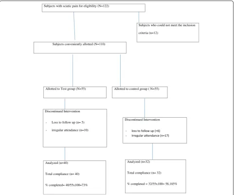
Moreover, deductions from the subjects’ recruitment flowchart (Fig.5) showed more subjects in the CG either absconded or were irregular with treatment compared to what obtained in the TG. These differences might be at-tributed not only to a single factor but a variety of pos-sible factors like not having the desired relief from pain, having good relief after few days of application of TENS, socio-economic and other considerations that are not
within the immediate capacity of the authors to discern. Significantly, however, 40 of 55 subjects (73%) conveni-ently allotted to test group completed the study. Also, 32 of 55 subjects (58%) conveniently allotted to the control group completed the study.
The strength of the study lies in the use of VAS which is still generally accepted as a good tool for measuring varia-tions in pain perception, the cost-effective nature of the measuring tool, the cooperation of the authors and the seemingly novel nature of the study which highlighted the application of TENS as a monotherapy treatment tool and one-hour application TENS for management of PISP.
The current study was however weakened by non-randomization of the samples, low sample size relative to the Subjects with sciatic pain for eligibility (N=122)
Allotted to Test group (N=55) Allotted to control group ( N=55)
Discontinued Intervention
- Loss to follow up (n= 5)
- irregular attendance (n=10)
Subjects who could not meet the inclusion
criteria (n=12)
Subjects conveniently allotted (N=110)
Discontinued Intervention
-Analyzed (n=40)
Total compliance (n= 40)
% completed= 40/55x100=73%
Analyzed (n=32)
Total compliance (n= 32)
% completed = 32/55x100= 58,185%
[image:8.595.58.542.87.488.2]calculated sample size; the possibility that subjects in the groups still ingested one form of analgesic medication or the other; the subjective nature of pain assessment tool used, and the fact that the treatment modes of intensity, frequency and pulse width which varied amongst participants, and time did not provide for equal treatment of participants using a uniformed parameter. Furthermore, the significant differ-ences in baseline VAS scores between age and duration of symptom could have influenced the outcome of the study.
Conclusions
The outcome of the study showed significant improvement in PISP after 10 weeks TENS application. It also shows that STENS also achieved varied pain relief to control subjects but not significant enough to compare the effect of TENS on the test group. This has shown the usefulness of TENS in managing PISP of sub-acute and chronic nature. The im-plication for management and rehabilitation is that TENS alone is beneficial in the management of injection-related nerve pain as demonstrated from the outcome of the current study. A future line of study is consideration of comparative effects of TENS and TENS in combination in the management of PISP. Also, future studies that should factor the limitations highlighted above are advocated by
the authors as it will help to strengthen quality, acceptabil-ity, and generalizability of the study outcome.
Abbreviations
CG:control group; NAUTHEC: Nnamdi Azikiwe University Teaching Hospital Ethics Committee; NII: Nerve Injection Injury; PISP: post injection sciatic pain; SD: Standard division; SNII: Sciatic Nerve Injection Injury; STENS: sham transcutaneous electrical nerve stimulation; TENS: transcutaneous electrical nerve stimulation; TG: treatment group; VAS: Visual Analog scale; X: Mean
Acknowledgments
All the staff and members of the Department of Physiotherapy, NAUTH, Nnewi, and Landmark Physiotherapy Services are well appreciated for their support and encouragement.
Funding
No form of funding was received from any source.
Availability of data and materials
The datasets used and/or analyzed during the current study are available from the corresponding author on reasonable request.
Authors’contributions
Authors UPO, SCI, and EYI were involved in the conception and design/ data collection and implementation; AVE, ICE, and AFM were involved in the analysis of data, interpretation of results and write up of this study, while ECO, OPI, and OAE were involved in the design and editing of the main paper. All the authors were involved in drafting the manuscript and critically revising it. The final version of the manuscript was read and approved by all the authors.
[image:9.595.59.537.86.370.2]Ethics approval and consent to participate
Ethics approval was obtained from the Nnamdi Azikiwe Univerity Teaching Hospital Ethics Committee and informed consent obtained from participants before the commencement of the study. The consent obtained from participants was written.
Consent for publication
Consent for publication was obtained from the patient that appeared in Figs.
1,2and3.
Competing interests
The authors declare that they have no competing interests.
Publisher’s Note
Springer Nature remains neutral with regard to jurisdictional claims in published maps and institutional affiliations.
Author details
1Department of Physiotherapy, Nnamdi Azikiwe University Teaching Hospital,
Nnewi, Anambra State PMB 5025, Nigeria.2Department of Medical Rehabilitation, Faculty of Health Sciences and Technology, University of Nigeria|, Enugu Campus, Enugu State, Nigeria.3Department of Medical Rehabilitation, Faculty of Health Sciences and Technology, Nnamdi Azikiwe University, Awka, Anambra State, Nigeria.
Received: 16 May 2018 Accepted: 7 November 2018
References
1. Kline, DG, Kim, D Midha, R. (1998). Management and results of sciatic nerve injuries: a 24-year experience. J Neurosurg 1998; 80; 13–23.
2. Tak SR, Dar GN, Halwai MA. Post-injection nerve injuries in kasmir: a menace overlooked. J Res Med Sci. 2008;13:244–7.
3. Arnozan, quoted by Wexburg E. Neuritis and polyneuritis (1835). In: Bumke O, Foerter O, editors. Handbuch der neurologie, vol 9. Berlin: Springer-Verlag; 1935. p. 87.
4. Turner GG. The site for intramuscular injections. Lancet. 1929;196:819. 5. Mishra P, Stringer MD. Sciatic nerve injury from intramuscular injection: a
persistent and global problem. Int J Clin Pract. 2010;64:1573–9. 6. Miller MA, Pisani E. The cost of unsafe injections. Bull World Health Organ.
1999;77:808–11.
7. Small SP. Preventing sciatic nerve injury from intramuscular injections literature review. J Adv Nurs. 2004;47:287–96.
8. Garfin S.R (2005). Sciatica: Description and diagnosishttps://www. spineuniverse.com/conditions/sciatica
9. Yeomans, S.G (2003). Sciatica and Sciatic Nerve.https://www.soinehealth.com
10. Sluka KA. The basic science of TENS and clinical implications. Article, pg 1; 2000.
11. Acute Pain Management Guideline Panel. Acute pain management: operative or medical procedures and trauma: a clinical practice guideline. No. 1. Rockville, Md.: Agency for Health Care Policy and Research, 1992. (AHCPR publication no. 92–0032.)
12. American Physical Therapy Association (1990). Transcutaneous Electrical Nerve Stimulation. (2009) Wikipedia, Free Encyclopedia.https://simple.m. wikipedia.org.wiki
13. Robison AJ, Snyder-macker L. Clinical electrophysiology: electrotherapy and electrophysiology testing. 3rd ed. Baltimore: Williamson Wilkins; 1995. 14. Katz J, Melzack. R. (1999). Measurement of pain. Surg Clin North Am. 1999
Apr; 79(2):231–252.
15. Scott J, Huskisson EC. Graphic representation of pain. Pain. 1976;2:175–84. 16. Carlsson AM. Assessment of chronic pain. Aspects of the Reliability and
Validity of the Visual Analogue Scale. Pain. 1983;16(1):87–101.
17. Kumar D, Marshall HJ. Diabetic peripheral neuropathy: amelioration of pain with transcutaneous electrostimulation. Diabetes Care. 1997;20(11):1702–5. 18. Kumar VN, Red Ford. TENS in rheumatoid arthritis. Arch Phys Rehabil. 1982;
63:595–6.
19. Finsen V, Persen L, Lovlien M, et al. Transcutaneous electrical nerve stimulation after major amputation. J Bone Joint Surg Br 1988. 1988;70(1): 109–12.
20. Katz J, Melzack R. Auricular transcutaneous electrical nerve stimulation (TENS) reduces phantom limb pain. J Pain Symptom Manag. 1991;6(2): 73–83.
21. Fatunde OJ, Familusi JB. Injection-induced sciatic nerve injury in Nigerian children. Cent Afr J Med. 2001;47(2):35–8.
22. Daniel WW. Biostatistics. A foundation for analysis in the health sciences 7th
edition. New York: John Wily and Sons; 1999.
23. Lwanga SK, Lemeshow S. Sample size determination in health studies: a practical manual. Geneva: World Health Organization; 1999.
24. Naing L, Winn T, Rusli BN. Practical issues in calculating the sample size for prevalence studies. Arch Orofac Sci. 2006;1:9–14.
25. Ali J, Yaffe CS, Sessle BJ. The effect of transcutaneous electrical nerve stimulation on postoperative pain and pulmonary function. Surgery. 1981;89:507–12. 26. Price DC, Megrath PA, Rafii A. The validation of visual analog scales as rational
scale measures for chronic and experimented pain. Pain. 1983;17:45–56. 27. Abelson K, Langley GB, Vilieg M, Wigley RD. Transcutaneous electrical nerve
stimulation in rheumatoid arthritis. N Z Med J. 1983;96:156–8. 28. Levy, A, Dalith, M, Abramovici, A, Pinkhand, J., Weinberger, A. (1997). TENS in
experimental acute arthritis of physical medicine and Rehabilitation pp 68, 75–78. 29. Mannheimer C, Lund S, Carlsson CA. The effect of TENS on joint pain in
patients with rheumatoid arthritis. Scand J Rheumatol. 1978;7(1):13–6. 30. Reitman C, Esses SI. Conservative options in the management of spinal
disorders. Part 1: bed rest, mechanical and energy transfer therapies. Am J Orthop. 1995;24:109–16.
31. Robinson AJ. TENS for the control of pain in musculoskeletal disorders journal of orthopedics and sports. Phys Ther. 1996;24:204–27. 32. Johnson MI, Ashton CH, Thompson JW. An in-depth study of long-term
uses of TENS. Implications for Clinical use of TENS on pain. 1991;44:221–9. 33. Oya T et al (2004). Journal of Back and Musculoskeletal Rehabilitation Vol. 17
number 34 pg. 127–133.
34. Fisbian V, Persen L, Lovlien M, et al. Transcutaneous electrical nerve stimulation after major amputation. J Bone Joint Surg Br. 1988;70(1):109–12. 35. White PF, Jennifer Philips BS, Timothy JP, William FC. Percutaneous Electrical
Nerve Stimulation (PENS) A Promising Alternative Medicine Approach to pain management; 1999.
36. Meechan JG, Gowans AJ, Welbury RR. (1998). The use of patient-controlled transcutaneous electrical nerve stimulation (TENS) to decrease the discomfort of regional anesthesia in dentistry: a randomized trial. J Dent 1998;26(5–6):417–420.
37. Soo-Ampon S, Vongsavan N, Apai W. (1997). Effect of TENS on experimentally induced dental pain. J Dent Res 1997; 76:59.
38. Harvey M, Elliott M. Transcutaneous electrical nerve stimulation (TENS) for pain management during cavity preparations in pediatric patients. ASDC J Dent Child. 1995;62(1):49–51.
39. Cheing GL, Hui-Chan CW, Chan KM. Do four weeks of TENS and/or isometric exercise produce a cumulative reduction of osteoarthritic knee pain? Clin Rehabil. 2002;16(7):749–60.
40. Mannheimer C, Emanuelsson H, Waagstein F, et al. Influence of naloxone on the effects of high frequency transcutaneous electrical nerve stimulation in angina pectoris induced by atrial pacing. Br Heart J. 1989;62(1):36–42. 41. Jarzem P, Harvey EJ, Arcaro N, Kazaronski J. Transcutaneous electrical nerve
stimulation fir nonacute low back pain: a non- randomized double-blind study of conventional, Nu- wave form acupuncture- type and sham therapies. 1997. Cochranelibrary- wily.com/ doi/epdf.
42. Carroll D, Moore RA, McQuay HJ, et al. Transcutaneous electrical nerve stimulation (TENS) for chronic pain; (3): (Cochrane review). Cochrane Database Syst Rev. 2001;4:CD003222.
43. Ellis J. A retrospective study of long-term users of TENS. Br J Ther Rehabil. 1996;3(2):88–93.
44. Loeser JD, Back RG, Christma A. Relief of by TENS. J Neurosurg. 1975;42:308–14. 45. Taylor, PI, Haliott, M, Faberty Faberty H. (1981). Treatment of OA of the knee
with TENS Pg. 233–246.Tinegate H, McLelland J (2002). Correspondence. Clin Lab Hematol 24(6):389–390.
46. Bates JAV, Nathan PW. Transcutaneous electrical nerve stimulation for chronic pain. Anesthesia. 1980;35:817.
47. Thompson JN. The role of TENS for the control of pain. In: Doyle D, editor. International symposium on pain control. London: Royal Society of Medical Services; 1987. p. 27–47.
49. Frampton VM. TENS Clayton’s electrotherapy 10thedition; 1998. p. 77–88.
50. Johnson MI, Ashton CH, Thompson JW. The consistency of pulse frequency and pulse patterns of transcutaneous electrical nerve stimulation (TENS) used by chronic pain patients. Pain. 1991;44:231–29.
51. Sjolund B, Erikesson MBH. The influence of naloxone analgesia produced by peripheral conditioning stimulation. Brain Res. 1979;173:295–301. 52. Woolfe SI, Gersh HR, Rao VR. Examination of electrodes placement and