LEABHARLANN CHOLAISTE NA TRIONOIDE, BAILE ATHA CLIATH TRINITY COLLEGE LIBRARY DUBLIN OUscoil Atha Cliath The University of Dublin
Terms and Conditions of Use of Digitised Theses from Trinity College Library Dublin
Copyright statement
All material supplied by Trinity College Library is protected by copyright (under the Copyright and Related Rights Act, 2000 as amended) and other relevant Intellectual Property Rights. By accessing and using a Digitised Thesis from Trinity College Library you acknowledge that all Intellectual Property Rights in any Works supplied are the sole and exclusive property of the copyright and/or other I PR holder. Specific copyright holders may not be explicitly identified. Use of materials from other sources within a thesis should not be construed as a claim over them.
A non-exclusive, non-transferable licence is hereby granted to those using or reproducing, in whole or in part, the material for valid purposes, providing the copyright owners are acknowledged using the normal conventions. Where specific permission to use material is required, this is identified and such permission must be sought from the copyright holder or agency cited.
Liability statement
By using a Digitised Thesis, I accept that Trinity College Dublin bears no legal responsibility for the accuracy, legality or comprehensiveness of materials contained within the thesis, and that Trinity College Dublin accepts no liability for indirect, consequential, or incidental, damages or losses arising from use of the thesis for whatever reason. Information located in a thesis may be subject to specific use constraints, details of which may not be explicitly described. It is the responsibility of potential and actual users to be aware of such constraints and to abide by them. By making use of material from a digitised thesis, you accept these copyright and disclaimer provisions. Where it is brought to the attention of Trinity College Library that there may be a breach of copyright or other restraint, it is the policy to withdraw or take down access to a thesis while the issue is being resolved.
Access Agreement
By using a Digitised Thesis from Trinity College Library you are bound by the following Terms & Conditions. Please read them carefully.
The effects of group II mGluR agonists on short-term
depression and neural coding in the rat dentate gyrus
in vitro.
by
John J. Kilbride BSc.
Thesis submitted for the degree of Doctor of Philosophy at the
University of Dublin, Trinity College.
Submitted October 1999
Department of Physiology
Trinity College
i Table of Contents
! Table of contents i
II Acknowledgements vii
III Declaration viii
IV Summary ix
V List of Abbreviations xi
Chapter 1 General Introduction
1.1 Preface 2
1.2 Synaptic transmission 3
1.3 Hippocampus 5
1.3.1 Principal hippocampal neurons 6
1.3.2 Trisynaptic circuitry 8
1.4 Glutamate receptors 9
1.4.1 AMPA / kainate receptors 9
1.4.2 NMDA receptors 10
1.4.3 Metabotropic glutamate receptors 12
1.5 Presynaptic Inhibition 16
1.6 Short-term plasticity 18
1.6.1 Short-term depression 19
1.6.2 Release statistics 21
1.6.3 Synaptic depression and neural coding 22
1.6.4 Short-term facilitation 23
1.6.5 Paired-pulse stimulation 24
1.7 Long-term plasticity 25
1.7.1 Long-term potentiation 25
Chapter 2 Materials and Methods
2.1 Animals 29
2.2 Artifical cerebro-spinal fluid (ACSF) 29
2.3 Preparation of slices 29
2.4 Electrophysiological recordings 30
2.4.1 Experimental set-up 30
2.4.2 Electrodes 31
2.4.3 EPSP recording 31
2.5 Data analysis and statistics 32
2.6 Compounds 33
Chapter 3 Neurophysiological studies of the presynaptic inhibitory action of the group II metabotropic glutamate receptor agonists, LY354740 and DCG-IV, in the rat hippocampus, in vitro.
3.1 Abstract 35
3.2 Introduction 36
3.3 Materials and methods 39
3.3.1 Slice preparation 39
3.3.2 Electrophysiology 39
3.3.3 Data analysis and statistics 40
3.3.4 Compounds 41
3.4 Results
3.4.1 LY354740 and DCG-IV inhibit the
3.4.2 LY354740 and DCG-IV inhibit the
EPSP in the lateral perforant path 43
3.4.3 LY354740 has no effect on EPSP
amplitude in CA1 44
3.5 Discussion 45
Chapter 4 Presynaptic group II mGluR modulation of short-term plasticity in the dentate gyrus
4.1 Abstract 49
4.2 Introduction 50
4.3 Materials and methods 54
4.3.1 Slice preparation 54
4.3.2 Electrophysiology 54
4.3.3 Data analysis and statistics 56
4.3.4 Compounds 57
4.4 Results 58
4.4.1 Short-term depression in the medial
and lateral perforant paths 58
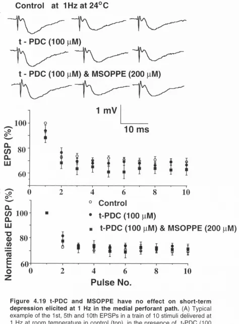
4.4.2 Short-term depression in the medial perforant path is relieved by activation of group II mGluR at high stimulation
4.4.3 Short-term depression in the medial perforant path is relieved by lowering extracellular calcium at high stimulation
frequencies 61
4.4.4 Short-term depression in the lateral perforant path is relieved by activation of group III mGluR at high stimulation
frequencies ' 63
4.4.5 Presynaptic mGluR autoreceptors are not involved in short-term depression
in the medial or lateral perforant paths 65
4.4.6 Cyclothiazide reduced high-frequency induced short-term depression in the
medial perforant path 67
4.5 Discussion 69
Chapter 5 Neural coding in the medial perforant path is modulated by presynaptic group II mGluR
5.1 Abstract 78
5.2 Introduction 79
5.3 Materials and methods 85
5.3.1 Slice preparation 85
5.3.2 Electrophysiology 85
5.3.3 Data analysis and statistics 86
5.3.4 Compounds 87
5.4 Results
5.4.1
5.4.2
5.4.3
5.4.4
5.4.5
The effect on synaptic transmission of a fixed percentage instantaneous increase in the presynaptic firing rate
of 50 % 88
The effect on the EPSP per unit time of a fixed percentage instantaneous increase in the presynaptic firing rate
of 50 % 90
The effect on synaptic transmission of a fixed percentage instantaneous increase in the presynaptic firing rate
of 1 0 0 % 91
The effect on the EPSP per unit time of a fixed percentage instantaneous increase in the presynaptic firing rate
of 1 0 0 % 92
The effect on synaptic transmission of a fixed increm ent instantaneous increase in the presynaptic firing
rate of 5 Hz 92
The effect on the EPSP per unit time of a fixed amplitude instantaneous increase in the presynaptic firing rate
5.4.7 The effect of the group II mGluR agonist LY354740 on the transient postsynaptic response induced by a fixed percentage
rate change (100 %) 94
5.4.8 The effect of the group II mGluR agonist LY354740 on the changes in the EPSP per unit time induced by fixed percentage
rate change (100 %) 96
5.4.9 The effect of the group II mGluR agonist LY354740 on the transient postsynaptic response induced by a fixed increment
rate change (5 Hz) 96
5.4.10 The effect of the group II mGluR agonist LY354740 on the changes in the EPSP per unit time induced by fixed increment
rate change (5 Hz) 97
5.4.11 LTD does not affect neural coding
in the medial perforant path 98
5.5 Discussion 100
5.5.1 Rate vs. temporal coding 100 5.5.2 Modulation of neural coding by
activation of presynaptic group II mGluR 103 5.5.3 LTD induction does not affect neural
coding in the medial perforant path 104
VI Bibliography 107
II Acknowledgements
In particular I would like to thank the following people: Professor Roger Anwyl, Professor Michael Rowan, Professor Marina Lynch, Mr. Kieran Walsh, Dr. Nick Breakwell, Dr. Jianqun Wu, and Dr. Tony Rush.
The Health Research Board of Ireland and Enterprise Ireland for financial support.
All the staff members of the Departm ent of Physiology, Trinity College.
Special thanks to all my friends, who have ensured it has been three years I will never forget.
Ill Declaration
I declare that this work has not been submitted previously as a thesis for a degree at this or any other institution and that it is entirely my own work. The Trinity College Dublin Library may lend this thesis without restriction.
^C)lu^
UctLry[J^
IV Summary
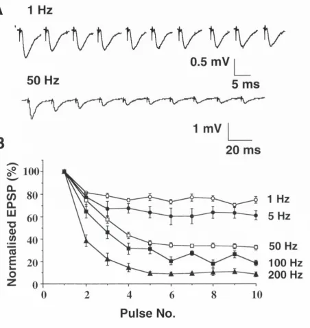
Short-term depression is an activity-dependent reduction in synaptic efficacy which has been observed at a wide variety of synapses in the CNS, and is often attributed to a depletion of the available store of neurotransm itter. At the medial and lateral perforant path-granule cell synapses, short trains of stimuli at a range of frequencies between 0.5 Hz and 200 Hz resulted in a reversible depression of the evoked EPSP amplitude. A steady-state level of short-term depression was attained after 5-10 stimuli. In both pathways the steady-state EPSP am plitude decreased with increasing stimulation frequencies. In the medial perforant path the onset of depression was well fitted (r2 = 0.935) with a single exponential function over all frequencies tested, with a time constant of decay ranging from 5 ms at 200 Hz to 1470 ms at 0.5 Hz.
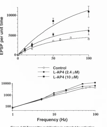
The tim e-averaged postsynaptic response (steady-state EPSP amplitude x frequency), which is a reliable estimate of transm itter mobilisation (Curtis and Eccles, 1960), increased linearly with frequency up to a limiting frequency of 10 Hz - 20 Hz in the medial and 20 Hz - 50 Hz in the lateral perforant paths. Short-term depression was found to impart a gain control m echanism on synapses in the medial perforant path. Specifically, the existence of a limiting frequency reduced the rate coding capabilities of these synapses, in so far as the postsynaptic response to high-frequency stim ulation was independent of stimulation frequency for rates above -2 0 Hz. This was accom panied by an increased sensitivity to transient rate changes which enables these synapses to respond to relative rather than absolute changes in firing rate and to discriminate preferentially between small fluctuations in the firing rate of slowly as opposed to rapidly firing afferents.
mGluR agonist LY354740, induced a reversible, dose-dependent depression of the EPSP amplitude in the medial perforant path. The EC5 0 value was found to be 115 ± 16 nM whilst the maximal depression observed was -8 0 % with a concentration of 10 pM. In the lateral perforant path, L-AP4 induced a dose-dependent reversible depression of synaptic transmission, with an EC5 0 value of 2.4 pM and a maximal depression of -8 0 % with 10 pM.
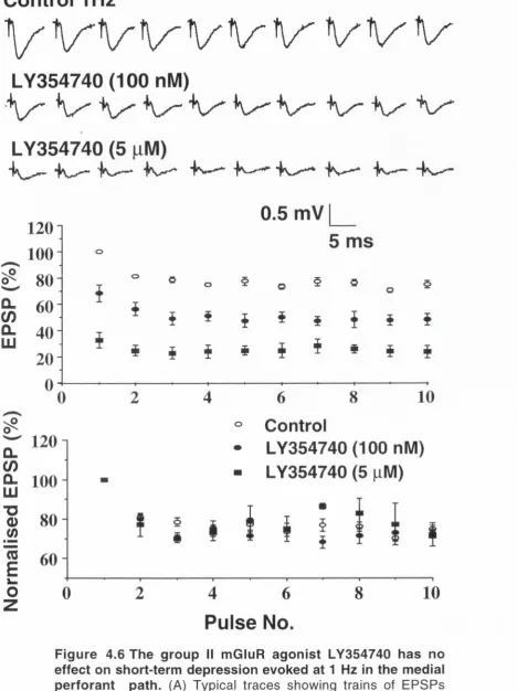
Modulation of short-term plasticity by selective activation of either group I! or group III mGluRs was exam ined over a range of frequencies between 1 and 200 Hz. Short-term depression was not altered by mGluR activation up to 20 Hz and 10 Hz in the medial and lateral perforant paths respectively. At frequencies greater than these, a dose-dependent inhibition of short-term depression was observed. The selective activation of group II and III m GluRs extended the linear range, over which transm itter mobilisation was independent of stim ulation frequency, and as such is further evidence of the attenuation of synaptic depression at high frequencies by LY354740 and L- AP4 respectively. The most likely mechanism through which short-term depression is attenuated by the activation of groups II and III mGluRs during HFS, is through a reduction in presynaptic Ca2+ influx which leads to a lessening of neurotransm itter depletion and a concom itant enhancem ent of facilitation. This theory is strengthened by the findings that lowering extracellular Ca2+ resulted in a sim ilar reduction of short-term depression to that resulting from activation of group II mGluR.
V Abbreviations
(1 S,3S)-ACPD (1 S,3S)-1 -Aminocyclopentane-1,3-dicarboxylic acid AMPA a-amino-3-hydroxy-5-methyl-4-isoxalone proprionic
acid
cAMP cyclic 3’,5'-adenosine monophosphate cGMP cyclic guanosine monophosphate CNS Central nervous system
Cyclothiazide 6-chloro-3,4-dihydro-3-(2-norbornen-5-yl)-2H-1,2,4-benzothiadiazine-7-sulphonamide-1,1-dioxide
DAG Diacylglycerol
DCG - IV (2S,2'R,3'R)-2-(2',3'-Dicarboxycyclopropyl)glycine EPSC Excitatory postsynaptic current
EPSP Excitatory postsynaptic potential
GABA Gamma-aminobutyric acid
IP3 inositol (1,4,5)-triphosphate
L-AP4 L(+)-2-Amino-4-phosphonobutyric acid LCCG-1 (2S,1 'S,2'S)-2-carboxycyclopropyl)glycine LFS Low frequency stimulation
L-SOP L - serine o - phosphate LTD Long - term depression LTP Long - term potentiation
L-frans-2,4,PDC frans-4-Carboxy-L-proline / L-^rans-pyrrolidine-2,4-dicarboxylic acid.
LY354740 (+)-2-aminobicyclo[3.1.0]hexane-2-6-dicarboxylic acid
mGluR Metabotropic glutamate receptor MSOP (RS)-alpha-Methylserine-O-phosphate
MSOPPE (RS)-alpha-Methylserine-O-phosphate monophenyl ester)
NMDA N-methyl-D-aspartic acid
NO Nitric Oxide
Picrotoxin PLC
Chapter 1
1.1 Preface
One of the m ost intriguing questions in science is how the brain
represents the environment around us. What we do know is that the brain
uses a com bination of electrical and biochemical signals to represent and
convey information internally. What remains more of a mystery is exactly
how these signals are encoded to represent an accurate picture of the
external world. It does appear that the intervals between the electrical
spikes are every bit as important as the spikes themselves. We can only
guess at the degree of accuracy which our brains achieve, because we
have nothing to compare them against. This illustrates part of the problem
facing the scientific community in so far as the instrument we are forced
to use in seeking to understand the nervous system, is the very object we
wish to com prehend and therefore it's possible we don't have the
processing power to understand our own hardware.
Synaptic transm ission involves dynamic, plastic processes. The fact that
some of these processes have been proposed as the cellular correlates of
learning and m em ory (Stevens, 1988; Bliss and C ollingridge, 1993;
M artinez and Derrick, 1996), in time, may or may not be proven correct.
The general public are largely interested in disorders of the mind or of
mental processes, that is, they only want to know why things go wrong
and how they can be fixed. The greatest incentive to study synaptic
transm ission lies not in finding out why it all goes wrong but rather in
discovering how it works so well for so long.
1.2 Synaptic transmission
(amplification, saturation, thresholding) processing once in the domain of
the postsynaptic cell (Koch, 1997). Nevertheless the net effect is to alter
the postsynaptic membrane potential such that at the cell soma when the
threshold fo r action potential generation is surpassed, a "spike" is
produced and propagated both down the axon and back into the dendrites.
In this way information is passed from neuron to neuron throughout the
neural network.
1.3 Hippocampus
The hippocampus is a curved mass of cortical tissue and it extends the
length of the floor of the temporal horn of the lateral ventricle in the limbic
system. The hippocampus consists of Ammon's horn, the dentate gyrus
and the subiculum. In all mammals two interlocking c-shaped principal cell
layers make up the hippocampus. They are the granule cell layer of the
dentate gyrus and the pyramidal cell layer of Ammon's horn. The latter
may be subdivided into four subfields: CA1 to CA4 (Cornu Ammonis)
(Witter, 1989; Patton and McNaughton, 1995)
1.3.1 Principal hippocampal neurons
Dentate granule cells
The granule cell layer consists of small (< 10 )j.M wide) spherically shaped
cell bodies, arranged 4 - 6 cells deep. In the rodent it has a distinctive U -
shaped structure (Fig. 1.1). Each granule cell gives rise to several
dendrites, which branch extensively and are densely covered with spines.
The dendritic tree extends from the apical portion of the cell body into the
m olecular layer where connections are made with many afferents from
several sources. The axon extends from the basal pole of the cell body
tov\^ards the hilar region, from where many collaterals emerge. The thin
unmyelinated axons are referred to as mossy fibres on account of the
characteristic appearance of their synaptic terminals, which exit the hilus
and enter stratum lucidum of CAS where they form synaptic connections.
(Witter, 1989; Patton and McNaughton, 1995).
CA3 pyramidal cells
# -1IS7AI
A bbreviations for Figures Il-VI cortical layers
ab angular bundle alv ajveus
CA1^,3 subfields of .‘'unm on's horn DG dentate gyrus
DG, infrapyramidal blade of the dentate gyrus DG, suprapyramldal blade of the dentate gjTus EC entorhinal cortex
n fimbria
HDG hilus o f the dentate gyrus hf hippocampal fissure PAR parasubiculum PC perirhinal cortex PRE presubiculum rs rhinaJ sulcus S subiculum sm molecular layer so stratum oriens sr stratum radiatum ve lateral ventrical
CA1 pyramidal cells
The cell bodies of the pyramidal layer are arranged 3 - 6 cells deep. They
are m u lti-pola r neurons with an elaborate dendritic tree extending
perpendicular to the cell body layer in both directions. Thick apical
dendrites pass through the cell sparse stratum oriens, stratum radiatum
and stra tu m la c u n o s u m -m o le c u la re , w here nu m erous syn a p tic
connections are made with afferent fibres. The axon and basal dendrites
originate from the base of the cell body, and after an initial spine-free
segment they become richly coated with spines. (Witter, 1989; Patton and
McNaughton, 1995).
Interneurons
1.3.2 Trisynaptic circuitry
The basic circuitry of the hippocampus has been known since the time of
Cajal at the turn of the century (Cajal, 1901). Arbitrarily, the entorhinal
cortex is considered to be the starting point in a unidirectional network of
excita tory connections through the dentate gyrus, CA3, CA1 and
subiculum. Functionally this means information originating in other cortical
areas can enter the hippocampus via the entorhinal cortex, be filtered and
processed throughout the hippocampal circuit before being returned to the
neocortex via the entorhinal cortex (Jones, 1993).
cells project to the subiculum and to the deep layers of the entorhinal
cortex. (Witter, 1989; Patton and McNaughton, 1995; Jones, 1993).
1.4 Glutamate receptors
1.4.1 AMPA / kainate receptors
Glutamate is the endogenous agonist at the AMPA / kainate, NMDA, and
metabotropic glutamate receptors. The AMPA receptor takes its name
from the s p e c ific a g o n ist, a-amino-3-hydroxy-5-methyl-4-isoxalone
antagonists at tine AMPA receptor include 6-cyano-7-nitroquinoxaline-2,3-
dione (CNQX) and 2,3-dihydroxy-6-nitro-7-sufamoyl-benzo (F) quinoxaline
(NBQX) (Honore et al., 1988; Sheardown et al., 1990).
The existence of a n oth er class of glutam ate receptor, w hich is
differentiated on the basis of its selectivity for kainate, has been known for
many years. However it is only recently that their functional significance
has become a little more clearer, due to the development of more specific
pharmacological compounds which enable the smaller kainate currents to
be teased out from the larger AMPA receptor mediated currents (Lerma et
al., 1997; Chittajallu et al., 1999). To date five kainate receptor subtypes
have been identified, namely GluR5,6,7 and KA1 and KA2. All share the
same topology, with an extracellular term inus, three transm em brane
domains (TM1, TM3 and TM4) leading to an intracellular C-terminal
domain. Tetanic stimulation delivered to the mossy fibre pathway into the
CAS region in the hippocampus elicits detectable kainate currents, which
suggests that the conductance through the channel is very small and is
not observable during low frequency test stimulation (Chittajallu et al.,
1999).
1.4,2 NMDA receptors
transm em brane dom ains with a loop between the first and second
domains. In comparison to AMPA receptors, the affinity for glutamate is
higher at NMDA receptors (EC50 ~ 0.4 - 1.7 ^M), and although they are
slower to open (> 2 ms), once open, they remain open for longer (Conti
and Weinberg, 1999). This receptor is coupled to cation channels which
allow the flux of Na+, K+ and most pertinently, Ca2+, across the cell
membrane. However, unlike the AMPA ligand-gated channels, these
channels are also voltage-gated, that is they must be depolarised before
the Mg2+ which blocks the channel at the resting membrane potential is
rem oved. T h e re fo re the se recepto rs are usu a lly a c tiv a te d by
depolarisation arising from AMPA receptor-coupled cation channels and
the NMDA receptor mediated current then enhances this excitatory
synaptic transmission. It is these two properties (high Ca^+ permeability
and voltage dependent channel block by Mg^+) that underlie its
involvement in the induction of LTP. (Collingridge et al., 1983a,b Malenka
and Nicoll, 1993).
In addition to the essential glycine binding site (Johnson & Asher, 1987),
several co-factors can modulate the channel conductance, for example
Zn2+and opioids. (D)-2-amino-5-phosphonopentanoic acid (D-AP5) and
(R )-3 -(2 -c a rb o x y p ip e ra z in -4 -y l)p ro p y l-l-p h o s p h o n ic acid ((R )-C P P )
(Watkins et al., 1990) act as effective competitive blockers at the NMDA
receptor, w hilst the p o lycyclic am ine, 5-m e th yl-1 0 ,1 1 -d ih yd ro -5 H -
dibenzo[a,d]cyclohepten-5,10-imine (MK-801) is a potent, non competitive,
1.4.3 Metabotropic glutamate receptors (mGluRs)
The cla ssica l m odel of fast e xcita to ry syna ptic tra n sm issio n at
glutamatergic synapses involves glutamate acting on lonotropic receptors
such as the AMPA and NMDA receptors. During the 1980s, it was
discovered that excitatory synaptic transmission could be modulated by
glutamate receptors linked directly to second messenger systems via GTP
-binding proteins with the discovery that of glutamate receptors coupled to
activation of phosphoinositide hydrolysis (Sladeczek, et al., 1985). Prior to
this, the prevailing wisdom held that such modulation of cell excitability
and synaptic transmission could only occur from extrinsic afferents such
as acetylcholine, serotonin and dopam ine, all of which activate G -
proteins. (Hille, 1994; Conn and Pin, 1997). As glutamate is the ubiquitous
p rin ciple e x cita to ry n e u ro tra n s m itte r in the brain, and syna ptic
transmission across glutamatergic circuits is at the heart of a number of
physiological processes such as learning and memory, then it is not
surprising that a mechanism exists whereby glutamate can regulate its
action on ionotropic receptors.
develop such compounds underlies much of the research interest in this
area.
Metabotropic glutamate receptors do not share any sequence homology
and are larger than all other G-protein coupled receptors (GPCR). As such
they may be regarded as a new family of GPCRs. All mGluRs consist of a
large extracellular dom ain (N-term inal) which houses the glutam ate
binding site. The intracellular loops separating the seven transmembrane
domains (TMDS) corresponds to the G-protein coupling sites. Within the
cytoplasm, there is a carboxyl-terminal domain whose function is as yet
unknown (Fig. 1.2) (Conn and Pin, 1997).
The fun ction al dive rsity of the mGluR fam ily is reflected by the
identification of 8
subtypes (mGluR 1-8) to date. They have been classified
into three groups (I, II, and III) based on their amino acid sequence
homology, transduction mechanisms and pharmacological profile.
(I) Group I mGluR
Group I mGluR (mGluR 1 and 5) are selectively activated by 3,5 -
dihydroxyphenylglycine (DHPG). They are coupled to phospholipase C
which induces the hydrolysis of ph osphoinositide (PI) and as a
consequence the intracellular levels of IP3
and DAG are elevated. IP3
acts
on receptors on the intracellular calcium stores, releasing sequestered
calcium into the cyctoplasm. DAG activates protein kinase C (PKC) which
in turn phosphorylates numerous substances.
cells the strongest labelling is for mGluR 1, which seem to be localised to
postsynaptic elements throughout the hippocampus. (Shigemoto et al.,
1997). In the hippocam pus, group I mGluRs mediate the inhibition of
excitatory synaptic transmission at the Schaffer collateral - CA1 synapses
(Gereau and Conn, 1995).
(II) Group II mGluR
Group II mGluR consists of the mGluR 2 and 3 subtypes, which are about
70 % identical to each other in their amino acid sequence. Both receptors
are negatively coupled to adenylate cyclase. Although the activation of
these receptors with pharmacologicai agonists is usually associated with
an inhibition in forskolin -stimulated cAMP levels (Pin and Duvoisin, 1995),
a number of studies have found that cAMP levels are elevated after group
II mGluR activation (Reid et al., 1996).
Immunocyctochemical studies have shown that in hippocampus the areas
most densely populated with group II mGluR include the neuropil of the
stratum lacunosum moleculare (CA1) and the medial perforant path of the
dentate gyrus (Shigemoto et al., 1997). Immunocytochemistry for mGluR 2
/ 3 has also been found in the neuropil at the mossy fibre terminal zone
(Yokai et al., 1996) and the inner layer of stratum lacunosum moleculare
of CA3. Immunoelectron microscopy suggests that of the two subtypes,
mGluR 3 is present at a lower density in the hippocampus. (Petralia et al.,
1996; Shigemoto et al., 1997). In contrast to group III mGluRs, group II
(and most likely mGluR 2) mGluRs are often found at the preterminal
region of small unmyelinated axons. Indeed they are rarely located at the
presynaptic membrane specialisation (Otterson and Landsend, 1997). The
Fig.1.2 metabotropic glutamate receptor
Cysteine rich
region
• f t
N - terminal binding site
7 Transmembrane
domains
>
44
extracellular
intracellular
C - terminal
activated by glutamate released from homologous release sites at low
firing rates. One possibly that has been suggested, is that glutamate
originating from heterologous presynaptic term inals "spills over" and
activates these receptors (Aztely et al., 1997; Scanzani et al., 1997). An
autoreceptor function has been ascribed to mGluR 2 in studies where
repeated single stimulation has led to an accumulation of glutamate over
and beyond the normal levels seen within the synaptic cleft and as a
consequence presynaptic group II mGluR in CAS are activated resulting
in either a reduction in excitatory synaptic transmission (Scanzani et al.,
1997) or long-term depression (Yokoi et al., 1996). The inhibition of
excitatory synaptic transmission by agonists selective for group II mGluR
is well documented (see Anwyl, 1999) and is discussed further in chapter
3.
(Ill) Group III mGluR
1.5 Presynaptic Inhibition
The term presynaptic receptor refers to a receptor located on the
presynaptic neuron which when activated by a neurotransmitter or a co
tra n sm itte r leads to a m odulation of the Ca2+ dependent release of
neurotransm itter from that nerve terminal. When it became apparent that
som e of these receptors were being activated by the endogenous
transm itter released from the terminal, the term "autoreceptor" was born
(L a n g e r, 1997). T o day
p re syn a p tic re cepto rs fo r a lm o st all
neurotransm itters have been identified throughout the central nervous
system . The hippocam pus has been shown to contain a wealth of
presynaptic autoreceptors including group II and III mGluRs, GABA
b,
Adenosine A i, NPY, Dopamine (D2 and D3), and the M2 Acetylcholine
receptor. These receptors modulate neurotransmission, insofar as they
decrease (or in some circumstances increase) transmitter release and as
a consequence alter synaptic transmission in response to activity-driven
dem ands (Wu and Saggau, 1997). The functional significance of
presynaptic receptors is not entirely understood although it is believed that
th e y se rve to a d ju st s yn a p tic stre ngth or p re ve n t e xce ssive
n e u ro tra n sm itte r release which m ight lead to cerebral injury. One
important consequence of the existence of presynaptic receptors is that it
challenged the traditional view that synaptic transmission occurs in one
direction only, that is from presynaptic to postsynaptic neuron (Langer,
1997). The mechanism underlying the action of presynaptic receptors has
been the subject of much debate over recent years. It appears that the
transduction m echanism s which are responsible for the inhibition of
neurotransmitter release involve either an inhibition of Ca^+ channels or of
the exocytotic machinery or alternatively a facilitation of presynaptic K+
usually inhibitory, that is, they exert a powerful negative feedback effect on
synaptic transm ission. However at higher stimulation frequencies, their
action is less certain. There is a growing body of evidence that suggests
that they are less effective at reducing synaptic transmission and may in
fact actually enhance it. The action of baclofen on GABA
breceptors at
the nMAG (B renow itz et al., 1998), cultured hippocam pal neurons
(Isaacson and Hille, 1997) and visual cortex (Varela et al., 1997) or the
action of acetylcholine at neocortical synapses (Tsodyks and Markram,
1997), and ventral striatum (Pennartz and Lopes da Silva, 1994) have all
been found to display this context - dependent activity.
1.6. Short-term plasticity
S ynaptic transm ission was once thought to be governed by static
processes of fixed strength. Today we know that this is not the case,
chemical synapses are dynamic structures whose post-synaptic output is
a variable function of both the strength or "weight" of the synaptic
connection, determined postsynaptically and the immediate history of the
presynaptic firing rate (Markram et al., 1998b) These processes are
regulated across both short and long-term timescales (Davis and Murphy,
1994).
Short- term plasticity refers to changes in synaptic efficacy that last from a
few milliseconds to tens of seconds and are induced by repetitive synaptic
stimulation (Zucker, 1989; Capogna, 1998). Short-term plasticity endows
synapses with the ability to make rapid transient alterations in their
information processing capabilities and it is for this reason that short-term
p la s tic ity is said to have a pivotal role in re gula ting synaptic
com m unication across the neural network (Koch, 1997; Abbott et al.,
1997; G erstner et al., 1997). At a behavioural level, many creatures
depend on short-term plasticity for their survival, for example, short-term
depression underlies sensory adaptation, contrast adaptation (Chance et
al., 1998) and the habituation of the escape response exhibited by many
fish and insects (Zucker, 1989).
Short-term plasticity may be differentiated into processes that cause either
a facilitation or a depression in the postsynaptic response to repeated
stim ulation (Zucker, 1989; Stevens and Wang, 1994). These activity
dependent changes occur essentially as a result of fluctuations in the
one or other of these processes to the exclusion of all others. In the main,
they do not, rather a given synapse is likely to display a heterogeneity of
release probabilities and therefore may exhibit a blend of facilitation and
depression (Rosenm und et al., 1993; Dobrunz and Stevens 1997;
Markram et al., 1998b). Depression of excitatory and inhibitory synapses
may occur simultaneously albeit to different extents (Varela et al., 1999,
Galarreta and Hestrin, 1998). In studies which have employed poisson-
distrib ute d spike trains which resem ble more closely the natural
stim ulation paradigm s, the dynam ic processes of fa cilita tion
and
depression co-exist, manifested as a postsynaptic potential which rises
and falls ove rtim e (Varela et al., 1997; Dobrunz and Stevens, 1999).
The balance between depression and facilitation has been shown to be
partly dependent on changes in the probability of neurotransmitter release
from presynaptic terminals. (Dobrunz and Stevens, 1997, 1999; Tsodyks
and Markram , 1997). Experim ental conditions which m anipulate the
release probability affects the time course and expression of short-term
plasticity. Reducing the probability of release by lowering extracellular
calcium (Varela et al., 1997; Barnes-Davies and Forsythe, 1995; Thies
1965) or by presynaptic inhibition of calcium entry has been shown to
reduce synaptic depression and reveal an underlying facilitation (Barnes-
Davies and Forsythe, 1995).
1.6.1 Short-term depression
Short-term depression or synaptic depression refers to a gradual decline
in the postsynaptic response that is observed during repetitive stimulation
(Zucker, 1989). A lthough depression was first observed at the
S chaeffer-collateral input to pyram idal cells in area CA1 of the rat
hippocampus (Dobrunz and Stevens, 1997; Debanne et al., 1996), CA3
(Scanzani, 1995), dentate gyrus (M cNaughton 1980, Debanne et al.,
1996), calyx of Held (Von Gersdorff et al., 1997), neocortex (Varela et al.,
1997; Abbott et al., 1997; Markram and Tsodyks, 1996; G alerreta and
Hestrin, 1998), ventrial striatum (Pennartz and Lopes da Silva, 1994),
nucleus magnocellularis (nMAG) (Brenowitz et al., 1998), medial nucleus
of the trapezoid body (MNTB) (Wang and Kaczmarek., 1998).
In the 1950s, Del Castillo and Katz (1954) discovered that during a train of
stimuli delivered to the neuromuscular junction there is a reduction in the
number of quanta or "packets" of neurotransmitter released which leads to
the observed short-term depression. This depletion model of synaptic
depression is still widely accepted today (Betz, 1970; Kasuno and Landau,
1975; Varela et al., 1997). Contemporary thinking holds that there is a
ready releasable pool or store of neurotransmitter available for release
after an approaching action potential signals the calcium influx necessary
to trigger release. The physical interpretation of this pool is as yet unclear,
although there is evidence to suggest that it corresponds to the
m o rph olog ica lly defined pool of docked vesicles (R osenm und and
Stevens, 1996; Dobrunz and Stevens, 1997). For depression to occur the
initial probability of neurotransmitter release from the presynaptic terminal
must be high, if repetitive stimulation
results in a depression of the
po stsyn a p tic response due to depletion of the available pool of
neurotransmitter, then it follows that the probability of release will decline
until a steady-state level of synaptic transmission is achieved where the
depletion and subsequent recovery processes are balanced (Betz, 1970;
whose time courses are dependent on the frequency of stimulation such
that replenishm ent of this store is faster at high frequencies where
depression is greater (Wang and Kaczmarek, 1998).
1.6.2 Release Statistics
At a presynaptic release zone, there are a given number of release sites
(N) and the probability that any given site will release a quantum of
ne uro tran sm itte r is often denoted by (P). It is believed that during
depletion, the fraction of release sites loaded with a quantum (one vesicle)
drops, the refore the probability of release depends not only on an
approaching action potential liberating a docked vesicle (Pa
) but also that
the quantum is actually filled with tra nsm itte r (Pp) (Zucker, 1973).
Therefore the probability of release may be expressed in the following
way: P = Pf
-Pa
- A
s such, P should fall during depletion as the fraction ofrelease sites which are primed for release falls. If the number of release
sites (N) falls, the explanation maybe that the action potential was either,
blocked (signal or conduction failure) or else it did not activate all the
release sites. Alternatively a reduction in the number of primed release
sites due to transm itter depletion might explain a reduction in the number
of release sites (Zucker, 1989; Copogna, 1998)
Other m echanism s which have been proposed to underlie short-term
depression include a calcium -dependent adaptation of neurotransm itter
release (Hsu et al., 1996) and autoreceptor activation (Forsythe and
Clements, 1990; Von Gersdorff et al., 1997). There is some evidence to
support postsynaptic AMPA receptor desensitisation (Otis et al., 1996) as
1.6.3 Synaptic depression and neural coding
In the cortex, there is a evidence that synaptic depression imparts a "gain
control" mechanism whereby afferents that are very active are handled at
a lower gain. This ensures that afferents that fire at low frequencies are
not drowned out by the noise created by more active afferents. In contrast
to inhibitory or adaptive mechanisms, synaptic depression is input specific
(Abbott et al., 1997; Koch, 1997; Gerstner et al., 1998). The postsynaptic
response increases linearly with presynaptic firing rate, up to a certain
limiting frequency, above which the postsynaptic response becomes
inversely proportional to the stimulation rate, so that at firing rates greater
than this limiting frequency, the time averaged steady-state postsynaptic
response becomes independent of firing rate (Tsodyks and Markram,
1997; Abbott et al., 1997; Markram et al., 1998a). Effectively this puts a
ceiling on the range of frequencies over which the postsynaptic neuron
can decode inform ation carried within the presynaptic firing rate (rate
coding). Even though the steady-state postsynaptic response to all firing
rates above the limiting frequency is equal, there exists a time-window
before synaptic depression takes effect, where the
change in firing rate is
signalled by a transient change in the postsynaptic response. Furthermore
the response to fixed percentage rate changes is equal for rates greater
than the limiting frequency, this bestows depressing synapses with the
ability to respond to fractional rather than just absolute changes in firing
rate, analogous to the way the auditory system codes changes in
loudness. Fixed amplitude changes are preferentially monitored on slowly
as opposed to rapidly firing afferents, which makes common sense as a 2
Hz change is more likely to be represent a significant change in content on
a neuron whose average input is 4 Hz then from one which is driven at 50
1.6.4 Short - term facilitation
release sites facilitates the fusion of vesicles and subsequent release of
neurctransmitter (Stanley, 1986).
1.6.5 Paired-pulse stimulation
1.7 Long-term plasticity
As communication between neurons occurs at synapses, then this would
appear to be the obvious place for neural plasticity to occur. The linkage of
synaptic strength to memory storage is an elaboration of the Hebbian
synapse: that is when an axon of one cell repeatedly excites another cell,
then some biochemical change or growth process occurs in one or both
cells, so as to make the first cell more efficient at firing the second cell
than before. (Hebb, 1949; Lisman, 1989; Koch, 1997). Over the past thirty
years, two experimental models; long - term potentiation (LTP) and long -
term depression (LTD) have become widely accepted as the cellular
correlates of learning and memory. For instance in the rat amygdala,
training by pairing a conditioned stim ulus (an audible tone) with an
unconditioned stimulus (a footshock) subsequently led to a potentiated
response following the conditioned stimulus alone, in a manner which was
similar to associate LTP (Rogan et al., 1997). Furthermore in slices taken
from the lateral amygdala, fear conditioning was found to induce LTP of
synaptic currents (McKenna and Shinnick - Gallagher, 1997).
1.7.1 Long-term potentiation
Long-term potentiation (LTP) is defined as a stable, relatively long - lasting
(hours
in vitro, days or w eeks
in vivo) enhancem ent in synaptic
transmission that occurs throughout the CNS, following a brief conditioning
period of presynaptic stimulation (Malenka et al., 1989; Malinow, 1994). In
1973 Bliss and Lom 0 were the first to induce this phenomenon in the
rabbit dentate gyrus
in vivo. Since then LTP has been induced in all areas
of the hippocampus as well as other brain regions (Nicoll et al., 1988; Bliss
and Collingridge, 1993; Robertson et al., 1996; Bear, 1995; Malenka and
1.7.2 Long-term depression
Chapter 2
2.1 Animals
The regulations as issued by the Department of Health on the use of
animals in scientific research were followed throughout the course of these
experiments. Male W istar rats weighing between 40-1 OOg (3 and 5 weeks)
were used throughout this study. The animals were supplied by the
BioResources Unit, Trinity College Dublin. Every effort was made to
ensure that the conditions under which the animals were kept, remained
stable on a day to day basis. The anim als were housed in a cage
containing 5-10 rats, with food and water available at all times. The
animal house in the BioResources unit was on a twelve hours light / dark
cycle, with a constant temperature of 21°C - 23 °C.
2.2 Artificial cerebro-spinal fluid (ACSF)
The incubation and experimental solution contained the following unless
otherwise stated (in mM): NaCI, 120; NaHCOa, 26; NaH
2P
0 4, 1.25; KCI,
2.5; Mg
2S
0 4, 2; CaCl
2, 2; glucose, 10. The pH was adjusted to 7.35 - 7.45
by gassing with 95 % O
2/ 5 % CO
2. The saline was prepared each
morning before experiments commenced.
2.3 Preparation of slices
Rapid decapitation was achieved using a guillotine without anaesthesia.
To begin with, the excess skin and muscle were cut away from the skull
using a pair of dissecting scissors. An incision was then made at the base
of the skull, which fa cilita te d its rem oval with a pair of forceps.
Subsequently a second incision along the midline of the skull was made to
allow the removal of the remaining bones with the forceps. Following this
the dura was cut using a pair of spring scissors before rem oving the
of the dissection. This part of the dissection was accomplished within 1.5
mins. The brain was trimmed to size and fixed on a stage with supergiue
before being placed in the cutting chamber of a Campden Vibroslice. The
vibroslice was set at a fast (8) vibration speed and a slow (2) advance
speed. On the first advance, the top 4mm of the brain was removed and
discarded, exposing the hippocampus. Subsequently several transverse
hippocampal slices, cut to a thickness of 350 |im, were removed. These
were then transferred to and maintained at room tem perature in an
submersion type chamber (Medical Systems Corporation, New York) with
normal ACSF, gassed with a mixture of 95 % O
2 / 5 % CO2 passing overthem.
2.4 Electrophysiologicai recording
2.4.1 Experimental set-up
Slices were placed in a purpose built submersion type recording chamber
with a volume of 0.5ml and secured down with a nylon mesh. The saline
was heated to 32°C, unless otherwise stated, via a stirring heater device
(Grant Instruments, Ltd. Cambridge, UK) and gassed with a mixture of 95
% O2 / 5 % C O
2 for 30 minutes before being delivered to the slice via agravity feed system at rates of up to 5ml per minute. The slices were
visualised under illumination, with a Nikon microscope with a magnification
of 25 X, which was set on a steel plate on a wooden table and surrounded
by a Faraday cage. The recording and stim ulation electrodes were
2.4.2 Electrodes
The bipolar stimulating electrodes were made by twisting two strands of
insulated tungsten wire (diam eter 0.01mm) together. The recording
electrodes were low resistance (~ 1MQ) glass microelectrodes which were
pulled using a Flam ing / Brown m icropipette puller, P-87 (Sutter
Instrument Corp. CA., U.S.A.). The recording electrodes were filled with
normal ACSF.
2.4.3 EPSP recording
Instruments, Quincy, Mass., U.S.A.), converted from analogue to digital
form (Axon instruments, Digidata 1200 or a MacLab/2e, AD/DA converter)
before being stored on either a Dell dimension 466 personal computer or a
Macintosh Performa 450 for subsequent off - line data processing.
2.5 Data analysis and Statistics
The "normalised" EPSP was calculated by normalising every value in a
particular condition to the
1
st EPSP amplitude in that condition.
However in chapters 4 and 5, where short trains of 10-20 stimuli were
delivered to the medial and lateral perforant paths of the dentate gyrus,
the EPSP (%) was calculated by expressing each EPSP as a
percentage of the
l^ t control EPSP.
Data was analysed using
student's paired t-test and analysis of variance (ANOVA) at the 5 %
2.6 Compounds
Unless otherwise stated the compounds used in this study were dissolved
into the ACSF directly. The vehicle used to dissolve picrotoxin was
dim ethyl sulphoxide (DMSO), however the final concentration of the
solvent was less than 0.05 % and no effect on synaptic transmission was
observed at that concentration. Cyclothiazide was dissolved in absolute
ethanol, LY354740, MSOP, MSOPPE, L-AP4 and L-trans-PDC were
dissolved in freshly made NaOH (0.1 N). After all final dilutions were made,
there was no observable change in the pH of the ACSF.
Cyclothiazide:
DCG - IV:
L-AP4:
L-frans-2,4,PDC:
LY354740:
MSOP:
MSOPPE:
Picrotoxin:
Tocris Cookson
Tocris Cookson
Tocris Cookson
Tocris Cookson
Eily Lily, (Indianapolis)
Tocris Cookson
Tocris Cookson
Chapter 3
Neurophysiological studies of the presynaptic inhibitory
action of the group II metabotropic glutamate receptor
agonists, LY354740 and DCG-IV, in the rat hippocampus,
3.1 Abstract
Electrophysiological studies were carried out on the presynaptic inhibitory
action of the group II metabotropic glutamate receptor (mGluR) agonists
(+)-2-am inobicyclo[3.1.0]hexane-2-6-dicarboxylic acid (LY354740) and
((2S ,2'R ,3'R )-2-(2',3'-D icarboxycyclopropyl)glycine) (DCG-IV) in three
paths of the rat hippocampus
in vitro, the medial and lateral perforant
paths to the dentate gyrus, and the Schaffer collateral/commissural path to
CA1. LY354740 caused a dose-dependent reversible inhibition of the field
excitatory postsynaptic potential (EPSP) in the medial and lateral perforant
paths, with an EC50 of 115 ± 16 nM and 230 ± 58 nM respectively.
Maximal inhibition by LY354740 was much greater in the medial path
(about 80 %) than in the lateral path (about 50 %). No inhibition was
observed in field recordings from the Schaffer collateral input to CA1. A
presynaptic locus of inhibition was confirmed by LY354740 inducing dose-
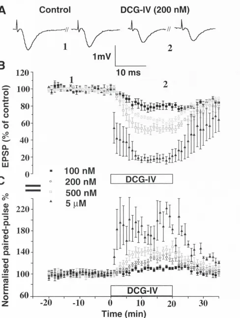
dependent changes in paired-pulse depression / facilitation. DCG-IV had a
3.2 Introduction
As the principal excitatory neurotransm itter in the mammalian central
nervous system , glutam ate plays a pivotal role in a num ber of
physiological and pathological conditions. Although glutamate has long
been known to activate the AMPA and NMDA ionotropic receptors
(iG luRs), thus altering transm em brane conductances through cation
channels, it was only in the 1980s that glutamate was observed to
activate a fam ily of receptors which are linked to second messenger
systems via g-proteins. These metabotropic glutamate receptors (mGluRs)
have been differentiated into 3 groups on the basis of their pharmacology,
signal transduction pathways and molecular biology.
G roup I (m G luRs 1 and 5) mGluR are linked to phosphoinositide
hydrolysis, group II ( mGluRs 2 and 3) and group III ( mGluRs 4,6,7 and 8)
are negatively coupled to adenylate cyclase and are further differentiated
on the basis of their sensitivity to pharmacological agonists. The activation
of m GluRs has been associated with a range of physiological and
pathological effects at both presynaptic and postsynaptic locations
throughout the CNS (reviewed by Conn and Pin, 1997). Presynaptic
inhibition of excitatory synaptic transm ission by m GluRs was first
established in hippocampal CA1 using the non-selective agonist trans-
aminocyclopentane-1,3-dicarboxylic acid (t-AGPD) (Baskys and Malenka,
1991; Desai and Conn, 1991). Since then a number of studies established
that presynaptically located mGluRs perform a sim ilar function in other
brain regions, such as the hippocampal dentate gyrus (Macek et al., 1996;
Bushell et al., 1996), Striatum (Lovinger, 1991) and the neocortex
The presynaptic locus of inhibition was established on the basis of the
following observations. Firstly no postsynaptic changes were associated
with the mGluR induced inhibition (Lovinger, 1991; Baskys and Malenka,
1991; Burke and Hablitz, 1994). Secondly, t-ACPD did not have a
diffe ren tial effect on either the AMPA or NMDA receptor mediated
components of excitatory synaptic transmission. This would suggest that
mGluR activation was depressing synaptic transmission by reducing the
level of glutam ate release from the presynaptic terminals (Baskys and
Malenka, 1991; Lovinger, 1991). Thirdly, a change in a form of short-term
plasticity known as paired-pulse depression / facilitation is indicative of a
presynaptic modulation of neurotransmitter release. Several studies have
shown that mGluR agonists enhanced paired-pulse facilitation in CA1 (
Baskys and Malenka, 1991); CAS (Kamiya, 1996) and the lateral perforant
path of the dentate gyrus (Kahle and Cotman, 1993; Dietrich et al., 1997)
whilst paired-pulse depression in the medial perforant path was reduced
(Brown and Reyman, 1995; O'Leary et al., 1997).
The developm ent of pharm acological agents selective for mGluR
subtypes has revealed the presence of presynaptic group II mGluR in
the hippocampus. In young CA1; (1S,3S)-1-am inocyclopentane-1,3-
dicarboxylic acid (IS , 3S) - ACPD (Vignes et al., 1995) and DCG-IV
(Yokai et al., 1996) have been shown to depress excitatory synaptic
transm ission, although these receptors appear to be absent in the
m ature CA1 (Kilbride et al., 1998). In the m edial perforant path,
(2S,1'S,2'S)-2-carboxycyclopropyl)glycine LCCG-1 (Ugolini and Bordi
1995); DCG - IV (Macek et al., 1996; Huang et al., 1997) and LY354740
(Kilbride et al., 1998) have all been shown to reduce excitatory synaptic
These findings have been supported by binding studies which have
localised mGluR 2 and 3 to presynaptic elements within the hippocampus
via subtype specific antibodies, in particular the medial perforant path of
the dentate gyrus was found to be densely populated with what appears to
be mGluR 2 (Shigemoto et al., 1997).
LY354740 is a recently synthesised high affinity efficacious and selective
group II mGluR agonist. (Bond et al., 1997: Monn et al., 1997; Schoepp et
al., 1997a,b). LY354740 suppressed forskolin-stimulated cAMP production
at group II mGluR with a nanomolar potency, but crucially it did not have
any agonist or antagonist action at groups I and III mGluRs. Interest in
LY354740 is partly because it has potential as a therapeutic tool in a
num ber of conditions. LY354740 counteracted the haloperidol-induced
muscle rigidity in an animal model of Parkinson's disease, (Konieczny et
al., 1998). An anti-convulsive action against ACPD-induced limbic seizures
was reported by Monn and his colleagues in 1997. Furthermore it has
been shown to have anxiolytic activity in the fear potentiated startle and
elevated plus maze models, without producing the unwanted secondary
effects such as memory impairment, that established agents such as
Diazepam produce. (Helton et al., 1998; Monn et al., 1997).
In this study, I have investigated the presynaptic inhibitory action of
LY354740 in the medial and lateral perforant paths of the hippocampal
dentate gyrus, as well as area GA1, comparing its action against that of
3.3 Materials and methods
3.3.1 Slice preparation
All experim ents were carried out on hippocampal slices obtained from
Wistar rats (50 -70g) (BioResources Unit, Trinity College, Dublin, Ireland).
Slices were obtained as described previously (Huang et al., 1997). Briefly,
the brain was rapidly removed after decapitation and placed in cold (5°C)
oxygenated (95 % O
2, 5 % CO
2) artificial cerebro-spinal fluid (ACSF)
containing in mM: NaCI, 120; NaHCOa, 26; NaH
2P
0 4, 1.25; KCI, 2.5;
Mg
2S
0 4, 2; CaCl
2, 2; glucose, 10). Hippocampal slices (350 fjM) were cut
using a Campden vibroslice (Campden Group Instruments, London, U.K.)
and transferred immediately to an incubation chamber, maintained at room
temperature, for a period of at least 60 minutes. Single slices were then
transferred to a submersion type recording chamber at 30-31 °C.
3.3.2 Electrophysiology
Field excitatory postsynaptic potentials (EPSPs) were recorded using
standard glass electrodes filled with ACSF. Both recording and stimulating
electrodes were placed in either the middle or outer third of the molecular
layer of the dentate gyrus in order to stimulate and record from either the
medial or lateral perforant path respectively, and in the Schaffer collateral /
com missural path in the stratum radiatum of CA1. Test EPSPs were
evoked using a Grass S48 stimulator (0.0166 Hz, pulse width 0.1 ms) via
a bipolar insulated tungsten wire electrode, adjusted to give about 30 % of
the maximal response (~1 mV). EPSP amplitude was measured using
Maclab Scope v3.5.6. Stimulation and recording from the medial or lateral
perforant pathway was verified by the use of paired-pulse stimulation
%) was indicative of the lateral perforant path. Further verification was
obtained by observing the switch from "source" to "sink" upon moving the
recording electrode from one pathway to the other, and the
pharmacological selectivity of each pathway was used as a post-
experimental cue to the identity of the recorded pathway.
3.3.3 Data analysis and statistics
For each EPSP pair, the second EPSP amplitude was divided by the first
and multiplied by
100to give the paired-pulse "percentage".
Apaired-
pulse percentage of less than 100 was indicative of paired-pulse
depression whereas a percentage greater than 100 was indicative of
paired-pulse facilitation. The effect of
LY354740in the lateral perforant
path and
CA1are presented in this way, in the text and in figures
3.3and
3.5.
However the effects of
LY354740and also DCG -
IVon paired-pulse
depression in the medial perforant pathway have been normalised to the
baseline or control period. This was done so that the dose-dependent
change in paired-pulse could be easily visualised in a figure displaying
four concentrations of an agonist. Summarised results are expressed as
normalised EPSP mean amplitude ± S.E.M. Data was analysed using
student's paired t-test, and repeated measures analysis of variance
(ANOVA)