Brain Tumour Detection and Segmentation
Techniques: A State-Of-The-Art Review
Mansi Lather1, Parvinder Singh2
1,2 CSE Department, DCRUST, Murthal, Sonipat, India
Abstract: Brain tumor is a disease difficult to cure. Therefore detection of brain tumor at an initial stage can help in easy and proper diagnosis. Image processing exercises a major role in analysis of the medical imagery. In medical image processing, brain tumor detection is considered as the most difficult and cha1lenging activity. Magnetic Resonance Imaging (MRI) is an advanced medical imaging approach for analyzing the body’s inner anatomy. MRI produces high quality images of human soft tissues that help in brain tumor diagnosis. Due to complex nature of brain MR images, the precise MRI image segmentation is necessary for brain tumor diagnosis. Next, the tumor classification into benign and ma1ignant is a tough job on account of differences in features of tissues of tumor such as gray level intensities, size, and structure. This paper addresses the potencies and weaknesses of the previously adduced classification strategies. The paper provides an insight into the reviewed literature to reveal new aspects of research and proposes a hybrid technique for brain tumor detection and segmentation.
Keywords: Magnetic Resonance Imaging (MRI); Brain tumor detection; Image processing; Denoising; Segmentation
I. INTRODUCTION
The brain is regarded as the command center of the nervous system, and it is the most complicated organ inside the body of human. It is a non- replaceable and soft and spongy mass of tissue. Human brain takes input from the sensory organs and forwards them as output to the muscles [1]. Intelligence, creativity, emotions, memory etc are governed by brain [2]. Therefore, any damage or harm in the brain will cause problems for personal health including mobility or cognition [1].
In diagnosis of brain, precise measurements are very difficult because of diversity in size, shape & appearance of tumors. A brain tumor is an aberrant and uncontro1led propagation of ce1ls [3]. A brain tumor does not only impact the immediate cells in its location but it also can cause damage to surrounding cells by causing inflammation. In medical image processing, brain tumor detection is considered as the most difficult & time absorbing activity. Medical imaging strategies exercise an important role in tumor detection. There are various imaging modalities such as X-ray Radiography, U1trasound imaging, Positron Emission Tomography (PET), Magnetic Resonance Imaging (MRI), Computed Tomography (CT). Among the various modalities MRI is regarded as the most proficient means for analyzing the body’s internal structure [4]. Early and accurate tumor detection is essential for efficient treatment planning.
The important goal of image processing application is to abstract from image data the main features, so that machine can gather an interpretative, descriptive, or reasonable plan.
Among the various steps of image processing the steps mainly considered for detection of brain tumor includes image denoising, morphological operation and image segmentation.
Image denoising is defined as the method of removing noise from the image. In medical imaging, for easy and proper diagnosis of diseases, denoising provides better clearance in the image [4]. Various schemes are available for removing noise from images [5], [6].The image denoising methods are broadly categorized as [5]:
A. Spatial filtering methods
Includes linear and non-linear filters
B. Transform domain filtering methods
Constitutes spatial frequency filtering methods and wavelet domain methods.
1) Morpho1ogical operators are non-1inear operators dealing with morphology & shape of images. They are related to pixel ordering and they don’t change the pixel’s numerical value. These operators are dependent on structuring e1ement, which is a small matrix of pixels each having va1ue one or zero and the choice of suitable structuring element plays an important role in the process. Various types of morphological operators include erosion, dilation, opening and closing [4].
Segmentation techniques used for analyzing the medical images are classified as [8]:
C. Region based methods
Includes thresholding and region growing methods.
D. CLASSIFICATION METHODS
Includes k-nearest neighbor and maximum likelihood methods.
E. CLUSTERING METHODS
Includes K-Means, FCM & expectation maximization methods.
In this paper work done by different scholars and researchers to assist in the problem of brain tumor segmentation has been reviewed along with their benefits and limitations. An improved hybrid technique for brain tumor detection and segmentation has also been proposed.
II. LITERATUREREVIEW
Ayed, Kharrat and Halima [9] In their paper proposed an approach that consists of five phases: In the first phase, feature extraction is done via 2D Discrete Wave1et Transform & Spatial Gray Level Dependence Matrix (DWT-SGLDM). In the second phase, to reduce features size, features are selected via SA. In the next phase, over fitting is avoided using Stratified K-fo1d Cross Va1idation. In the fourth phase to optimize SVM parameters, GA-SVM model is used. Finally SVM is used for creating the classifier.
On the T2-weighted brain MR image datasets, this method obtained high c1assification accuracy. This strategy could further be used for image classification with differences in pathological conditions, types and disease status.
Ramya and Sasirekha [4] in their paper proposed a segmentation technique consisting of three phases: fourth order partial differential equation is used to denoise the image; then the morphological operators are used to remove the skull part and finally segmentation is done using region growing segmentation. The precision of this method is high than the watershed segmentation algorithm.
Its future work includes use of Neural Network c1assifier or Support Vector Machine (SVM) c1assifier for classifying the stages of tumor and tumor size calculation for better analysis of tumor.
El-Khamy, El-Khoreby and Sadek [10].In this paper they introduced a hybrid technique of FCM and conformed threshold. The proposed technique consists of five stages: the first stage involves preprocessing for enhancing the intensity of input brain MR image for next stages. The second stage involves the use of a rectangular window for image histogram in order to calculate the number of clusters for FCM input. The third stage is to use FCM to find the center of clusters. The fourth stage is to use the conformed threshold value in order to segment the tumor. The final stage is tumor detection from the segmented image. This method gives better results for correctness and processing time than the global threshold method of segmentation, but the completeness is better in global threshold method than the proposed method.
Its future work includes tumor diameter calculation in three dimensional brain MRI images for accurately planning the treatment. Dadheech, Gupta and Mathur [11] in their paper presented a fuzzy dependent detection of edges via K-means c1ustering technique. The K-means clustering technique is used to create different chunks to be fed as input to the mamdani fuzzy inference system. The result of this is the formation of threshold attribute to be then fed into the classical sobel edge detector which enhances its capability of detecting the edges using the fuzzy logic.
The result presents that fuzzy derived k-means c1ustering increases the effectiveness of c1assical sobel edge detector besides holding most of the relevant details. Its future work includes the use of proposed technique on different edge detectors.
Gupta, Khare and Srivastava [12] In this paper they introduced a new method using Genetic Algorithm (GA), Curve Fitting and SVM. Image segments are created using GA. After application of GA, the resultant segments might be relinquishing some of the details in their adjoining segments. Curve fitting is applied to properly segment the image without the loss of information. After segmenting the image, features are extracted from the segments. SVM is then used to classify these extracted features. The c1assified data then assists in determining the tumor using the extracted features. This method is more accurate and precise than the method using Mahalanobis distance.
necessary. Its future work includes image segmentation, classification and performance analysis.
Selkar and Thakare [14] in their paper presented watershed and thresholding algorithm that consists of three stages. Firstly quality of the scanned image is enhanced by removing noise. Secondly thresholding and watershed segmentation is applied to get a high intensity portion called tumor from the whole image. Finally, edge detection operator is applied for extracting the boundary and for finding the tumor size. The result shows efficient tumor detection by using thresholding algorithm rather than watershed algorithm and canny edge operator gives efficient boundary extraction results rather than prewitt and Robert operator.
Abid, BenMessaoud and Kharrat [15] In their paper presented an automatic brain tumor segmentation method in MRI images. The proposed method constitutes four steps – The first step is image pre-processing. The second step involves extraction of features using wave1et tansform-spatial gray 1evel dependence matrix (WT-SGLDM). In third step dimensionality reduction is done using GA and the final step involves classification of reduced features using SVM.
This method surpasses manual segmentation as well as FCM algorithm.
Beham and Gurulakshmi [16] In this paper they proposed a technique comprising of three phases: The first phase is image enhancement in which outer elliptical shaped object is eliminated. The second phase is morphological processing, conducted for extracting the needed region. The final phase is the segmentation using K-means clustering algorithm. This unsupervised method is efficient and less prone to error and can be carried out with lesser amount of data giving accurate output compared to supervised methods.
Gopal and Karnan [17] In this paper they presented a hybrid approach such as FCM with GA and PSO for detecting the tumor. The tumor detection is done in two phases. The first phase involves pre-processing & enhancement using the tracking algorithm for elimination of fi1m relics and median filter to eliminate the high frequency components. The second phase involves segmentation and c1assification using GA with FCM and PSO with FCM. PSO with FCM outperforms GA with FCM.
A critical review of the studied literature is summarized in table I.
Table I Comparison of Different Papers Reviewed
Author Paper Title Methods Used Advantages Limitations
Ayed,
Kharrat and
Halima (2016)
MRI Brain Tumor
Classification using
Support Vector
Machines and
Meta-Heuristic Method [9].
DWT-SGLDM for feature
extraction. Simulated Annealing (SA) for reducing the size of features. Stratified K-fo1d Cross Va1idation to avoid over fitting. GA-SVM for SVM parameters optimization. SVM for c1assifier construction.
Minimum number of features for classifying
patho1ogical and normal brain reduces the cost of classifier.
SA and GA requires greater computational time which rises with the growth in generation number.
Ramya and
Sasirekha (2015)
A Robust Segmentation
A1gorithm using
Morphological Operators for Detection of Tumor in MRI [4].
Image denoising: fourth order
Partial Differential Equation
(PDE).
Skull Removal: Morphological Operators (erosion and dilation).
Segmentation: Seed point
selection based region growing segmentation.
Fourth order PDE
removes noise
effectively and
favors better edge
preservation. The
detection accuracy
is high in
comparison to
watershed segmentation.
Initial seed point
selection depends on user ability.
El-Khamy, El-Khoreby
and Sadek
(2015)
An Efficient Brain Mass Detection with Adaptive C1ustered based Fuzzy
C-Mean and
Thresholding [10].
Fuzzy C-Mean (FCM) and conformed threshold.
Improvement in
correctness and
reduction in
operational time
than the g1obal
threshold segmentation method.
Completeness result
better in global
Dadheech,
Gupta and
Mathur (2015)
The K-means C1ustering
Based Fuzzy Edge
Detection Technique on MRI Images [11].
Fuzzy based sobel edge
detection using K-means
c1ustering approach.
Performance
enhancement of
classical sobel edge
detector besides
seizing most of the important details.
Computational cost
complexity is high
Gupta, Khare and
Srivastava (2014)
Optimization Technique,
Curve Fitting and
Machine Learning used to Detect Brain Tumor in MRI [12].
GA to create image segments.
Curve fitting to properly
segment the image without loss of information.
SVM to classify extracted
features.
More accurate and precise results than the method using Mahalanobis distance
Variation in image database demands new training set.
Arivoli, Lakshmi and Vinupriyadha rshini (2014)
Noise and Skull removal
of Brain Magnetic
Resonance Image using Curvelet transform and Mathematical
Morphology [13].
Noise removal: curvelet
transform
Skull removal: mathematical morphology
Segmentation: spatial FCM
Curvelet transform is an efficient noise removal method that considers both faint
linear and curvy
linear features.
Results presented are
preliminary and
requires clinical
evaluation.
Selkar and
Thakare (2014)
Brain Tumor Detection and Segmentation By Using Thresholding and
Watershed Algorithm
[14].
Image enhancement: Noise
removal
Segmentation: Thresholding and watershed method
Edge detection: Prewitt, Sobel, Canny edge detection operator
Thresholding
algorithm detects
tumor more
efficiently than
watershed algorithm
and canny edge
operator gives
efficient boundary
extraction results
rather than prewitt and robert operator.
Watershed method
results in
over-segmentation.
Abid, BenMessaoud
and Kharrat
(2014)
Brain Tumor Diagnostic Segmentation based on
Optima1 Texture
Features and Support
Vector Machine
C1assifier [15].
Preprocessing of image,
extraction of feature using
wave1et tansform-spatial gray 1evel dependence matrix
(WT-SGLDM), GA to reduce
dimensionality and reduced
feature classification using
SVM.
Using the optimal features, ma1ignant and benign tumors are segmented with high classification precision.
Applicative where the parameters must be updated.
Beham and
Gurulakshmi (2012)
Morpho1ogical Image
Processing Approach On The Detection Of Tumor and Cancer Cells [16].
Image enhancement to remove outer elliptical shaped object.
Morphological processing to
extract the required region and
K-means clustering
segmentation method
Less error sensitive and can be applied to minimal amount of data with reliable results compared to supervised
segmentation methods.
K-means clustering
does not work well
with non-globular
Gopal and Karnan (2010)
Diagnose Brain Tumor
through MRI using
Image Processing
Clustering Algorithms
such as Fuzzy C-Means along with Intelligent Optimization Techniques [17].
Pre-processing and enhancement using the tracking algorithm and median filter. Segmentation and classification using PSO with FCM.
PSO with FCM has lower classification
error rate and
execution time and better accuracy than GA with FCM.
Median filter
significantly denoises the image but the image appears with blurred boundaries.
III.PROPOSEDSYSTEM
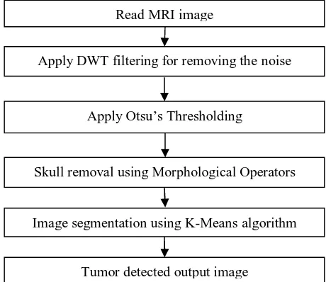
[image:6.612.187.417.337.536.2]In the previous section, we discussed various techniques for brain tumor detection and segmentation. There are various difficulties such as noise, non-cerebral tissues etc that results in poor segmentation and improper tumor detection. To overcome these issues, we are proposing an improved hybrid method for brain tumor detection and segmentation, so as to assist neurosurgeons in identifying the boundary of diagnostic region so that tumor can be precisely removed in surgical operation. Early and accurate detection of tumor is vital for proper diagnostics. The proposed system will consist of mainly three steps. First is image denoising (removal of noise from image) using Discrete Wavelet Transform (DWT). Next is skull removal (removal of brain’s non-cerebral tissues) using morphological operators and finally image segmentation (extracting the region of interest) using K-Means and Otsu’s thresholding method. The proposed technique will be as shown in fig. 1.
Fig. 1 Steps for Tumor Detection
IV.CONCLUSIONS
Image processing is widespread in analyzing the biomedical images and is vital for studying anatomical structures, computing tissue volume, and aberration scrutiny, pathology, planning treatment and computer-assisted surgery. A brain tumor is an aberrant and uncontrolled propagation of ce1ls. Finding the accurate border of the area comprising an identified brain tumor is a difficult task and needs to be addressed as it is applicable to many medical moda1ities and tumor types. In this investigation various automatic and semi-automatic methods for the detection of brain tumor through MRI has been studied. There are various difficulties such as noise, non-cerebral tissues etc that results in poor segmentation and improper tumor detection. To overcome these issues, we are proposing an improved hybrid method for brain tumor detection and segmentation.
V. ACKNOWLEDGMENT
The authors are thankful to all the reviewers for their important comments and suggestions. Read MRI image
Apply DWT filtering for removing thenoise
Apply Otsu’s Thresholding
Skull removal using Morphological Operators
Image segmentation using K-Meansalgorithm
REFERENCES
[1] Lewis Tanya and Writer Staff, “Human Brain: Facts, Functions & Anatomy,” 2016. [Online]. Available: http://www.livescience.com/29365-human-brain.html. [Accessed: 24-May-2017].
[2] “Anatomy of the Brain,” 2016. [Online]. Available: https://www.mayfieldclinic.com/PDF/PE-AnatBrain.pdf. [Accessed: 24-May-2017].
[3] Mandal Ananya, “What is a Brain Tumor?,” 2014. [Online]. Available: http://www.news-medical.net/health/What-is-a-Brain-Tumor.aspx. [Accessed: 24-May-2017].
[4] L. Ramya and N. Sasirekha, “A Robust Segmentation Algorithm using Morphological Operators for Detection of Tumor in MRI,” in IEEE International Conference on Innovations in Information Embedded and Communication Systems ICIIECS 2015, 2015, pp. 2–5.
[5] F. C. Harris, M. C. Gadiya, M. C. Motwani, and R. C. Motwani, “Survey of Image Denoising Techniques,” Proc. GSPX, pp. 27–30, 2004. [6] R. C. Gonzalez and R. E. Woods, Digital Image Processing, 2nd ed. Upper Saddle River, New Jersey: Prentice Hall, 2001.
[7] “Image Segmentation,” Wikipedia, The Free Encyclopedia. 2017. [Online]. Available:
https://en.wikipedia.org/w/index.php?title=Image_segmentation&oldid=781506915. [Accessed: 24-May-2017].
[8] A. Altameem, A. A.Norouzi, A. E. Rad, A. Rehman, M. S. M. Rahim, M. Uddin, and T. Saba, “Medical Image Segmentation Methods , Algorithms , and Applications,” IETE Tech. Rev., vol. 31, no. 3, pp. 199–213, 2014.
[9] A. Kharrat, M. B. Ayed, and M. B. Halima, “MRI Brain Tumor Classification using Support Vector Machines and Meta-Heuristic Method,” 2015 Int. Conf. Intell. Syst. Des. Appl., pp. 446–451, 2016.
[10] M. A. El-Khoreby, R. A. Sadek, and S. E. El-Khamy, “An Efficient Brain Mass Detection with Adaptive Clustered based Fuzzy C-Mean and Thresholding,” in 2015 IEEE International Conference on Signal and Image Processing Applications (ICSIPA), 2015, pp. 429–433.
[11] N. Mathur, P. Dadheech, and M. K. Gupta, “The K-means Clustering Based Fuzzy Edge Detection Technique on MRI Images,” 2015 Fifth Int. Conf. Adv. Comput. Commun., pp. 330–333, 2015.
[12] S. Khare, N. Gupta, and V. Srivastava, “Optimization Technique, Curve Fitting and Machine Learning used to Detect Brain Tumor in MRI,” in IEEE International Conference on Computer Communication ans Systems (ICCCS 2014), 2014, pp. 254–259.
[13] A. Lakshmi, R. Vinupriyadharshini, and T. Arivoli, “Noise and Skull removal of Brain Magnetic Resonance Image using Curvelet transform and Mathematical Morphology,” Int. Conf. Electron. Commun. Syst. ICECS 2014, pp. 1–4, 2014.
[14] M. N. Thakare and R. G. Selkar, “Brain Tumor Detection and Segmentation By Using Thresholding and Watershed Algorithm,” Int. J. Adv. Inf. Commun. Technol. IJAICT 2014, vol. 1, no. 3, pp. 321–324, 2014.
[15] A. Kharrat, M. Abid, and M. BenMessaoud, “Brain tumour diagnostic segmentation based on optimal texture features and support vector machine classifier,” Int. J. Signal Imaging Syst. Eng., vol. 7, no. 2, pp. 65–74, 2014.
[16] M. P. Beham and A. . Gurulakshmi, “Morphological Image Processing Approach On The Detection Of Tumor,” Int. Conf. Devices, Circuits Syst. (ICDCS), 2012, pp. 350–354, 2012.