Technology (IJRASET)
Comparison of Morphological and Edge detection,
Line Operator based Retinal Blood Vessel
Detection Algorithm
Kokel Hetal N.#1, Bhavin Mehta*2 #
*Biomedical Department, Gujarat Technological University
Abstract— Diabetic Retinopathy has turned out to be the most commonly occurring disease in patients with uncontrolled level of diabetes. So, it has become necessary to diagnose it at preliminary stage. Blood Vessels and optic disc are normal features of any retinal image. They are very helpful in detecting abnormal features of diabetic retinopathy. In detection of non-proliferative diabetic retinopathy (NPDR), sometimes, the abnormal features look like normal features. Hence, to avoid false positives, these features are removed before processing. Also, in the analysis of proliferative diabetic retinopathy (PDR), for neo-vascularization detection, the blood vessels are studied. So, after the blood vessels have been segmented, the features can be extracted and then the existence of neo-vascularization can be found out. There are various methods for blood vessel segmentation. This paper reviews two different methods for blood vessels segmentation: morphological and edge detection, line operator based methods. The algorithms were implemented on a set of images from different standard databases, ‘diaretdb’ and ‘High Resolution Fundus Images’; and non-standard database collected from ‘Bankers Retina Clinic and Laser Centre, Ahmedabad’.
Keywords
—
blood vessel segmentation, diabetic retinopathy, edge detection, line operator, morphological, neo-vascularization, non-proliferative diabetic retinopathy, NPDR, PDR, proliferative diabetic retinopathyI.INTRODUCTION
Diabetes mellitus (denoted diabetes from now on) is a chronic, life-threatening disease. Diabetic Retinopathy (DR) is the medical
condition where the retina is damaged because of the fluid leaks from the blood vessels into the retina. It has been found that during first 20 years of diabetes, all patients with type-I diabetes develop DR; whereas >60% patients with type-II diabetes have DR [3]. Diabetic Retinopathy is an ophthalmic disease that results due to prolonged existence of uncontrolled diabetes mellitus. Also for the detection of diabetic retinopathy, the primary diagnosis is done for non-proliferative kind, which is done by detecting features like exudate, microaneurysms, cotton wool spots and haemorrhages. Later, if such features are not indicated, further diagnosis involves studying the vascular structure on the retinal surface. This motivation leads to detection and analysis of blood vessels using various segmentation algorithms. Blood vessel and optic disc are normal features of any fundus image. So, if features have similar properties or look like any other feature of diabetic retinopathy, it becomes necessary to detect them first and exclude them from the image. There are many different approaches for segmenting blood vessels from fundus images. Some use two-dimensional matched filter, fuzzy c-means classifier, tracker algorithms, using edge detection and line operator, texture based segmentation, simple subtraction after morphological operations and many more [1,2,4,6,7,8]. Unsupervised texture based segmentation was also proposed [7]. In this method, the blood vessel segmentation is carried out by analysing texture properties and non-vessel parts of the image. Firstly, the image is converted from original RGB image to Gaussian and L*a*b perpetually uniform colour spaces. Then pre-processing is done on Luminance from L*a*b and green band from RGB image which involves adaptive histogram equalization for contrast enhancement. Later, Gabor filters with twenty four different orientations and three different wavelengths are applied on the enhanced image for texture feature extraction and a feature vector is formed for each pixel. Using Fuzzy C-means clustering algorithm, this feature vector can be classified as vessel or non-vessel based on the texture properties. Finally, we obtain the blood vessels. The main advantage of this method is that it works efficiently to segment both minor as well as major blood vessels. In this paper, a method based on edge detection and line operator has been reviewed and compared with the one that involved morphological operations.
II.COMPARISON
Technology (IJRASET)
Figure 1Steps to be performed for Segmentation method based on Edge Detection & Line Operator
A novel approach that involves usage of line as well as edge detector operators has been reviewed [2]. It proposed a method named RBVSLE i.e. Retinal Blood Vessel Segmentation method based on Line operator and Edge detectors. RBVSLE extracts the highest intensity value and its corresponding pattern. All the edge seeds in the selected pattern are called edge candidate seeds. The proposed method segments the edge candidates by using line operator and classifies whether it is a blood vessel pixel or not. It then finds the next vessel growth seed and continues the same procedure again. The flow of the algorithm is as shown in figure 1. Firstly, pre-processing was done on the acquired retinal fundus image. Pre-processing includes Green channel segmentation and contrast enhancement. Green Channel of the retinal image has the maximum information available in it and hence the green channel of the fundus image has been extracted. Following green channel segmentation, contrast levels of the image were adjusted using ‘adapthisteq’ function in Matlab with window size [30 30] and contrast ratio 0.03. After pre-processing, edge detection was carried out and for this purpose canny edge detector was used with threshold value between range 0.1 and 0.45. Thereafter, pixel intensities in 10 different directions were calculated (0°, 20°, 40°, 60°, 80°, 100°, 120°, 140°, 160° and 180°). Using these results a mask was created. Blood vessels can be segmented using this method with higher accuracy. However, it may fail to detect some minor vessel branches.
B. Morphological Based Segmentation
A review of morphological based method for blood vessels detection is presented [1]. In this method, firstly green channel segmentation was done; then it was closed using structured element of disc type with two different disc radii. For experimental purpose, these radii were kept as 4 and 10. Then the obtained results were subtracted from each other i.e. results obtained with higher radius were subtracted from that of lower radius. After the subtraction, thresholding was applied on the image using Otsu’s algorithm. This gave quite a convincing detection of blood vessels. This method worked efficiently for most of the images and gives faster results.
Figure 2Steps for Morphological based Blood Vessel Segmentation
Figure 3
III.RESULTS & DISCUSSIONS
[image:3.612.94.495.461.618.2]Technology (IJRASET)
from 3 seconds to 8 seconds; whereas for edge detection, line operator method it ranges from 4 seconds to 12 seconds. Thus, we can conclude that morphological method gives more precise output.
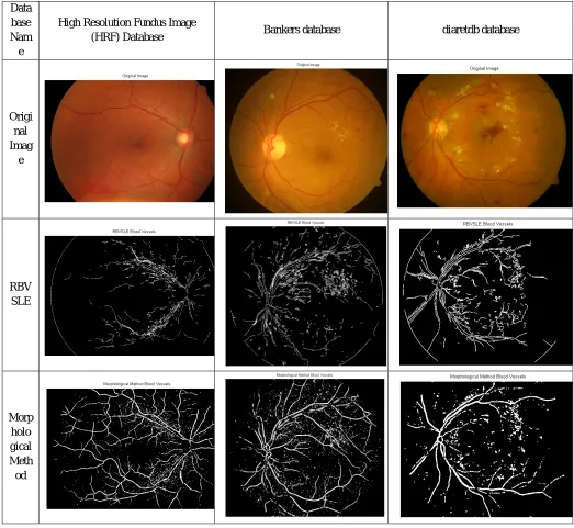
Data base Nam
e
High Resolution Fundus Image
(HRF) Database Bankers database diaretdb database
Origi nal Imag e
RBV SLE
[image:4.612.44.567.112.593.2]Morp holo gical Meth od
Table 1 Results obtained for images from High Resolution Fundus (HRF) database, bankers database and ‘diaretdb’ database
Though, a major drawback using this method is that when the blood vessel are extracted using morphological operations, some dot like objects are also obtained so, if by any chance, that dot is an implication of any abnormal features like microaneurysm or exudates, it might also lead to false detection. Though, chances of this happening are very low as our main aim behind this is to study the features and analyse the blood vessels on the basis of these features.
IV.CONCLUSIONS
Technology (IJRASET)
the physical characteristics of blood vessels when diagnosing proliferative diabetic retinopathy as neo-vascularization is the preliminary stage where abnormal growth of blood vessels takes place. Also, it might also be of use for detecting non-proliferative diabetic retinopathy seeing that intensity of some abnormal features might be same as that of blood vessels like that of microaneurysms; which poses hindrance if the segmentation is done using thresholding or intensity values. Also, from reviewing both the methods, it leads to the conclusion that blood vessels are obtained very efficiently and rapidly using morphological operations.
V.ACKNOWLEDGMENTS
The author shows deepest gratitude to guide, Prof. Bhavin Mehta, Assistant Professor, Biomedical Department, Government Engineering College, Gandhinagar for his constant support, encouragement and co-operation throughout the progress of the work. The author also acknowledges Dr. Dhaval Modi, Ophthalmologist at “Modi Eye Care” for his clinical guidance related to the field of Ophthalmology. The author would also like to thank Dr. Alay Banker of “Bankers Retina Clinic & Laser Centre, Ahmedabad” for providing good quality retinal fundus image for forming a dataset of non-standard and unanalysed retinal fundus images.
REFERENCES
[1] Lydia Glory Priyadarshini M., J. Anitha, A Region Growing Method of Optic Disc Segmentation in Retinal Images, 2014 International Conference on Electronics and Communication Systems (ICECS-2014), pp 1-5,February 13-14, 2014.
[2] Chin-Chen Chang, Chia-ChenLin, Pei-Yan Pai and Yen-Chang Chen, A Novel Retinal Blood Vessel Segmentation Method Based on Line Operator and Edge Detector, IEEE, 2009 Fifth International Conference on Intelligent Information Hiding and Multimedia Signal Processing, 978-0-7695-3762-7/09, pp 299-302, 2009.
[3] Watkins, J. P., ABC of diabetes retinopathy, British Medical Journal, 326:924–926, 2003.
[4] Subhasis Chaudhuri, Shankar Chatterjee, Norman Katz, Mark Nelson, and Michael Goldbaum, Detection of Blood Vessels in Retinal Images Using Two-Dimensional Matched filters, IEEE Transactions on Medical Imaging, Volume 8, Issue no. 3, pp 263-269, September 1989.
[5] Enrico Grisan, Alessandro Pesce, Alfredo Giani, Marco Foracchia and Alfredo Ruggeri, A new tracking system for the robust extraction of retinal vessel structure, Proceeding of the 26th Annual International Conference of the IEE EMBS San Francisco, CA, USA, pp 1620-1623, September 1-5, 2004.
[6] Soopotharom Supot, Chaichana Thanapong, Pintavirooj Chuchart, and Sangworasil Manas, Automatic Segmentation of Blood Vessels in Retinal Image Based on Fuzzy K-Median Clustering, Proceedings of the 2007 IEEE International Conference on Integration Technology, Shenzhan, China, pp 584-588, March 20-24, 2007.
[7] Alauddin Bhuiyan, Baikunth nath, Joselito Chua and Ramamohanarao Kotagiri, Blood Vessel Segmentation from Color Retinal Images using Unsupervised Texture Classification, IEEE, ICIP, Volume 5, pp 521-524, 2007.
[8] Tamir Yedidya and Richard Hartley, Tracking of Blood Vessels in Retinal Images Using Kalman Filter, Digital Image Computing: Techniques and Application (DICTA) 2008, IEEE Computer Society, pp 52-58, 2008
[9] H.Sassan, S.F. Aris@Azis, A Simple Approach of Blood Vessels Detection in Retinal Images using MATLAB, 2012 IEEE Student Conference on Research and Development, pp 245-249, 2012.
[10] Ana Salazar-Gonzalez, Djibril Kaba, Yongmin Li, and Xiaohui Liu, Segmentation of Blood Vessels and Optic Disk in Retinal Images, IEEE Journal of Biomedical and Health Informatics, Volume 18 Issue No. 6, pp 1874-1886, November 2014.
[11] High Resolution Fundus Image Database https://www5.cs.fau.de/research/data/fundus-images/