N A N O E X P R E S S
Open Access
Monodisperse upconversion GdF
3
:Yb, Er rhombi
by microwave-assisted synthesis
Haiqiao Wang
1*and Thomas Nann
2Abstract
We have synthesized a variety of monodisperse colloidal GdF3:Yb, Er upconversion nanocrystals with different shape, size, and dopants by microwave-assisted synthesis. Typical upconversion emission from Er3+was observed. In addition to highly monodisperse spherical particles, we were able to prepare monodispersed rhombic-shaped slices that showed a tendency for self-assembly into stacks.
Introduction
In recent publications we have shown that microwave-assisted synthesis allows for the preparation of highly monodisperse, spherical upconversion nanocrystals [1], as well as nanocrystals with unusual morphologies [2]. In this research letter, we report on the microwave-assisted synthesis of monodispersed spherical and rhombic GdF3-based nanoparticles, which show a high tendency for self-assembly in one- and two-dimensional superstructures.
Research on upconversion nanocrystals increased expo-nentially over the past several years (e.g., in 2000, one arti-cle on upconversion nanomaterials was published, in 2009, it have been 57) as the extremely attractive prospects for applications of these materials in bioanalytics [3], (cancer) therapy [4], and electro-optics [5]. And subsequently, it is concluded that the most efficient infrared-to-visible upconversion phosphors are Yb/Er or Yb/Tm co-doped fluorides, such as hexagonal phase NaYF4[6,7], LaF3[8,9], and orthorhombic phase YF3[10], GdF3[11]. Especially in the past few years, the NaYF4-based phosphors, as the highest efficient upconversion phosphors, with different morphologies and different dopants have been widely investigated, based on various synthesis procedures. How-ever, GdF3as one of the efficient upconversion phosphor host [8], not too much work has been reported focusing on Yb/Er codoped fluorecence upconversion. Although Tm, Dy, Ho, and Yb/Tm doped GdF3has been reported
[11-14]. As far as we know, in 1971, the preparation of GdF3:Yb, Er phosphor was reported firstly by Major et al. In their procedure, the oxide precursors were dissolved in high purity nitric acid and precipitated with excess hydro-fluoric acid, and finally experienced calcination. They found that the color of the anti-Stokes luminescence of the Yb/Er doped GdF3phosphors was controllable by pre-paration processes, and was associated with the crystal structure of the host lattices. And they gave a dominant green emission when excited by 940 nm infrared light [10]. In 2006, Fan et al. employed a hydrothermal synth-esis procedure to produced Yb/Er codoped GdF3 nanopar-ticles. For their prepared sample, typical upconversion emission was observed but with much weaker intensity than that of bulk crystal [15].
Microwave-assisted synthesis of nanomaterials offers several interesting synthetic opportunities which are based on the specific microwave effects: (i) microwave irradiation is absorbed by polar and ionic substances only (dielectric heating); (ii) enhanced reaction rates can be observed; (iii) heterogeneous heating (viz.“hot spots”) and wall effects can be suppressed [16]. Details on the microwave-assisted synthesis of AYF4(A = Na, Li) nano-crystals can be found elsewhere [1]. This research letter is concerned with the effects of microwave irradiation on the shape evolution of GdF3nanocrystals.
In this paper, we firstly presented the preparation of colloidal upconversion GdF3:Yb, Er nanocrystals, based on a new microwave-assisted synthesis method. We have synthesized a variety of GdF3-based upconversion nanocrystals with different shape, size, and dopants by microwave-assisted synthesis. In addition to highly monodisperse spherical particles, we were able to
* Correspondence: [email protected]
1Institute Materials for Electronics and Energy Technology (I-MEET), Friedrich
Alexander Universität Erlangen-Nürnberg, Marternsstraße 7, 91058, Erlangen, Germany.
Full list of author information is available at the end of the article
prepare monodisperse rhombic-shaped slices that showed a tendency for self-assembly into stacks.
Experimental
According to our previous work [1], introduction of Li+ can help to enhance the upconversion efficiency. So here in the procedure, Li+ was used as well. In a typical synthesis (standard conditions), 0.115 mmol (13.8 mg) lithium trifluoroacetate (TFA), 0.083 mmol (41.2 mg) gadolinium TFA, 17μmol (8.5 mg) ytterbium TFA, and 1.7μmol (0.86 mg) erbium TFA were dissolved in 6 ml of a 1:1 (v:v) mixture of oleic acid and octadecene in a nitrogen atmosphere. The solution was thoroughly degassed at 120°C and transferred into a microwave reaction vessel. Then, the mixture was heated for 10 min at 290°C by microwave irradiation. The resulting nanocrystals were precipitated by addition of 3 ml of ethanol to the cold reaction solution and subsequent centrifugation. The supernatant was discarded and the nanocrystals were repeatedly washed with ethanol. Even-tually, the particles were re-dissolved in chloroform or toluene for further studies.
Transmission electron micrographs (TEMs) were recorded on a JEOL 2000EX and a JEOL 2010 micro-scope at 200 kV. Selected area electron diffraction (SAED) patterns were measured on the same instru-ment. The X-ray diffraction (XRD) patterns were mea-sured with a thermo ARL XTRA equipped with a Cu X-ray tube (l= 1.5418 Å). Upconversion spectra were recorded on an Oceanoptics USB spectrometer and a home-made cuvette holder and excitation source (980 nm, 100 mW laser diode).
[image:2.595.305.538.524.681.2]Results and discussion
Figure 1 shows TEMs of the resulting nanocrystals. It was observed that the particles were regularly shaped rhombic plates with approximately 3.2 nm thickness, approximately 45 nm in length and with a roughly cal-culated aspect ratio of 1:3. Both micrographs show
clearly that the nanoplates have a strong tendency to form two-dimensional aggregates. This effect can be attributed to the minimization in energy, which is achieved by hydrophobic interaction of the surface ligands of the nanoparticles at their largest faces.
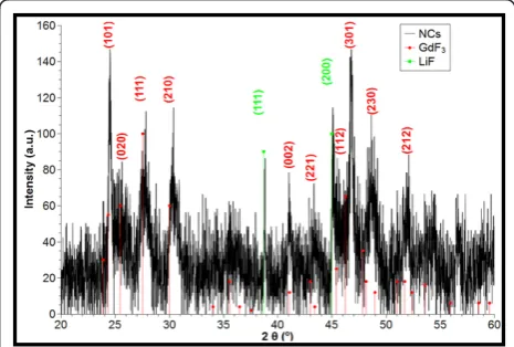
The XRD pattern of these particles (Figure 2) which shows orthorhombic phase GdF3was obtained. Almost all the diffraction peaks of the XRD pattern can be assigned, respectively, to the planes of orthorhombic GdF3 crystalline (JCPDS-file 012-0788), as indicated (101), (020), (111), (210), (002), (221), (112), (301), (230), (212) in Figure 2 (red, round dot). However, two more weak diffraction peaks can also be observed at 2θ= 38.7° and 45°, which cannot be assigned to GdF3. We consid-ered that these two weak peaks arose from the diffraction of LiF (JCPDS-file 045-1460) [17]. And it suggests that the Li+was not only introduced into the expected phos-phor GdF3crystal to replace some Gd3+sites as impurity but also a few LiF was formed. A further confirmation of a predominant GdF3lattice can be found by measuring distances of the lattice fringes in the high resolution transmission electron microscopy (HRTEM). Lattice fringes were found with distances of 3.29 and 2.94 Å (cf. Figure 3B), corresponding well to the theoretically calculated distance of the {111} and {210} planes of the orthorhombic-YF3 space group GdF3 (orthorhombic phase JCPDS-file 012-0788), respectively. It is noteworthy that the same XRD patterns were observed for decreased and increased reactant concentrations.
In our previous work, we found that changing the reaction parameters (especially the concentration of reactants) in the synthesis of NaYF4-based upconversion nanocrystals, has crucial influence on the morphology of the resulting particles [2]. In this work, we observed that, based on our precursors, GdF3 nanocrystals were
Figure 1 TEMs of GdF3nanocrystals.(A)Overview graph of
[image:2.595.58.292.572.691.2]single nanocrystals and“stacks”.(B)Blow-up of self-assembled nanocrystals.
obtained and always adopt a rhombic shape under microwave irradiation, even when other synthesis para-meters were changed.
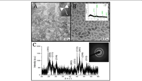
Figure 3 shows TEMs of GdF3 nanocrystals that were synthesized under the above conditions, but with 25% of the original concentration of reactants. The particles are smaller than the ones displayed in Figure 1 (approxi-mately 15-18 nm in length), but have roughly the same thickness and aspect ratio. Therefore, the rate of growth along the “edges”of the particles has to be much faster
as compared to the primary faces ({111} and {210}). Fig-ure 3B shows a HRTEM of the same particles. It can be observed that the lattice fringes are always aligned with one edge of the rhombi. This observation and the over-all shape of the nanocrystals are in agreement with the anticipated orthorhombic-YF3space group [18].
[image:3.595.61.537.407.679.2]When the concentration of reactants was increased by a factor of five, compared with the standard conditions, the nanocrystals were still predominantly rhombic in shape (in addition, some spherical particles were observed), but approximately 150 nm in length and 5 nm in thickness (cf. Figure 4A). This finding confirms further that the nanocrystals grow preferentially in two dimensions. Again, the XRD pattern is associated well with the orthorhombic phase GdF3, and without diffrac-tion peaks of LiF any more (Figure 4C). Figure 4C inset shows the SAED pattern of this prepared sample. Figure 4B shows the TEM of the obtained nanocrystals that were prepared under identical conditions as the ones in Figure 4A, but using traditional conductive heating. Shape and size of these particles are roughly in the order of magnitude of the standard conditions described above. However, the XRD pattern (inset Figure 4B) of these nanocrystals shows that mainly cubic LiF nano-crystals have been synthesized.
Figure 3TEMs of GdF3 nanocrystals prepared with 25% of the original concentration of reactants.(A)Overview picture.(B)
High-resolution TEM of single nanocrystals.
Figure 4TEM, XRD and SAED characterization of obtained samples.(A)TEM micrograph of GdF3nanocrystals synthesized at five times the
standard concentration.(B)LiF nanocrystals synthesized by traditional conductive heating at the same conditions like A. Inset: XRD pattern of the obtained nanocrystals.(C)XRD pattern of GdF3nanocrystals synthesized at five times the standard concentration. Inset: SAED pattern of the
Figure 5 shows the upconversion emission spectrum of the nanocrystals from Figure 4A, under 980 nm near infra-red (NIR) excitation. Mainly two emission bands were observed, with emission peaks at 521, 545, and 660 nm. These emission peaks can be attributed to the 4f-4f transitions of the Er3+ions. The green emission accounts for the2H(11/2),4S(3/2)®4I(15/2)transition, the red emis-sion is caused by the4F(9/2)®4I(15/2)transition. The dif-ference compared with typical reported Yb/Er emission spectrum is that a weak red emission at approximately
628 nm was observed, which could be attributed to the transition4I(9/2)® 4I(15/2). Based on Yb/Er codopants, the observed upconversion efficiency of the GdF3-based nanocrystals is relatively lower than that of the NaYF4 -based nanocrystals in our lab [1]. Comparing these values with data from the literature shows that the microwave-assisted synthesis does not influence the optical proper-ties of the nanocrystalsper se.
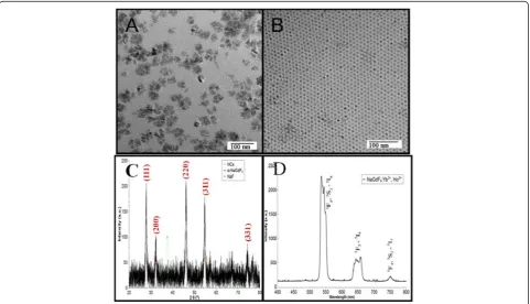
[image:4.595.57.290.89.248.2]In order to establish the reason for the strict rhombic shape of the nanocrystals, we replaced lithium with sodium in the above synthesis method. Yb/Ho codoped NaGdF4 nanocrystals were synthesized under the same conditions as above. Figure 6A shows the TEMs of the resulting particles with high and low reactant concentra-tion, respectively. It can be seen that monodisperse, spherical particles were synthesized at low reactant con-centration, and a bimodal distribution of monodisperse, spherical, and irregular larger nanocrystals at higher concentrations. Hence, these particles adopted a com-pletely different morphology compared with GdF3. Figure 6C shows a XRD pattern of these particles, which confirms that the nanocrystals have crystallized in the cubica-NaGdF4 phase (JCPDS 27-697). Therefore, we can conclude that the rhombic morphology of the GdF3 nanocrystals was primarily driven by the crystal lattice. Under the 980 nm excitation, mainly three emis-sion bands were observed (Figure 6D). Predominantly Figure 5Upconversion spectrum of GdF3:Yb, Er nanocrystals
[image:4.595.58.540.426.702.2]under excitation of 980 nm.
green upconversion luminescence was observed at 542 nm, corresponding to the transition from the5F4 and 5
S2 to the 5I8 ground state. A weaker red and NIR upconversion luminescence was observed at 650 and 751 nm, which could be attributed to the transition from the 5F5 ® 5I8 and 5F4, 5S2 ® 5I7 states, respec-tively, which is in agreement with data from the literature.
Conclusions
Our experimental results allow for three major conclu-sions:
1. The presence of lithium does not impair the pre-dominant orthorhombic-YF3 space group of GdF3at all concentrations tested.
2. The crystallization in the orthorhombic-YF3space group and the rhombic shape of the nanoparticles are specific microwave effects. Conductive heating leads to completely different nanocrystals, although rhombic in shape.
3. The optical properties (viz. upconversion) of the nanocrystals seem to be unaffected by the micro-wave-assisted synthesis method.
Thus, beyond being rapid and easy to use, microwave-assisted synthesis of upconversion nanocrystals allows for the crystallization of new nanocrystals and morphol-ogies. Rhombic plates, like the ones synthesized in our study, might be key to self-assembly or supramolecular strategies towards an improvement of the upconversion quantum yield.
Abbreviations
SAED: selected area electron diffraction; TFA: trifluoroacetate; TEM: transmission electron microscopy; XRD: X-ray diffraction; HRTEM: high resolution transmission electron microscopy; NIR: near infra-red.
Acknowledgements
We acknowledge gratefully the financial support of the Deutsche Forschungsgemeinschaft (DFG project NA 373/7-1) for this work. Furthermore, we would like to thank Dr Richard D Tilley of the Victoria University of Wellington, NZ, for the HRTEM measurements. And we also thank Dr Miroslaw Batentschuk of the University Erlangen for proof reading of the manuscript.
Author details
1Institute Materials for Electronics and Energy Technology (I-MEET), Friedrich
Alexander Universität Erlangen-Nürnberg, Marternsstraße 7, 91058, Erlangen, Germany.2Ian Wark Research Institute, University of South Australia, Mawson
Lakes, South Australia 5095, Australia.
Authors’contributions
WH participated in the design of the study, carried out the synthesis, analyzed the data, and drafted the manuscript. NT participated in the design of the study and helped to draft the manuscript All authors read and approved the final manuscript.
Competing interests
The authors declare that they have no competing interests.
Received: 3 November 2010 Accepted: 29 March 2011 Published: 29 March 2011
References
1. Wang H, Nann T:Monodisperse Upconverting Nanocrystals by Microwave-Assisted Synthesis.ACS Nano2009,3:3804.
2. Wang H, Tilley RD, Nann T:Size and shape evolution of upconverting nanoparticles using microwave assisted synthesis.CrystEngComm2010,
12:1993.
3. Wang M, Mi C, Wang W, Liu C, Wu Y, Xu Z, Mao C, Xu S:Immunolabeling and NIR-Excited Fluorescent Imaging of HeLa Cells by Using NaYF4:Yb,Er Upconversion Nanoparticles.ACS Nano2009,3:1580.
4. Chatterjee DK, Yong Z:Upconverting nanoparticles as nanotransducers for photodynamic therapy in cancer cells.Nanomedicine2008,3:73. 5. Downing E, Hesselink L, Ralston J, Macfarlane R:A Three-Color, Solid-State,
Three-Dimensional Display.Science1996,273:1185.
6. Auzel F:Upconversion and Anti-Stokes Processes with f and d Ions in Solids.Chem Rev2004,104:139.
7. Wang F, Liu XG:Recent advances in the chemistry of lanthanide-doped upconversion nanocrystals.Chem Soc Rev2009,38:976.
8. Yi GS, Chow GM:Colloidal LaF3:Yb,Er, LaF3:Yb,Ho and LaF3:Yb,Tm nanocrystals with multicolor upconversion fluorescence.J Mater Chem
2005,15:4460.
9. Shen HX, Wang F, Fan XP, Wang MQ:Synthesis of LaF3: Yb3+,Ln3+ nanoparticles with improved upconversion luminescence.J Exp Nanosci
2007,2:303.
10. Low NMP, Major AL:Effects of preparation on the anti-stokes luminescence of Er-activated rare-earth phosphors.J Lumin1971,4:357. 11. Cao CY, Qin WP, Zhang JS:Study on Upconversion Luminescence and
Luminescent Dynamics of 20%Yb3+, 0.5%Tm3+ Co-Doped YF3 and GdF3 Nanocrystals.J Nanosci Nanotechnol2010,10:1900.
12. Chen DQ, Wang YS, Yu YL, Huang P:Structure and Optical Spectroscopy of Eu-Doped Glass Ceramics Containing GdF3 Nanocrystals.J Phys Chem C2008,112:18943.
13. Shan ZF, Chen DQ, Yu YL, Huang P, Lin H, Wan YS:Luminescence in rare earth-doped transparent glass ceramics containing GdF3 nanocrystals for lighting applications.J Mater Sci2010,45:2775.
14. Wong HT, Chan HLW, Hao JH:Towards pure infrared to near-infrared upconversion of multifunctional GdF3:Yb3+,Tm3+ nanoparticles.
Opt Exp2010,18:6123.
15. Fan XP, Pi DB, Wang F, Qiu JR, Wang MQ:Hydrothermal synthesis and luminescence behavior of lanthanide-doped GdF3 nanoparticles.IEEE Trans Nanotechnol2006,5:123.
16. Kappe CO:Controlled Microwave Heating in Modern Organic Synthesis.
Angew Chem Int Ed2004,43:6250.
17. Ranieri IM, Bressiani AHA, Morato SP, Baldochi SL:The phase diagram of the system LiF-GdF3.J Alloys Compd2004,379:95.
18. Zalkin A, Templeton DH:The Crystal Structures of YF3 and Related Compounds.J Am Chem Soc1953,75:2453.
doi:10.1186/1556-276X-6-267
Cite this article as:Wang and Nann:Monodisperse upconversion GdF3:
Yb, Er rhombi by microwave-assisted synthesis.Nanoscale Research
Letters20116:267.
Submit your manuscript to a
journal and benefi t from:
7Convenient online submission
7Rigorous peer review
7Immediate publication on acceptance
7Open access: articles freely available online
7High visibility within the fi eld
7Retaining the copyright to your article