Cozens, C. and Pinheiro, Vitor B. (2018) Darwin Assembly: fast, efficient,
multi-site bespoke mutagenesis. Nucleic Acids Research 46 (8), e51. ISSN
0305-1048.
Downloaded from:
Usage Guidelines:
Please refer to usage guidelines at
or alternatively
Darwin Assembly: fast, efficient, multi-site bespoke
mutagenesis
Christopher Cozens
1and Vitor B. Pinheiro
1,2,*1University College London, Gower Street, London WC1E 6BT, UK and2Institute of Structural and Molecular Biology,
Birkbeck College, University of London, Malet Street WC1E 7HX, UK
Received October 23, 2017; Revised December 21, 2017; Editorial Decision January 20, 2018
ABSTRACT
Engineering proteins for designer functions and biotechnological applications almost invariably re-quires (or at least benefits from) multiple mutations to non-contiguous residues. Several methods for multiple site-directed mutagenesis exist, but there remains a need for fast and simple methods to ef-ficiently introduce such mutations – particularly for generating large, high quality libraries for directed evolution. Here, we present Darwin Assembly, which can deliver high quality libraries of >108
transfor-mants, targeting multiple (>19) distal sites with mini-mal wild-type contamination (<0.25% of total popula-tion) and which takes a single working day from pu-rified plasmid to library transformation. We demon-strate its efficacy with whole gene codon reassign-ment of chloramphenicol acetyl transferase, mutat-ing 19 codons in a smutat-ingle reaction in KOD DNA poly-merase and generating high quality, multiple-site li-braries in T7 RNA polymerase and Tgo DNA poly-merase. Darwin Assembly uses commercially avail-able enzymes, can be readily automated, and offers a cost-effective route to highly complex and customiz-able library generation.
INTRODUCTION
Biology, through natural enzymes, has explored and ex-ploited a vast repertoire of chemical reactions, including many that can be harnessed for the manufacture of clini-cally, biotechnologically and culturally relevant molecules (1). Nevertheless, natural enzymes are optimised to theirin vivosetting and are often unsuited for the synthesis of bio-logical or synthetic compoundsin vitroor in heterologous host organisms. Consequently, expression and functional optimization or more radical engineering is often required to generate the desired enzymatic activity, whether boosting an existing activity or changing enzyme function altogether. Those needs have led to the flourishing of directed evolu-tion and protein engineering and it has repeatedly proven
possible to enhance a number of protein properties, includ-ing expression, foldinclud-ing (2), thermostability (3), substrate specificity (4–6) and catalytic efficiency (7,8). While sin-gle amino acid mutations can have profound effects, they are rarely sufficient to generate the desired function and often multiple, distal mutations are required in the pro-tein of interest, e.g. T7 RNA polymerase mutants with al-tered promoter recognition (R96L, K98R, E207K, E222K, N748D, P759L (9)), archaeal DNA polymerase variants ca-pable of efficiently synthesising RNA (Y409G E664K (10)), and aminoacyl transferases used for the expansion of the genetic code (T107C, P254T, C255A (11)).
Sequential cycles of individual site mutagenesis and screening can be effective in navigating from natural to engineered catalyst (4,5,7,12,13) but such an approach re-quires a sequence landscape where improvement can be de-tected in each of the intermediates and pre-defines the evo-lutionary path followed (14,15). Significantly, such iterative approaches cannot uncover phenotypes requiring epistatic mutations as multiple mutations are never made in the same round (16–18). Simultaneous multiple site mutagenesis by-passes this limitation but shifts the bottleneck to the gen-eration of such enzyme libraries. PCR-based methods for introducing mutations to single codons, or a short patch of contiguous codons, in a gene of interest are efficient and well-established, for example iPCR (inverse PCR (19)). In iPCR, a single back-to-back primer pair is used to amplify the whole plasmid containing the gene of interest, and the PCR product is then ligated to regenerate the full-length circular plasmid. Mutations can easily be incorporated into the primers to change the sequence in the PCR product, but this method is limited to the number of mutations that can be incorporated into a single iPCR primer pair.
Simultaneous introduction of multiple mutations at dis-tal sites in a gene is significantly more challenging. A num-ber of strategies have been developed to introduce multiple mutations into plasmid DNA (Table 1) that cover a wide spectrum in specificity (precision over which sites are tar-geted for mutagenesis), efficacy (fraction of mutagenized population), efficiency (number of transformants modified at all targeted positions) and complexity (number of
experi-*To whom correspondence should be addressed. Tel: +44 20 7679 4481; Email: [email protected]
C
The Author(s) 2018. Published by Oxford University Press on behalf of Nucleic Acids Research.
mental steps involved per mutagenesis cycle)––with at least one of these being inevitably compromised.
We set out to develop a new library assembly method that could uncompromisingly address all four quality bot-tlenecks of simultaneous multiple site saturation mutagen-esis. Here, we describe Darwin Assembly––a simple, fast, low-cost and flexible platform capable of delivering large (>108 transformants), user-defined libraries with any
de-sired combination of mutations anywhere in a gene of in-terest, or indeed in multiple genes/features in a plasmid. Darwin Assembly uses commercially available reagents, is automation-compatible and the protocol is not affected by changes in the number or density of mutation sites of a given target. It takes a single working day (from plasmid isola-tion to library transformaisola-tion) and is both highly effective (<0.25% wild-type sequences in the generated libraries) and efficient (all clones mutated at all positions targeted).
MATERIALS AND METHODS
Reagents
Enzymes were from New England Biolabs (NEB; Ipswich, MA, USA) unless otherwise stated. MyTaq HS DNA Poly-merase was from Bioline (London, UK). KOD Xtreme Hot Start DNA Polymerase was from EMD Millipore (Wat-ford, UK). All oligonucleotides were from Integrated DNA Technologies (IDT; Coralville, IA, USA). ATP and NAD were from NEB. DTT was from Promega (Madison, WI, USA). PEG 8000 was from Fisher Scientific (Loughbor-ough, UK). dNTPs were from Bioline. PCR products were purified using GeneJET PCR Purification Kits (Thermo Fisher Scientific, Waltham MA, USA), Nucleospin Gel and PCR Clean-up (Machery-Nagel GmbH, D ¨uren, Germany) or Monarch PCR and DNA Cleanup kits (NEB). Gel pu-rification was carried out using Monarch DNA Gel Extrac-tion Kit (NEB).
Unless otherwise stated, all site-directed mutagenesis was carried out by iPCR (19) followed by PCR purification col-umn cleanup and blunt end ligation. All oligonucleotide se-quences are provided in Supplementary Table S1 and all plasmids sequences are provided as Supplementary Infor-mation.
Plasmids and cells
Plasmid pSB1C3A2 was made from pSB1C3 (http://parts. igem.org/Part:pSB1C3) by inserting a beta-lactamase gene for ampicillin resistance (plasmid pSB1C3A) and removing an unwanted Nb.BtsI restriction site by site-directed muta-genesis, so that there were only nicking sites on one strand. Plasmid pRST.AS11B.AS3.4 (11), encoding Sac-charomyces cerevisiae tryptophanyl tRNA synthetase, and pUC T7RSS (20) were kind gifts from Andrew Ellington (University of Texas at Austin, Texas, USA). pRST.AS11B.AS3.4 was mutated to remove BsaI sites (plasmid pRST.11C.AS3.4), which would interfere in the cloning post-assembly. T7RSS (encoding T7 RNA polymerase ‘reduced secondary structure’) was subcloned into a pBAD30 (21) modified by site-directed mutagenesis to remove its BsaI sites. Site-directed mutagenesis was also carried out to remove a BspQI site within the T7RSS
open reading frame by introducing a silent mutation (T7RSS gene A1671G), and to introduce 2 BspQI sites in the pBAD30 vector immediately downstream of the T7RSS gene (plasmid pBAD3-b2 T7SS in Supplementary Information).
Plasmid pET29aTgoT encoded a codon-optimised engineered archaeal B-family DNA polymerase from Ther-mococcus gorgonarius (22) (GeneWiz, UK) in a modified pET29a(+) vector lackingropand harbouring a modified translation initiation sequence engineered to remove S-tag and thrombin cleavage sites. Plasmid pGDR11-KOD, en-coding KOD (archaeal B-family DNA polymerase from
Thermococcus kodakarensis), was the kind gift of John Cha-put (University of California, Irvine, USA). pGDR11-KOD was mutated to remove a BsaI site in the beta-lactamase gene (plasmid pGDR11b-KOD), which would interfere in the cloning post-assembly.
Escherichia coli NEB 10-beta or T7 Express lysY/Iq
(NEB) were used in all experiments, with transformation using electrocompetent cells and following manufacturer’s recommendations. Plates were LB agar and liquid media was 2xTY. Antibiotics were used at 100g/ml (ampicillin), 33g/ml (chloramphenicol) or 50g/ml (kanamycin).
Oligonucleotide design
The oligonucleotides used for Darwin Assembly can be di-vided into three groups: mutagenic primers (‘inner’ assem-bly oligonucleotides), assemassem-bly boundary oligonucleotides and outnest PCR primers––see Figure 1 for details. All oligonucleotide sequences used in this work are summarized in Supplementary Table S1.
‘Inner’ mutagenic primers were designed to have anneal-ing temperatures between 55 and 60◦C (assuming no mis-matches) and to have at least 11 nucleotides to each side of a mutation (in some cases extended until a G or C was reached) to ensure efficient primer binding to the single-stranded template and efficient ligation by Taq DNA ligase. Boundary oligonucleotides form the 5- and 3-ends of the strand generated during isothermal assembly (Figure
1). The boundary oligonucleotides were designed to an-neal to the same strand at each end of the region being assembled and to harbor overhangs. Mutations can be in-troduced in the annealing sequences (same design as above for ‘inner’ mutagenic primers), but usually were not. Over-hangs typically include restriction sites and outnesting PCR priming sites. Three variants of assembly boundary primers were used in the work. The first consisted of two oligonu-cleotides: the 5-oligonucleotide containing no modifica-tions and the 3-oligonucleotide having a protected 3-end (3inverted-dT) to protect the single stranded 3 overhang from exonuclease degradation during the assembly reac-tion. This method was efficient for short assemblies (<1.0 kb) but longer assemblies (2–3 kb) required modifications.
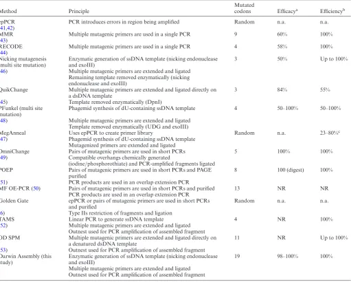
Table 1. Common mutagenesis methods for the introduction of multiple distal mutations
Method Principle
Mutated
codons Efficacya Efficiencyb
epPCR PCR introduces errors in region being amplified Random n.a. n.a.
(41,42)
MMR Multiple mutagenic primers are used in a single PCR 9 60% 100%
(43)
RECODE Multiple mutagenic primers are used in a single PCR 4 58% 100%
(44)
Nicking mutagenesis (multi site mutation)
Enzymatic generation of ssDNA template (nicking endonuclease and exoIII)
3 50% Up to 100%
(46) Multiple mutagenic primers are extended and ligated Remaining template removed enzymatically (nicking endonuclease and exoIII)
QuikChange Multiple mutagenic primers are extended and ligated directly on a dsDNA template
3 84% 55%
(45) Template removed enzymatically (DpnI) PFunkel (multi site
mutation)
Phagemid synthesis of dU-containing ssDNA template 4 50–100% 50–100%
(48) Multiple mutagenic primers are extended and ligated Template removed enzymatically (UDG and exoIII)
MegAnneal Uses epPCR to create primer library Random n.a. 23–80%c
(47) Phagemid synthesis of dU-containing ssDNA template Mutagenized primers are extended and ligated
OmniChange Pairs of mutagenic primers are used in short PCRs 5 100% 100%
(49) Compatible overhangs chemically generated
(iodine/phosphorothiate) and PCR-amplified fragments ligated POEP Pairs of mutagenic primers are used in short PCRs and PAGE
purified
8 100 (digest) 100%
(51) PCR products are used in an overlap extension PCR
MF OE-PCR (50) Pairs of mutagenic primers are used in short PCRs and purified 13 NR NR PCR products are used in an overlap extension PCR
Golden Gate epPCR or pairs of mutagenic primers are used in short PCRs and purified
Random n.a. n.a.
(6) Type IIs restriction of fragments and ligation
TAMS Linear PCR to generate ssDNA template 4 NR 100%
(52) Multiple mutagenic primers are extended and ligated Outnest used for PCR amplification of assembled fragment OD SPM Multiple mutagenic primers are extended and ligated directly on
a denatured dsDNA template
11 NR Up to 100%
(53) Outnest used for PCR amplification of assembled fragment Darwin Assembly (this
study)
Enzymatic generation of ssDNA template (nicking endonuclease and exoIII)
19 98–100% 100%
Multiple mutagenic primers are extended and ligated Outnest used for PCR amplification of assembled fragment
Error-prone PCR (41) is simple but mutations introduced cannot be targeted to particular codons and its effectiveness is dependent on amplification biases and random incorporation of errors (42). In these circumstances efficacy and efficiency are not applicable concepts (n.a.). Some methods are not effective at mutating the whole population (43,44) and/or incorporating all mutations (45,46). Methods that can introduce mutations effectively and efficiently tend to require complex and time-consuming steps, such as phagemid propagation to generate dU-containing DNA (47,48), chemical degradation steps (49), multiple PCR reactions followed by overlap extension PCR reactions to assemble the mutated gene (44,50,51) or modularization of the library into PCR-tractable libraries that can be later re-assembled via Golden Gate assembly (6). Other methods, such as TAMS (52) and OD SPM (53), are simple and effective. Both introduce diversity by using primers containing the target mutations for primer extension and ligation against single-stranded templates followed by PCR and cloning (52,53). However, neither of these methods remove the second strand of the original plasmid and both use T4 DNA ligase, which can ligate across gaps, mispairs and cannot tolerate high temperatures favourable for specific annealing (28)––with resulting compromises in efficiency of library assembly.
aEfficacy is defined as the fraction of the population containing mutations.
bEfficiency is defined as the fraction of mutated clones where all targeted sites are mutated. Some methods do not report (NR) their efficacy or efficiency.
due to improved purification of the assembled DNA from mutagenic primers and single-stranded plasmid template.
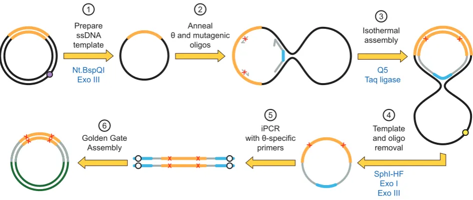
The third variant tested used a single, long oligonu-cleotide harboring both 5 and 3 binding sites, akin to a padlock probe (23), with sites for post-assembly amplifica-tions (i.e. restriction sites and outnest PCR priming sites) linked by a short flexible linker (dT5). This topology,
re-ferred to as the oligonucleotide, generates a closed cir-cle after isothermal assembly, allowing exonucir-clease cir-cleanup and hence removal of partial assembly products,
unincorpo-rated oligonucleotides and original plasmid (Figure2 and Supplementary Figure S1).
Nt.BspQI Exo III Prepare
ssDNA template
1
Anneal boundary and inner
oligonucleotides
2 Isothermal
assembly
Q5 Taq ligase 3
Assembly purification 4
PCR with outnest
primers 5
X X
Golden Gate Assembly
6
X X X X
X X
3’-3’dT
3’-3’dT
X X
X
X
dT3’-3’
X X
Boundary oligonucleotide
Inner oligonucleotide
[image:5.612.58.536.70.412.2]Outnest primer
Figure 1. Principles of Darwin Assembly. Plasmid DNA (black, with the gene of interest in orange) is nicked by a nicking endonuclease (at the purple dot)
and the cut strand degraded by exonuclease III (1). Boundary and inner (mutagenic) oligonucleotides are annealed to the ssDNA plasmid (2). Key features of the oligonucleotides are highlighted: 5-boundary oligonucleotide is 5-biotinylated; non-complementary overhangs are shown in blue with Type IIs endonuclease recognition sites shown in white; mutations are shown as red X in the inner oligonucleotides; 3-boundary oligonucleotide is protected at its 3-end. After annealing, primers are extended and ligated in an isothermal assembly reaction (3). The assembled strand can be isolated by paramagnetic streptavidin-coated beads (4) and purified by alkali washing prior to PCR using outnested priming sites (5) and cloning (6) using the type IIS restriction sites (white dots). The purification step (4) is not always necessary but we found it improved PCR performance, especially for long assembly reactions (>1 kb).
melting/melt.php), or oligonucleotides for PCR with Q5 Hot Start DNA polymerase, which were calculated using NEB Tm calculator (http://tmcalculator.neb.com/).
Single strand plasmid generation
Plasmid DNA, isolated from overnight bacterial cultures using GeneJET Plasmid Miniprep Kits (Thermo Fisher Sci-entific), was made single stranded by co-incubation with a nicking endonuclease and exonuclease III (exoIII). Typ-ically, DNA was digested at 50–60 nM (90–235 ng/l, de-pending on plasmid size) with 3–10 U of nicking enzyme and 40–60 U of exoIII perg of DNA. Plasmid DNA was vacuum concentrated (SpeedVac, ThermoFisher Scientific) if necessary prior to digestion. Reactions, in appropriate re-striction enzyme buffer (typically 1×CutSmart buffer or 1x NEBuffer 3.1), were carried out for 2 h at 37◦C, followed by 20 min at 80◦C to inactivate the enzymes. Nt.BspQI and Nb.BtsI were used successfully but Nt.BbvCI was con-sistently less active under the conditions used. No other nicking endonucleases have been tested. Digestion was
con-firmed by agarose gel electrophoresis followed by SYBR Gold (Thermo Fisher Scientific) staining. ssDNA was used for assembly without further purification and was not re-quantified.
Oligonucleotide phosphorylation
Nt.BspQI Exo III Prepare
ssDNA template
1
Anneal θ and mutagenic
oligos 2
X
X
Isothermal assembly
Q5 Taq ligase
3
X X
Template and oligo removal
SphI-HF Exo I Exo III
4
X X
iPCR with θ-specific
primers 5
X X
Golden Gate Assembly
6
X X X X
[image:6.612.76.550.67.268.2]X X
Figure 2. Darwin Assembly using aoligonucleotide. Here, a singleoligonucleotide is used in place of the two boundary oligonucleotides allowing
enzymatic cleanup after the assembly reaction. Plasmid DNA (black, with the gene of interest in orange) is nicked by a nicking endonuclease (at the purple dot) and the nicked strand degraded by exonuclease III (1). Inner oligonucleotides and a singleoligonucleotide are annealed to the ssDNA plasmid (2). Theoligonucleotide encodes both assembly priming and termination sequences linked by a flexible linker such that successful assembly of the mutated strand results in a closed circle (3). The template plasmid can now be linearized (e.g. at the yellow dot, by adding a targeting oligonucleotide and appropriate restriction endonuclease) and both exonuclease I and exonuclease III added to degrade any non-circular DNA (4). The mutated gene can now be amplified from the closed circle by PCR (5) and cloned into a fresh vector (6) using the type IIS restriction sites (white dots).
Isothermal assembly
Firstly, single-stranded plasmid DNA (typically 0.1–0.2 pmol per reaction) was mixed with mutagenic oligonu-cleotides (at 100–250-fold molar excess) and boundary as-sembly oligonucleotides (at 10–50-fold molar excess over plasmid DNA). Plasmid and oligonucleotide mixtures, typ-ically between 3 and 5 l, were then annealed by freezing samples at –20◦C for at least 15 min and subsequently re-turning to room temperature. Alternatively, efficient anneal-ing was also obtained by heatanneal-ing samples for 5 min to 95◦C and cooling them at 0.1◦C/s to 4◦C.
After annealing, 1 volume 2×Darwin Assembly enzyme mix (2×DA mix) was added and the reaction incubated at 50◦C for 10 min to 1 h, depending on the length of the as-sembly (60◦C was also successfully tested, data not shown). Ten minutes is sufficient even for long assemblies (e.g. T7 RNA polymerase; 2.7 kb) although slightly higher PCR yields are seen after longer reaction times. 2×DA mix con-sists of: 0.05 U/l Q5 High-Fidelity DNA polymerase (not hot start), 8 U/l Taq DNA ligase, 2 mM NAD+, 0.4 mM each dNTP, 10% (w/v) PEG 8000, 2 mM DTT and 1× CutSmart buffer (recipe available in SI). This reaction mix is closely modeled on the Gibson Assembly (25), with Phusion replaced by Q5 DNA polymerase and the reaction buffer replaced with CutSmart buffer. It is stored frozen (and like commercially available Gibson Assembly mixes, will freeze solid).
Assembly purification with streptavidin-coated paramagnetic beads
Streptavidin-coated paramagnetic beads were used to clean up assemblies carried out using 5-biotinylated 5-boundary oligonucleotides.
Initially, 5l Dynabeads MyOne Streptavidin C1 beads (Thermo Fisher Scientific) were blocked in 2×BWBS-T (20 mM Tris–HCl pH 7.4, 2 M NaCl, 0.2% (v/v) Tween-20, 2 mM EDTA) for over an hour on a spinning wheel rotator at room temperature. The beads were isolated on a magnetic stand and resuspended in 50l 2×BWBS-T.
Once the assembly reaction was completed, its volume was adjusted to 50l with PCR-grade water and transferred to a 1.5 ml tube. The pre-blocked beads were added to the reaction and mixed thoroughly. Assembly binding to beads was carried out for 3 h at room temperature, although it was later found that 10 min were sufficient (albeit at slight cost to yield). After capture, beads were isolated on a mag-netic stand and washed twice in 200l 37◦C 30 mM NaOH and once in 200l EB-T (10 mM Tris–HCl pH 8.8, 0.1 mM EDTA, 0.01% Tween-20). Alkali denaturation is carried out at 37◦C to improve bead handling – this is not essential for plasmid denaturation. The wash with EB-T is included to neutralise the beads and to reintroduce Tween-20, to pre-vent the beads adhering to tube walls or aggregating. After washing, the beads were resuspended in 10l EB (10 mM Tris–HCl pH 8.8) and used directly for PCR. A more de-tailed version of this approach is provided in the accompa-nying Supplementary Information (Supplementary Proto-col 1).
Assembly purification by exonuclease
en-donuclease restriction site present in the ssDNA template (e.g. SphI in pET29), so that the chosen endonuclease can cleave the local dsDNA template generated. This creates free 3- and 5-ends enabling exoIII and exoI to degrade the starting template, along with any partial assembly products and free oligonucleotides. A more detailed version of this approach is provided in the accompanying Supplementary Information (Supplementary Protocol 2)
For the TgoT experiment, a 5x master mix consisting 4 U/l exoI, 2 U/l exoIII, 1 U/l SphI-HF and 50 M oligonucleotide pET SphIcut was prepared, of which 4l were added to a 16l isothermal assembly reaction, giving final concentrations of 0.8 U/l exoI, 0.4 U/l exoIII, 0.2 U/l SphI-HF and 10M pET SphIcut. Reactions were carried out for 40 min at 37◦C, followed by incubation for 20 min at 80◦C to inactivate endo- and exonucleases.
PCR amplification of assembled DNA
PCR amplification was carried out directly from the as-sembly reactions, or after its purification (whether exonu-clease or beads). When carried out directed from assem-bly reactions, typically 1l assembly was used as template per 25l PCR. Primers outnest 1 and outnest 2, targeting sites on the boundary assembly oligonucleotides, were used to specifically amplify the assembled libraries (as their se-quences were chosen to not be present in the original plas-mid DNA template).
Multiple enzymes [MyTaq (Bioline), Q5 Hot Start DNA polymerase (NEB) and KOD Xtreme (EMD Millipore)] were successfully used for post-assembly PCR. In general, KOD Xtreme was found to be the most robust regard-ing successful PCR amplification. Reactions consisted 1× KOD Xtreme buffer, 0.3M outnest 1, 0.3M outnest 2, 0.4 mM each dNTP and 0.01 U/l KOD Xtreme DNA polymerase. The cycling parameters used were 2 min 95◦C, followed by 20–28 cycles of 15 s at 98◦C, 15 s at 64◦C and 2 min 30 s at 68◦C, with a final polishing step of 2 min at 72◦C. These conditions were typical for 2–3 kb PCR prod-ucts, and extension time was adjusted as per manufacturer recommendations for shorter assemblies.
After 25l pilot PCRs, PCRs were scaled up (usually to 2×50l reactions) and gel purified using Monarch PCR and DNA cleanup kits (NEB). These were preferred as they allow low elution volumes and result in clean DNA (low A230 contamination). The purified products were digested with BsaI-HF to remove outnesting priming sites and gen-erate suitable overhangs for cloning, and again purified to remove enzyme, salts and cleaved primers. DNA concentra-tion was determined by spectrophotometry (SPECTROstar Nano, BMG Labtech, UK).
Vector amplification and cloning
Vectors were made by iPCR using Q5 Hot Start High Fidelity DNA Polymerase. Reactions consisted of 1x Q5 buffer, 1x Combinatorial Enhancer Solution (CES) (26), 0.2 mM each dNTP, 0.5M each primer and 0.01 U/l Q5 HS DNA polymerase. The PCR conditions used were 1 min at 98◦C, followed by 30 cycles of 15 s at 98◦C, 15 s at 60◦C and 3 min at 72◦C. A final polishing step extension of 1 min
at 72◦C was routinely included. Annealing temperature and extension time varied according to the primer pair and the size of vector being prepared.
Amplification reactions were purified, and treated con-comitantly with BsaI-HF, rSAP and DpnI to create the compatible overhangs, dephosphorylate the vector and de-grade any contaminating original template. The amplified vector DNA was then purified using PCR purification columns (Nucleospin, Machery Nagel) and quantified as above. For point mutations, 20l ligations containing 50 fmol insert, 12.5 fmol vector and 0.5 U/l T4 DNA ligase were ligated for 10 min to 2 h at room temperature in 1×T4 DNA ligase buffer. For libraries, typically 300 fmol insert was ligated into 75 fmol vector at room temperature for 2 h in 100l reactions, using 0.8 U/l T4 DNA ligase and supplemented with 0.5 U/l of 5deadenylase to maximise ligation efficiency.
All ligations were phenol:chloroform:isoamyl alcohol purified, isopropanol precipitated, resuspended in 5–10l EB and transformed into electrocompetent NEB 10beta (CAT, KOD and T7RSS) or NEB T7 Express LysY/Iq (TgoT)E. coli.
Illumina sequencing and data analysis
All deep sequencing was carried out on an Illumina MiSeq at the UCL Genomics Centre using a 150 cycle MiSeq Reagent Kit v3 (Illumina). Libraries were prepared by PCR using KOD Xtreme Hot Start DNA Polymerase (EMD Millipore) to append barcodes and sequencing primers. Libraries were gel purified on agarose gels stained with SYBR safe (Life Technologies), visualised using a blue light source and purified using Monarch DNA Gel Extraction kits (NEB). Libraries were then quantified using a Qubit Fluorometer (Thermo Fisher Scientifc).
Codon frequencies were counted in R version 3.3.0 (27) using RStudio version 1.0.136 for Mac OS X after data processing. Raw FASTQ files were obtained from the Miseq and quality-filtered, trimmed, barcode-split and converted to FASTA using the FASTX-Toolkit (http:// hannonlab.cshl.edu/fastx toolkit/index.html). If necessary, Prinseq (http://prinseq.souceforge.net/) or TAGCleaner (http://tagcleaner.sourceforge.net/)were used to omit reads below a minimum or above a maximum length. FASTA headers were removed, reading frames aligned and the DNA sequence split into triplet codons to facilitate reading into R. More detail on the commands used is available in the Supplementary Methods (Supplementary Protocol 3).
RESULTS
Principles of Darwin Assembly––fast, efficient, multi-site be-spoke mutagenesis
from plasmid DNA to library within a day and it is com-patible with automation.
Single-stranded template is generated from purified plas-mid DNA by the coupled reaction of a nicking endonucle-ase and exonucleendonucle-ase III (exoIII). The endonucleendonucle-ase specifi-cally cuts one strand of plasmid DNA and exoIII selectively and efficiently degrades the nicked DNA strand, leaving be-hind a circular single-stranded template for assembly. Nev-ertheless, we found it increased the robustness of the assem-blies to position a nicking site shortly after the 3-end of the assembly reaction in some instances (data not shown). Nicking endonuclease and exoIII are heat inactivated and the single-stranded template can be used directly in assem-bly, as all enzymes used have activity in a common buffer.
For assembly, primers (except for the 5 boundary as-sembly oligonucleotide) are phosphorylated enzymatically and added to the assembly reaction in the desired molar ra-tios. Mutations are introduced by the oligonucleotides and primer excess ensures efficient binding to ssDNA template, contributing to the high efficiency of Darwin Assembly. Boundary assembly primers are used at lower concentra-tions than mutagenic primers (but still in excess of ssDNA template) to limit side reactions that could undermine a suc-cessful assembly. Primer concentration was not extensively optimised as the concentrations used proved robust for sembly and generated minimal background or unwanted as-semblies.
Once oligonucleotides and ssDNA template are an-nealed, assembly is carried out with a non-strand displac-ing thermostable DNA polymerase (Q5) and a thermostable DNA ligase (Taq DNA ligase), akin to Gibson Assembly (25). The high reaction temperature and the choice of DNA ligase both contribute to the specificity of the assembly, lim-iting off-target annealing of the primers and limlim-iting liga-tion to nicks on the nascent DNA strand (28), respectively. Depending on the complexity of the assembly, no purifi-cation may be necessary, with assembly reactions used di-rectly as PCR templates. Purification, whether based on nu-cleases or isolation of biotinylated DNA post-assembly, im-proves the quality of subsequent PCR amplification mak-ing it less likely to contain secondary amplification prod-ucts (data not shown). We assume the main source of secondary PCR products is carryover of inner assembly oligonucleotides to the PCR, where they act as primers. Re-ducing the concentration used in the assembly reaction may remove the need for purification but at the possible risk of missing out some mutations.
PCR amplification post-assembly generates sufficient material for Golden Gate cloning (29), ensuring highly ef-ficient and scarless cloning of the assembled fragment. Our deep sequencing results show that 98–100% of targeted codons are mutated as expected. No wild-type clones were detected by Sanger sequencing, even in the absence of any purification steps, and deep sequencing of biotin-purified assembled libraries showed wild-type contamination was always<0.25%, a level comparable with sequencing error rates.
In developing the methodology and to explore its poten-tial applications, we have applied Darwin Assembly to dif-ferent genes, including chloramphenicol acetyl transferase (CAT), Saccharomyces cerevisiae tryptohanyl tRNA
syn-A
B
Oligo excess (boundary / inner)
Assembly (min)
1000 bp 500 bp 300 bp
25 / 125 50 / 250
0 10 60 NT
MW 0 10 60 45
(TTA) 63 (CTT)
105 (CTC)
117 (CTA)
158 (TTA)
205 (CTT)
[image:8.612.324.567.72.240.2]208 (TTA)
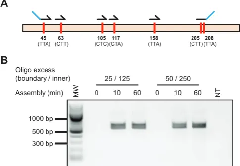
Figure 3. Whole gene codon reassignment. Codons targeted for
reassign-ment and oligonucleotides used in Darwin assembly (A). Amino acid num-bers are original codon sequences are shown. Outnest PCR showing am-plification of the mutated CAT gene following the Darwin Assembly re-action across a range of assembly times (0, 10 and 60 min) and assembly oligonucleotide concentrations (B). Concentrations shown represent the molar excess of boundary (e.g. 25) and inner oligonucleotides (e.g. 125) over plasmid concentration used. Expected assembly amplicon is 568 bp. MW: 100 bp ladder (NEB).
thetase (ScWRS), Thermococcus gorgonarius DNA poly-merase (TgoT), Thermococcus kodakaraenis DNA poly-merase (KOD) and T7 RNA polypoly-merase (T7RSS), which differ in length as well as composition. We demonstrate Darwin Assembly can be used to wholesale reassign codons in a gene of interest, create amino acid scanning and insertion/deletion (indel) libraries, as well as complex mu-tational libraries – where diversity can be highly customis-able (e.g. non-redundant equimolar incorporation of triplet codons).
Whole gene codon reassignment
We chose chloramphenicol acetyl transferase (CAT) as a suitable initial target to develop and optimise Darwin As-sembly, as it is a small (660 bp), well-characterized gene. Our initial proof of principle was to reassign all leucine codons in CAT to CTG. Six oligonucleotides were used to introduce the required 10 point mutations (in seven differ-ent codons spread over 512 nucleotides, Figure3A). Assem-blies were carried out using two boundary oligonucleotides and without any pre-PCR purification. Two assembly con-ditions were tested, with either 125×inner and 25× bound-ary oligonucleotide molar excess over plasmid, or 250× in-ner and 50×boundary oligonucleotide molar excess over plasmid.
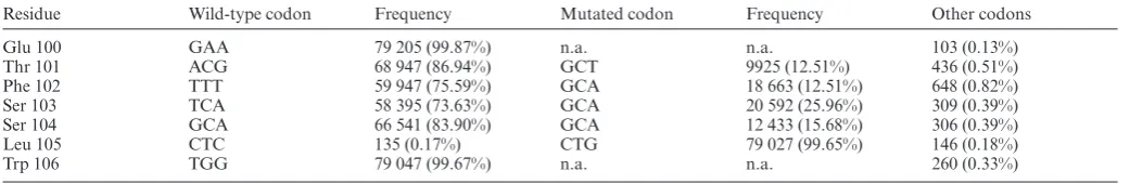
Materi-Table 2. Deep sequencing of CAT gene alanine scanning library
Residue Wild-type codon Frequency Mutated codon Frequency Other codons
Glu 100 GAA 79 205 (99.87%) n.a. n.a. 103 (0.13%)
Thr 101 ACG 68 947 (86.94%) GCT 9925 (12.51%) 436 (0.51%)
Phe 102 TTT 59 947 (75.59%) GCA 18 663 (12.51%) 648 (0.82%)
Ser 103 TCA 58 395 (73.63%) GCA 20 592 (25.96%) 309 (0.39%)
Ser 104 GCA 66 541 (83.90%) GCA 12 433 (15.68%) 306 (0.39%)
Leu 105 CTC 135 (0.17%) CTG 79 027 (99.65%) 146 (0.18%)
Trp 106 TGG 79 047 (99.67%) n.a. n.a. 260 (0.33%)
Near complete mutation of Leu105 confirms the efficiency of the approach with alanine represented in all target sites with frequencies between 12.51% and 25.96% (expected frequency: 20%). Sites not targeted by mutagenesis (Glu100 and Trp106)––and where mutations are not applicable (n.a.)–– show the underlying error rate of sequencing (0.13–0.33%) is comparable with the error obtained in the engineered positions (0.18–0.82%).
als and Methods (shown in Figure1) to remove the initial ssDNA template and unincorporated oligonucleotides.
Encouraged by the high efficiency of the initial assembly reactions, we decided to investigate if the method was suf-ficiently robust and efficient for the generation of libraries of increasing complexity: site saturation (all possible mu-tations at a single position), partial saturation (mutually exclusive mutations targeting different sites), and indel li-braries (deleting or inserting whole codons).
We tested the introduction of diversity by making a small library in Saccharomyces cerevisiae tryptophanyl tRNA synthetase, targeting positions important in substrate speci-ficity (Thr107, Pro254 and Cys255 (11)) with limited degen-eracy (KKC, KKC and RYA, respectively). Assembly was successful and a small number of transformants were se-quenced. All three sites were successfully targeted in the five sequenced transformants and no off-site mutations were observed (data not shown).
Alanine scanning library generation
Scanning libraries introduce a single point mutation at dif-ferent sites in a target gene. Alanine scanning is a tradi-tional approach to identify functradi-tionally important residues in enzymes (30) but scanning libraries can also be used to map the local functional neighbourhood of an enzyme (31) or even to generate datasets for deep mutational scanning-guided rational protein design (32). Mutant generation can be laborious when mutations are introduced individually by site-directed mutagenesis.
We reasoned that Darwin Assembly would efficiently generate scanning libraries by combining oligonucleotides targeting the same binding site but each introducing a different mutation (Supplementary Figure S3). Using the CAT gene as our model, we assembled an alanine scan li-brary around residues Thr101 and Ser104, using Leu105 (CTC→CTG) recoding as an assembly control. Five in-ner oligonucleotides were designed: four introducing an alanine mutation (Thr101Ala, Phe102Ala, Ser103Ala or Ser104Ala) and one wild-type. All five oligonucleotides in-troduced the CTC→CTG control mutation at the Leu105 codon.
The five oligonucleotides were mixed in a 1:1:1:1:1 ra-tio in the assembly reacra-tion to create a library where each variant was expected to be 20% of the final population. As-sembly was successful and over 103transformants were
iso-lated. Transformants were pooled, their plasmid DNA pu-rified and deep sequenced––generating nearly 80 000 reads.
The control Leu105 (CTC→CTG) mutation was present in 99.7% of the samples, a frequency comparable to sites that had not been targeted for mutagenesis (see Table2).
Crucially, an expected proportion of reads (16 739 or 21.1%) were wild-type (containing only the Leu105 recod-ing). This demonstrates that even though there may be a de-gree of incorporation bias, with alanine incorporation rang-ing from Thr101 (12.5%) to Ser103 (26.0%), these are un-likely to have been caused by the mismatches alone.
Generation of deletion and insertion (indel) libraries
Although insertions and deletions are known to play crucial roles in protein function (e.g. antibodies), there are few tools available to explore length as a parameter in protein engi-neering (BioRxiv:https://doi.org.10.1101/127829) (33). We therefore decided to investigate if Darwin Assembly could be used as a tool to explore indels for protein engineering.
We generated two libraries in CAT: one exploring sin-gle and double deletions and one exploring sinsin-gle and double insertions (Supplementary Figure S3). As with the scanning libraries, the indel libraries were assembled using an equimolar mixture of inner oligonucleotides designed against a common binding site. In addition, similar controls were included, using a wild-type oligonucleotide and the Leu105 (CTC→CTG) controls. The deletion library was as-sembled with four inner oligonucleotides, maintaining the wild-type sequence, deleting Phe102 or Ser104, or deleting both sites. The insertion library was generated with three oligonucleotides, maintaining the wild-type sequence, or in-troducing one or two degenerate codons (NNS) between Phe102 and Ser103.
Assemblies of both libraries were successful and sev-eral thousand transformants were obtained for each library. Libraries were pooled and sequenced obtaining approxi-mately 2.1×10⁵ reads for analysis. As both libraries
con-tained a wild-type assembly control and were pooled for se-quencing, analysis of assembly biases cannot easily be de-termined.
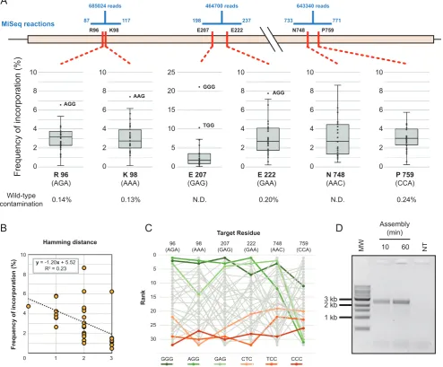
[image:9.612.41.562.82.167.2]R96 K98 E207 E222 N748 P759
Frequency of incorporation (%) 0
2 4 6 8 10 0 2 4 6 8 10 0 2 4 6 8 10 0 2 4 6 8 10 0 2 4 6 8 10 0 0 2 4 6 8 0 0 2 4 6 8 10 0 2 4 6 8 10 0 2 4 6 8 10 0 2 4 6 8 10 0 5 10 15 20 25 R 96 (AGA) K 98 (AAA) E 207 (GAG) E 222 (GAA) N 748 (AAC) P 759 (CCA) AGG AAG GGG TGG AGG Wild-type
contamination 0.14% 0.13% N.D. 0.20% N.D. 0.24%
A
Assembly (min) 3 kb 2 kb 1 kb NTMW 10 60
D
MiSeq reactionsB
Hamming distance
87 117 198 237 733 771
685024 reads 464700 reads 643340 reads
C
Target ResidueRank 96 (AGA) 98 (AAA) 207 (GAG) 222 (GAA) 748 (AAC) 759 (CCA) 5 10 15 20 25 30 0
GGG AGG GAG CTC TCC CCC
Frequency of incorporation (%)
0 1 2 3
2 4 6 8 10
y = -1.20x + 5.52
[image:10.612.66.561.75.484.2]R2 = 0.23
Figure 4.Darwin assembled T7 RNA polymerase library. Sequencing of three separate regions of the assembled gene were used to determine the assembled
diversity in the targeted codons (A). Number of reads obtained for each segment are shown as well as the range of each reaction (shown in blue). Frequency of incorporation of the 32 possible codons (NNS) is shown as a box plot, with overrepresented outliers explicitly labelled. Wild-type contamination was determined where possible (N.D.: not determined). Incorporation trends were detected in all positions and best correlated to the number of mismatches they introduced. A typical correlation is shown (B) but other positions varied (0.03–0.37), always indicating that the higher the number of mutations being concomitantly introduced (i.e. larger Hamming distance), the lower incorporation of the variant in the population. Incorporation trends were also analysed to determine systematic biases of incorporation. Incorporation frequencies were ranked at each position and rank order analysed (C)––the top three highest ranked (greens) and lowest ranked (oranges) codons are highlighted for clarity. Incorporation biases were detected but it was not clear whether that was the result of the very imbalanced sequence composition of the wild-type residues targeted. Outnest PCR of the T7 RNA polymerase library (expected product of 2840 bp) confirms that as little as 10 minutes of isothermal assembly were sufficient for successful amplification of a library (D). MW: 1 kb ladder (NEB). NT: no template PCR control.
Together, the insertion and deletion libraries confirm that Darwin Assembly can be used to investigate small changes in length in a target gene, but may require further optimisa-tion to minimise assembly biases of deleoptimisa-tion libraries. Dele-tions and inserDele-tions decrease the melting temperature of the mutagenic primers, with thePhe102Ser104 oligonu-cleotide having a predicted melting temperature of nearly 15◦C lower than the Leu105CTG controls (Supplementary Table S5). As assembly was carried out with both oligos present in a single reaction, kinetic and thermodynamic fac-tors can account for the observed frequency variation. We
expect that extending mutagenic primers that introduce in-sertions and deletions to normalise melting temperatures in the assembly reaction would result in a more even distribu-tion across the different lengths.
Darwin Assembly enables the synthesis of custom complex libraries ideal for directed evolution
(2.4 kb). These are more typical of enzymes targeted for en-gineering and represented a greater challenge.
As expected, assemblies longer than 1.2 kb were ini-tially less robust and invariably required considerable PCR optimisation (data not shown). We concluded the PCR problems were caused by carryover of unincorporated in-ner oligonucleotides and/or partial assembly products from the assembly reaction to PCR. Therefore, we investigated two strategies to remove template and unextended (or partially assembled) oligonucleotides: either purifying as-sembled molecules (Figure 1) or by selectively degrading template and unassembled products (Figure 2). Both ap-proaches were successful and enabled assembly of libraries spanning more than 2 kb with T7RSS and TgoT respec-tively.
For T7RSS, we designed a library targeting residues pre-viously implicated in switching promoter specificity (9): Arg96, Lys98, Glu207, Glu222, Asn748, Pro759. The 2724 bp assembly, targeting six codons, used five inner oligonu-cleotides introducing degeneracy (NNS) at each of the tar-get positions (Figure4A). Assembled libraries were ampli-fied, cloned and transformed, with approximately 1×10⁷
transformants isolated.
Sequencing of the pooled transformants confirmed that at each position, all possible variants were introduced (Fig-ure4, Supplementary Table S6). Wild-type contamination, in codons where that could be detected, was always<0.24% and comparable to sequencing errors detected in non-targeted positions. Incorporation biases were small and cor-related primarily with the number of mismatches between template and target codon. Overall, there were also de-tectable biases over the whole assembly (Kruskal–Wallis ANOVA on ranked target codons, P = 0.001), but since the template residues were clustered in sequence space (5/6 were 67% purine or above), it is possible that the overall in-corporation bias observed is a consequence of the individ-ual mismatch biases, rather than a systematic bias.
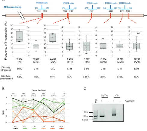
A known limitation of targeting residues with NNS (or NNK) degenerate oligonucleotides is the bias they intro-duce in translation: oversampling residues that have mul-tiple codons (e.g. Leu and Arg) and increasing the min-imum library size required to adequately sample the se-quence space introduced. Alternatives to NNS have been developed for site-directed mutagenesis, such as ‘small intel-ligent’ libraries that mix four oligonucleotides with different degrees of degeneracy (NDT, VMA, ATG and TGG) and in a suitable ratio (12:6:1:1, respectively) to generate equal cov-erage of all 20 amino acids (34). Such libraries are impossi-ble when targeting multiple sites using simple iPCR. Never-theless, as Darwin Assembly targets distal sites using inde-pendent inner oligonucleotides, it is well suited to generat-ing multi-site intelligent libraries – where nucleic acid varia-tion matches protein variavaria-tion, maximising library quality. We therefore designed the TgoT library to target eight residues implicated in template recognition and DNA du-plex affinity modulation (Tyr384, Val389, Ala490, Tyr493, Phe587, Glu664, Gly711 and Asn735 (10,35)), with a mix-ture of limited or with small intelligent degeneracies (Figure
5).
The assembly spanned 2389 bp and targeted the eight residues with six inner oligonucleotides and a
non-mutagenic ‘’ boundary. The resulting library was success-fully amplified, cloned and transformed with an estimated 2.25×10⁸transformants generated. Pooled transformants
were sequenced, confirming that the designed diversity was introduced in all targeted residues (Figure5). As with the T7 library, a positional bias is detectable and correlates weakly to the number of mismatches between template and target codons. Overall, there is a detectable bias in the final as-sembly beyond a potential systematic error in the mixing of the oligonucleotides (Kruskal-Wallis ANOVA on ranked target NDT codons,P=0.005), but it is less clear whether the bias is a consequence of the individual template/target codon mismatches––e.g. systematic underrepresentation of GGT even at residue 711 (GGC). (Figure5B, Supplemen-tary Tables S6–S8).
Overall, both T7RSS and TgoT libraries do effectively sample the introduced diversity, given transformants iso-lated and the measured per site diversity. Both libraries have the expected diversity at multiple distal sites, have minimal contamination of wild-type sequences and are free of as-sembly errors, despite the size of the genes. Moreover, Dar-win Assembly enabled generation of those libraries within a single working day and at a lower cost than current poten-tial commercial suppliers.
DISCUSSION
Library quality is a major contributing factor to directed evolution (36). It impacts the minimum library size that has to be generated to adequately sample a given sequence land-scape (16,37) and it impacts the choice of selection strat-egy (depending on the relative rarity of a ‘successful’ vari-ant and gain of function over wild-type). Generating high-quality libraries that target multiple distal sites in a gene is challenging, with commercial library synthesis and modu-lar assembly of the library being available only recently.
Darwin Assembly is a highly efficient method for si-multaneous mutagenesis of multiple sites. It is capable of introducing point mutations, bespoke diversity, insertions and/or deletions in complete or partial replacement of wild-type. It is fast, requires no specialised strains or chemical steps and libraries are easily generated in a single working day (from plasmid miniprep to transformation) using com-mercially available enzymes. We also observed very low lev-els of wild-type sequence contamination (peaking in TgoT E664 at 1.95% but generally <0.5%). Overall, this makes Darwin Assembly uniquely suited for generating high qual-ity libraries for directed evolution experiments (36,38).
Frequency of incorporation (%) Y 384 (TAT) V 389 (GTG) A 490 (GCA) F 493 (TTT) F 587 (TTC) E 664 (GAG) AAT Diversity introduced
N.A. 0.66% 0.32%
1.3% 2.0% N.A.
A
MyTaq
6 kb 3 kb 1.5 kb
MW + –
C
B
Target ResidueRank 389 (GTG) 493 (TTT) 587 (TTC) 664 (GAG) 711 (GGC) 735 (AAT) 5 10 15 20 0
ATG GAT CAT CTT TTT GGT
N 735 (AAT) 0.4% G 711 (GGC) 1.6% AAT Wild-type contamination
YWC S-Int SRC S-Int S-Int S-Int S-Int S-Int
Y384 V389 A490 F493 F587 E664 G711 N735
MiSeq reactions 0 2 4 6 8 10 12 14 0 2 4 6 8 10 12 14 0 2 4 6 8 10 12 14 0 2 4 6 8 10 12 14 0 2 4 6 8 10 12 14 0 2 4 6 8 10 12 14 0 5 10 15 20 25 30 35 40 0 5 10 15 20 25 30 35 40
371 415 476 524 574 624 640 684 700 746
4756352 reads 4756352 reads 612345 reads 2153533 reads 2806200 reads
MyTaq
MW + –
Q5
[image:12.612.68.559.76.513.2]Assembly + –
Figure 5. Darwin assembled TgoT DNA polymerase library. (A) Five separate sequencing reactions (range and reads shown in blue) were required to
sample the diversity introduced across the eight target residues (shown in red along the TgoT gene). Mutations included focused degeneracies (e.g. YWC used against Y384) or ‘small intelligent’ (S-int) diversity (NDT, VMA, ATG and TGG oligonucleotides mixed in a 12:6:1:1 ratio). Resulting incorporation is shown in box plots with outliers explicitly labelled. Wild-type contamination was determined from positions where diversity excluded those sequences (N.A.: not applicable). As with the T7 RNA polymerase library, incorporation trends and biases were analysed to identify any biases in assembly. Ranked incorporation frequencies are shown for the residues targeted with ‘small intelligent’ diversity, and the top three highest (greens) and lowest (oranges) ranked codons (based on a straight sum of ranks) are highlighted (B). Outnest PCR of the TgoT DNA polymerase library (expected product of 2501 bp) showing that the final PCR can be carried out with either A- (MyTaq) or B-family (Q5) polymerases (C). MW: 1 kb ladder (NEB). NT: no template PCR control.
(e.g. tRNA and aminoacyl tRNA synthetase) on the same plasmid could be targeted with no protocol modifications, providing the boundary oligonucleotides are sited appropri-ately.
Post assembly cleanup was not necessary for short frag-ments, making a one-pot assembly feasible. Longer assem-blies greatly benefited from the removal of wild-type tem-plate and unincorporated oligonucleotides and both meth-ods we present here were successful. Post-assembly amplifi-cation was marginally better for T7RSS, suggesting that
38 point mutations covering 19 codons in a single reaction and see no reason why this is the limit of the technology.
SUPPLEMENTARY DATA
Supplementary Dataare available at NAR Online.
ACKNOWLEDGMENTS
The authors thank Jared Ellefson and Andy Ellington (Uni-versity of Texas) for the kind gift of pRST.11B.AS3.4 (en-coding ScWRS) and the T7RSS gene. We also thank John Chaput (University of California, Irvine) for the kind gift of the pGDR11-KOD plasmid harbouring the KOD DNA polymerase gene.
FUNDING
Biotechnology and Biosciences Research Council [BB/N01023X/1, BB/N010221/1]; European Research Council [ERC-2013-StG project 336936 (HNAepisome)]. Funding for open access charge: BBSRC [BB/N01023X/1].
Conflict of interest statement.None declared.
REFERENCES
1. Turner,N.J. (2009) Directed evolution drives the next generation of biocatalysts.Nat. Chem. Biol.,5, 567–573.
2. P´edelacq,J.-D., Cabantous,S., Tran,T., Terwilliger,T.C. and Waldo,G.S. (2006) Engineering and characterization of a superfolder green fluorescent protein.Nat. Biotechnol.,24, 79–88.
3. Bloom,J.D., Labthavikul,S.T., Otey,C.R. and Arnold,F.H. (2006) Protein stability promotes evolvability.Proc. Natl. Acad. Sci. U.S.A., 103, 5869–5874.
4. Kan,S.B.J., Lewis,R.D., Chen,K. and Arnold,F.H. (2016) Directed evolution of cytochrome c for carbon–silicon bond formation: Bringing silicon to life.Science,354, 1048–1051.
5. Reetz,M.T., Wang,L.W. and Bocola,M. (2006) Directed evolution of enantioselective enzymes: Iterative cycles of CASTing for probing protein-sequence space.Angew. Chem. Int. Ed.,45, 1236–1241. 6. Quaglia,D., Ebert,M.C.C.J.C., Mugford,P.F. and Pelletier,J.N. (2017)
Enzyme engineering: a synthetic biology approach for more effective library generation and automated high-throughput screening.PLoS
One,12, e0171741.
7. Fox,R.J., Davis,S.C., Mundorff,E.C., Newman,L.M., Gavrilovic,V., Ma,S.K., Chung,L.M., Ching,C., Tam,S., Muley,S.et al.(2007) Improving catalytic function by ProSAR-driven enzyme evolution. Nat. Biotechnol.,25, 338–344.
8. Khersonsky,O., R ¨othlisberger,D., Dym,O., Albeck,S., Jackson,C.J., Baker,D. and Tawfik,D.S. (2010) Evolutionary optimization of computationally designed enzymes: Kemp eliminases of the ke07 series.J. Mol. Biol.,396, 1025–1042.
9. Carlson,J.C., Badran,A.H., Guggiana-Nilo,D.A. and Liu,D.R. (2014) Negative selection and stringency modulation in phage-assisted continuous evolution.Nat. Chem. Biol.,10, 216–222.
10. Cozens,C., Pinheiro,V.B., Vaisman,A., Woodgate,R. and Holliger,P. (2012) A short adaptive path from DNA to RNA polymerases.Proc. Natl. Acad. Sci. U.S.A.,109, 8067–8072.
11. Ellefson,J.W., Meyer,A.J., Hughes,R.A., Cannon,J.R., Brodbelt,J.S. and Ellington,A.D. (2014) Directed evolution of genetic parts and circuits by compartmentalized partnered replication.Nat. Biotechnol.,32, 97–101.
12. Shapiro,M.G., Westmeyer,G.G., Romero,P.A., Szablowski,J.O., K ¨uster,B., Shah,A., Otey,C.R., Langer,R., Arnold,F.H. and Jasanoff,A. (2010) Directed evolution of a magnetic resonance imaging contrast agent for noninvasive imaging of dopamine.Nat.. Biotechnol.,28, 264–270.
13. Jeschek,M., Reuter,R., Heinisch,T., Trindler,C., Klehr,J., Panke,S. and Ward,T.R. (2016) Directed evolution of artificial metalloenzymes for in vivo metathesis.Nature,537, 661–665.
14. Leconte,A.M., Dickinson,B.C., Yang,D.D., Chen,I.A., Allen,B. and Liu,D.R. (2013) A population-based experimental model for protein evolution: effects of mutation rate and selection stringency on evolutionary outcomes,Biochemistry,52, 1490-1499.
15. Currin,A., Swainston,N., Day,P.J. and Kell,D.B. (2015) Synthetic biology for the directed evolution of protein biocatalysts: navigating sequence space intelligently.Chem. Soc. Rev.,44, 1172–1239. 16. Packer,M.S. and Liu,D.R. (2015) Methods for the directed evolution
of proteins.Nat. Rev. Genet.,16, 379–394.
17. Dickinson,B.C., Leconte,A.M., Allen,B., Esvelt,K.M. and Liu,D.R. (2013) Experimental interrogation of the path dependence and stochasticity of protein evolution using phage-assisted continuous evolution.Proc. Natl. Acad. Sci. U.S.A.,110, 9007–9012. 18. Podgornaia,A.I. and Laub,M.T. (2015) Pervasive degeneracy and
epistasis in a protein-protein interface.Science,347, 673–677. 19. Stemmer,W.P.C. and Morris,S.K. (1992) Enzymatic inverse PCR: A
restriction site independent, single-fragment method for
high-efficiency, site-directed mutagenesis.Biotechniques,13, 214-220. 20. Davidson,E.A., Meyer,A.J., Ellefson,J.W., Levy,M. and
Ellington,A.D. (2012) An in vitro autogene.ACS Synth. Biol.,1, 190–196.
21. Guzman,L.M., Belin,D., Carson,M.J. and Beckwith,J. (1995) Tight regulation, modulation, and high-level expression by vectors containing the arabinose P(BAD) promoter.J. Bacteriol.,177, 4121–4130.
22. Pinheiro,V.B., Taylor,A.I., Cozens,C., Abramov,M., Renders,M., Zhang,S., Chaput,J.C., Wengel,J., Peak-Chew,S.-Y., McLaughlin,S.H. et al.(2012) Synthetic genetic polymers capable of heredity and evolution.Science,336, 341–344.
23. Baner,J., Nilsson,M., Mendel-Hartvig,M. and Landegren,U. (1998) Signal amplification of padlock probes by rolling circle replication. Nucleic Acids Res.,26, 5073–5078.
24. Lee,J.H., Skowron,P.M., Rutkowska,S.M., Hong,S.S. and Kim,S.C. (1996) Sequential amplification of cloned DNA as tandem multimers using class- IIS restriction enzymes.Genet. Anal. - Biomol. Eng.,13, 139–145.
25. Gibson,D.G., Young,L., Chuang,R.-Y., Venter,J.C., Hutchison,C.A. and Smith,H.O. (2009) Enzymatic assembly of DNA molecules up to several hundred kilobases.Nat. Methods,6, 343–345.
26. Ralser,M., Querfurth,R., Warnatz,H.-J., Lehrach,H., Yaspo,M.-L. and Krobitsch,S. (2006) An efficient and economic enhancer mix for PCR.Biochem. Biophys. Res. Commun.,347, 747–751.
27. Core Team,R (2016)R: A Language and Environment for Statistical Computing. R Foundation for Statistical Computing, R Foundation for Statistical Computing, Vienna, Austria Vienna, Austria. 28. Lohman,G.J.S., Bauer,R.J., Nichols,N.M., Mazzola,L., Bybee,J.,
Rivizzigno,D., Cantin,E. and Evans Jr,T.C. (2016) A
high-throughput assay for the comprehensive profiling of DNA ligase fidelity.Nucleic Acids Res.,44, e14.
29. Engler,C., Kandzia,R. and Marillonnet,S. (2008) A one pot, one step, precision cloning method with high throughput capability.PLoS
One,3, e3647.
30. Cunningham,B.C. and Wells,J.A. (1989) High-resolution epitope mapping of hGH-receptor interactions by alanine-scanning mutagenesis.Science,244, 1081–1085.
31. Sunden,F., Peck,A., Salzman,J., Ressl,S. and Herschlag,D. (2015) Extensive site-directed mutagenesis reveals interconnected functional units in the alkaline phosphatase active site.Elife,2015, 1–76. 32. Shin,H. and Cho,B.K. (2015) Rational protein engineering guided by
deep mutational scanning.Int. J. Mol. Sci.,16, 23094–23110. 33. T ´oth-Petr ´oczy, ´A. and Tawfik,D.S. (2014) Hopeful (protein InDel)
monsters?Structure,22, 803–804.
34. Tang,L., Gao,H., Zhu,X., Wang,X., Zhou,M. and Jiang,R. (2012) Construction of ‘small-intelligent’ focused mutagenesis libraries using well-designed combinatorial degenerate primers.Biotechniques,52, 149–158.
35. Ellefson,J.W., Gollihar,J., Shroff,R., Shivram,H., Iyer,V.R. and Ellington,A.D. (2016) Synthetic evolutionary origin of a proofreading reverse transcriptase.Science,352, 1590–1593. 36. Tee,K.L. and Wong,T.S. (2013) Polishing the craft of genetic diversity
creation in directed evolution.Biotechnol. Adv.,31, 1707–1721. 37. Nov,Y. (2013) Fitness loss and library size determination in
38. Reetz,M.T., Kahakeaw,D. and Lohmer,R. (2008) Addressing the numbers problem in directed evolution.ChemBioChem,9, 1797–1804. 39. Yang,Z., Chen,F., Chamberlin,S.G. and Benner,S.A. (2010)
Expanded genetic alphabets in the polymerase chain reaction.Angew. Chem. - Int. Ed.,49, 177–180.
40. Zhang,Y., Lamb,B.M., Feldman,A.W., Zhou,A.X., Lavergne,T., Li,L. and Romesberg,F.E. (2017) A semisynthetic organism engineered for the stable expansion of the genetic alphabet.Proc. Natl. Acad. Sci. U.S.A.,114, 1317–1322.
41. Cadwell,R.C. and Joyce,G.F. (1992) Randomization of genes by PCR mutagenesis.Genome Res.,2, 28–33.
42. Copp,J.N., Hanson-Manful,P., Ackerley,D.F. and Patrick,W.M. (2014) Error-prone PCR and effective generation of gene variant libraries for directed evolution.Methods Mol. Biol.,1179, 3–22. 43. Hames,C., Halbedel,S., Schilling,O. and Stulke,J. (2005)
Multiple-mutation reaction: a method for simultaneous introduction of multiple mutations into the glpK gene of Mycoplasma
pneumoniae.Appl. Environ. Microbiol.,71, 4097–4100. 44. Jin,P., Kang,Z., Zhang,J., Zhang,L., Du,G. and Chen,J. (2016)
Combinatorial evolution of enzymes and synthetic pathways using one-step PCR.ACS Synth. Biol.,5, 259–268.
45. Hogrefe,H.H., Cline,J., Youngblood,G.L. and Allen,R.M. (2002) Creating randomized amino acid libraries with the QuikChange (R) Multi Site-Directed Mutagenesis Kit.Biotechniques,33, 1158. 46. Wrenbeck,E.E., Klesmith,J.R., Stapleton,J.A., Adeniran,A.,
Tyo,K.E.J. and Whitehead,T.A. (2016) Plasmid-based one-pot
saturation mutagenesis saturation mutagenesis.Nat. Methods,13, 1–5.
47. Pai,J.C., Entzminger,K.C. and Maynard,J.A. (2012) Restriction enzyme-free construction of random gene mutagenesis libraries in Escherichia coli.Anal. Biochem.,421, 640–648.
48. Jones,D.D., Firnberg,E. and Ostermeier,M. (2012) PFunkel: efficient, expansive, user-defined mutagenesis.PLoS One,7, e52031.
49. Dennig,A., Shivange,A. V, Marienhagen,J. and Schwaneberg,U. (2011) OmniChange: the sequence independent method for simultaneous site-saturation of five codons.PLoS One,6, e26222. 50. W¨aneskog,M. and Bjerling,P. (2014) Multi-fragment site-directed
mutagenic overlap extension polymerase chain reaction as a competitive alternative to the enzymatic assembly method.Anal. Biochem.,444, 32–37.
51. Peng,R.-H., Xiong,A.-S. and Yao,Q.-H. (2006) A direct and efficient PAGE-mediated overlap extension PCR method for gene
multiple-site mutagenesis.Appl. Microbiol. Biotechnol.,73, 234–240. 52. Young,L. and Dong,Q.H. (2003) TAMS technology for simple and
efficient in vitro site-directed mutagenesis and mutant screening. Nucleic Acids Res.,31.