Review
Exosome Release of Drugs: Coupling with
Epithelial-Mesenchymal Transition
Takanori Eguchi 1,2,*, Kisho Ono 3, Stuart K. Calderwood4 and Kuniaki Okamoto 1
1 Department of Dental Pharmacology, Graduate School of Medicine, Dentistry and Pharmaceutical Sciences, Okayama University, Okayama 700-8525, Japan; [email protected] (T.E.), [email protected] (K.O.)
2 Advanced Research Center for Oral and Craniofacial Sciences, Graduate School of Medicine, Dentistry and Pharmaceutical Sciences, Okayama University, Okayama, Japan.
3 Department of Oral and Maxillofacial Surgery, Okayama University Hospital, Okayama 700-0914, Japan; [email protected] (K.O.)
4 Department of Radiation Oncology, Beth Israel Deaconess Medical Center, Harvard Medical School, Boston, MA 02215; [email protected] (S.K.C.)
* Correspondence: E-mail: [email protected] (T.E.); Tel.: +81-86-235-6662
Abstract: Extracellular vesicles (EVs), such as exosomes or oncosomes are released with molecules unfavorable for survival from cells. In addition, accumulating evidence has shown that tumor cells often eject anti-cancer drugs such as chemotherapeutics and targeted drugs within EVs, a novel mechanism of drug resistance. The EV-releasing, drug resistance phenotype is often coupled with cellular dedifferentiation and transformation, cells undergoing epithelial-mesenchymal transition (EMT) and taking on a cancer stem cell phenotype. Recent studies have shown that the release of EVs is also involved in immunosuppression. The concept of the resistance-associated secretory phenotype (RASP) is reviewed herein.
Keywords: resistance-associated secretory phenotype (RASP); extracellular vesicle (EV); exosome; oncosome; drug resistance; epithelial-mesenchymal transition (EMT); heat shock protein (HSP); cell stress response; hypoxia; acidosis; tumor immunology
1. Introduction
Recent studies have unveiled the existence of and significant biological roles for extracellular vesicles (EVs) such as exosomes. EVs are nano-particles surrounded by lipid membranes, containing a variety of molecular cargos such as proteins, small and large RNAs, DNA, lipids, glycans, minerals, and metabolites that are thus secreted by cells [1-5]. Earlier studies have classified the range of EVs into exosomes (50-200 nm), ectosomes (100-1000 nm; also known as microvesicles) [6-8], and apoptotic bodies (1-10 μm) based on their mechanisms of generation and release, while additional types of EVs have been reported, consisting of oncosomes (oncogenic EVs) [9-11], large oncosomes (1-10 μm) [12,13], matrix vesicles [14-16], migrasomes (50 nm to 3 μm) [17,18], exopheres (~4 μm), exomeres (~35 nm), and bacterial outer membrane vesicles (OMV) [19,20] [4,21]. EVs are also classified by their size into small EVs (s-EVs; 30-500 nm) and large EVs (L-EVs; > 1 µm). These vesicles play roles in discarding unfavorable molecules from cells, while also mediating cell-to-cell communication by transferring their cargo molecules to recipient cells or organs in local and/or distant tissues [22]. Recent studies have shown that anti-cancer drugs, including chemotherapeutics and targeted drugs, can be released from cells within EVs, suggesting a novel mechanism of drug resistance. EV-mediated drug efflux is often coupled with cellular dedifferentiation involving the so-called epithelial-to-mesenchymal transition (EMT) [23].
EMT involves a cellular transformation or dedifferentiation from an epithelial phenotype into the mesenchymal phenotype and is important in many aspects of cell biology, including development, inflammation, and cancer [24-26]. Epithelial cells are usually tightly connected to each other through intercellular adhesion and cell junction including adherence junction, desmosome, gap junction, synaptic junction, occluding/tight junction, whereas loss of these connections/adhesions is accompanied by altered cellular shapes, increased motility and migratory activities of the cells. Pre-cancerous cells often exhibit EMT, increased migration and invasion of the cells within the tumor milieu [27]. EMT is a complex process consisting of multiple sequential steps and pathways, promoted by extracellular prompts such as transforming growth factor β (TGFβ) signaling [28], epidermal growth factor (EGF) signaling [23,29], matrix metalloproteinases (MMPs)[30], intracellular signals, and transcription factors [27]. It has been shown that EMT increases the properties of cancer stem cells (CSC) or cancer-initiating cells (CIC), which are highly resistant, recurrent, and metastatic [31-33].
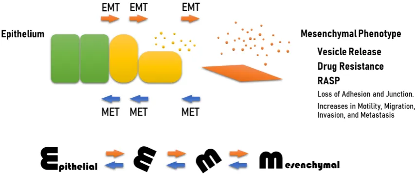
Recent studies have shown that increased exosome release is coupled with EMT (Figure 1). EMT enhances the exosome-releasing phenotype of cells, while, conversely, tumor-derived exosomes initiate EMT in epithelial cells as well as driving EMT in cancer cells [23]. Moreover, it has been also shown that anti-cancer drugs were released with exosomes from tumor cells, suggesting a mechanism of cancer drug resistance. The vesicle-releasing and drug-releasing phenotypes can be an aspect of the resistant-associated secretory phenotype (RASP). Studies showing EMT-coupled exosome release are reviewed as a mechanism of drug resistance and immunosuppression in cancer.
Figure 1. EMT coupled with vesicle release, drug resistance, and RASP. EMT is cellular dedifferentiation, transformation or reprogramming in which epithelial cell phenotype is switched into mesenchymal phenotype. Epithelial cells adhere to each other by epithelial intercellular adhesion molecules (i.e. E-cadherin and claudins) and desmosomes, which are often lost in EMT. The cells conferring EMT acquire increased motility, migration, invasion, and metastasis. EMT is a multi-step event and reversible, so-called plasticity. This review wraps up that EMT is often coupled with vesicle release, drug resistance, and resistance-associated secretory phenotype (RASP).
Oncosomes have been defined as oncogenic EVs or oncogenic exosomes that molecularly transfer tumor-promoting factors such as oncoproteins, oncomiR, and circulating tumor DNA (ctDNA) [11,12,34,35], while a number of studies have reported that tumor-derived exosomes played similar oncogenic roles without using the term “oncosome”. In 2008, Janus Rak et al first defined oncosomes as they reported intercellular transfer of EGF receptor variant III (EGFRvIII), an oncogenic receptor, by microvesicles derived from brain tumor cells [11]. In the next year, Di Vizio et al reported that oncosome formation in prostate cancer is associated with a region of frequent chromosomal deletion in metastatic disease [35]. This group thereafter reported that large oncosomes, larger than 1 µm could be selectively sorted by flow cytometry in human prostate cancer tissues and in the circulation of mice with metastatic disease and contained MMPs, RNA, caveolin-1, and the GTPase ADP-ribosylation factor 6 [13]. The large oncosomes (or defined as L-EVs) carried most of the tumor DNA circulating in prostate cancer patient plasma [12]. In this study, whole-genome sequencing revealed that the DNA in L-EVs reflects genetic aberrations of the cell of origin, including copy number variations (CNV) of genes frequently altered in metastatic prostate cancer, i.e. MYC, AKT1, PTK2, KLF10, and PTEN. Later studies have shown that a number of additional oncogenic factors were contained in oncosomes, such as oncomiR miR-520g [36], 14-3-3 and beta-catenin [37].
A proteomic study revealed that oral cancer-derived oncosomes contain heat shock protein (HSP) family members, a number of extracellular matrix (ECM) proteins, and transcriptional regulators [38]. HSPs have been shown to assist in folding of oncoproteins essential for cancer cell survival and resistance [39-41]. Therefore, HSP-rich oncosomes and their molecular transfer can be crucial in tumor progression and resistance.
2.2. EV-mediated chaperone transfer
Human tumor cells are progressively exposed to stresses such as hypoxic stress, immune and inflammatory stress, and therapeutic stress. Such cell stresses trigger the expression of HSPs, stress-resistant cytoprotective proteins with anti-apoptotic and senescence deterring activity. Intracellular HSPs are molecular chaperones playing key roles in protein folding of normal and oncogenic proteins and balancing between proteostasis and proteolysis [39,42-44]. In addition, extracellular HSPs play key roles in cell-cell communication in cancer and immunity [45]. Extracellular HSPs and HSP-rich EVs can promote cancer progression by enhancing EMT, migration, invasion, heterogeneity, metastasis, CSC/CIC properties, and drug resistance in cancer cells and angiogenesis [46-52]. Proteomic analysis of oral cancer-derived oncosomes revealed that a number of HSP family members are contained within EVs (i.e. HSP90 homologs, large HSP members, and HSP70 family members) [38]. HSPs and oncoproteins contained within EVs could be involved in RASP, cotransferred to recipient cells leading to cancer expansion and malignant conversion of the tumor microenvironment (Figure 2).
2.3. Stromal signal-driven tumor progression
Tumor cells are surrounded by tumor stroma composed of various types of cells including cancer-associated fibroblasts (CAFs) with properties of mesenchymal stem cells (MSCs), tumor-associated immune cells including tumor-tumor-associated macrophages (TAMs), T cells, B cells, and dendritic cells (DCs), tumor endothelial cells (TECs), adipocytes, and normal epithelial cells. These stromal cells communicate with each other using cytokines, growth factors, MMPs [53], ECM, microRNAs, and EVs [54,55]. Earlier studies suggested that tumor stroma was tumor-suppressing, although recent studies have unveiled that stroma signals often drive tumor progression. Among the various stromal cells, we here review the crucial roles of CAFs, TAMs, and TECs.
immune evasion by producing IL-6, COX-2/PGE2, and SDF-1/CXCL12. CAFs control ECM deposition and remodeling by producing fibronectin, collagen 1A1, tenascin C, osteopontin, and MMPs [62]. It has been shown that these secretory phenotypes of CAFs were often carried by EVs, including TGFβ, MMPs, microRNA, and ECMs, which alter epithelial cells, tumor cells, and tumor milieu [53,55,62-64]. Proteomics of stroma-derived EVs is important to elucidate the mechanism of tumor progression. Proteomics of secretory factors (EV and soluble) derived from CAFs identified 4247 proteins, among which a new cancer biomarker MFAP5 was discovered [65]. TAMs produce multiple immunomodulatory lipids, and several proteins involved in lipid metabolism were enriched in TAM-EVs compared to source TAMs [66].
Stromal cells, including TAMs and CAFs, are involved in drug resistance. It has been shown that exosomal miR-196a derived from CAFs conferred cisplatin resistance in head and neck cancer (HNC) through targeting CDKN1B and ING5 [63]. A concept of macrophage interference on chemotherapy was also recently suggested [67]. A new study showed that targeted elimination of macrophages elicited a type I interferon response in the tumor milieu that enhances the efficacy of platinum, but not taxane-based chemotherapy, underlining complicated regulatory roles for macrophages in chemotherapy-treated tumors [67]. TECs with high aldehyde dehydrogenase activity showed drug resistance [68]. TECs and TAMs can play key roles in drug resistance inasmuch as these tumor-associated cells often express drug resistance genes [69-74].
Figure 2. Exosome-mediated carcinogenic transfer. Cancer cells (orange diamonds) and/or CSCs also known as CICs can express oncoproteins (red bars) e.g. mutant or amplified receptor tyrosine kinases
(RTKs) such as EGFR family members, which are functionalized by molecular chaperones HSPs (blue
balls). The carcinogenic and resistance factors of EVs can be transferred to epithelial cells (green hexagons) and initiate carcinogenic EMT [23]. These factors carried by EVs can be transferred to and alter cancer-associated fibroblasts (CAFs) (gray diamond) and immune cells (shown in purple) such as TAMs. The EV-mediated transfer of carcinogenic factors and HSPs is a novel manner of cancer expansion and malignant conversion of tumor microenvironment with resistant phenotype.
3.1. HSP as mediators of RASP
HSPs are often carried by EVs, including exosomes, ectosomes, and oncosomes as cargos and are also potentially associated on the membrane surface of EVs [38,45,75]. Since HSPs are stress-responsive and promote stress-resistance, extracellularly released HSPs are a major aspect of RASP. Exosomal HSPs can promote the folding of oncoproteins upon molecular co-transfer to recipient cells and resultant increases in chaperoning power. High metastatic oral cancer-derived s-EVs contained significant levels of HSPs, including HSP90α, HSP90β, TRAP1, HSP110/HSPH1, and HSP70, which were coordinately increased with EGFR and EpCAM/CD326 as compared with low metastatic ones [38]. Oncosomal molecular cotransfer of oncoproteins such as mutant EGFR and amplified HSPs [42] can thus promote oncogenesis and resistance in cancer cells themselves and in the recipient cells at the local and distant milieu [23,34,38].
3.2. Exosomal ejection of drugs
There are currently two types of EV-mediated (or exosomal) mechanisms of ejection of anti-cancer drugs: (i) EV-mediated ejection of drugs targeting cell surface molecules such as EGFR -targeted cetuximab resistance, and (ii) exosomal ejection of chemotherapeutics as in cisplatin resistance. Cell surface oncoproteins, such as CD326/EpCAM, EGFR, and PD-L1, are often released from cancer cells by two mechanisms including the release of EVs and protein shedding by proteinases. The oncosomes containing such cell surface molecules can play roles as decoys against molecularly targeted drugs. Indeed, the targeted anti-EGFR antibody medication cetuximab binds to EGFR on the cell surface and inhibits EMT [23], although cetuximab was ejected by oral cancer cells within EVs containing EGFR in response to the therapeutic stress [29]. Known as a mechanism of antibody-dependent cellular cytotoxicity (ADCC), antibody drugs can recruit Fragment crystallizable region receptor (FcR)-expressed immune cells leading to cytolysis by cytotoxic T lymphocytes (CTLs) or by natural killer (NK) cells and phagocytosis by macrophages, although these antitumor immune cells can be released with EVs from cancer cells (Figure 3). The EV-mediated ejection of drugs is a new form of drug resistance in cancer cells as well as a novel aspect of RASP. Immune check point inhibitors target cell surface molecules such as programmed cell death-1 (PD-1) and Programmed cell death-ligand 1 and permit tumor cell killing by tumor-specific CTL. However, recent studies have shown that PD-L1 is often found on exosomes, playing key roles in spreading immunosuppression [76-81]. Chemotherapeutics are also reported to be secreted with exosomes. Cisplatin was secreted with exosome from ovarian cancer cells [82], melanoma cells [83], and A549 lung cancer cells [84] (see later table as well).
3.3. Ejection of toxic lipids and lipophilic drugs
Figure 3. Exosome release in immune evasion. (a) Cancer cells and CSCs/CICs express oncoproteins
such as mutant or amplified EGFR, whose signal promotes the progression of cancer cells e.g. EMT
[23]. (b) Activities of cell-surface oncoproteins can be neutralized and inhibited by host-derived endogenous antibodies, molecularly targeted antibody drugs and/or small molecule inhibitors [29]. (c) The antibodies are also recognized by fragment crystallizable region receptors (FcR) of immune cells,
including phagocytes and NK cells. (d, e) Cancer cells can eject molecularly targeted drugs (d, blue stars) and antibodies (e) by releasing EV containing oncoproteins as RASP [29]. Meanwhile, cancer cells can further transform and acquire resistant phenotype. (f) Cancer cells can also eject immune cells
by releasing EVs. (g) The immune checkpoint enables cancer cells to evade immune cells. For example,
4. Exosomal drug resistance
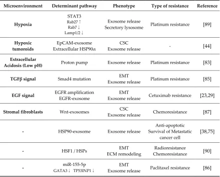
A number of studies have reported that platinum drugs such as cisplatin and carboplatin were released with exosomes as a mechanism of chemoresistance in cancer cells (Table 1). In addition, the antibody drug cetuximab was also released with oncosomes [29]. Cancer cells found in oral squamous cell carcinoma (OSCC), colorectal carcinoma (CRC), and non-small cell lung carcinoma (NSCLC) often acquire genetic amplification of EGFR, while EGFR-containing EVs are released from these cancer cells. Cetuximab bound to EGFR-EVs was co-released from OSCC cells, suggesting a mechanism of cancer drug resistance [29]. Interestingly, recent studies have shown that vesicle-releasing properties were often coupled with cellular transformation phenotypes including EMT [23,29,85,86] and CSC [44,87]. Thus, it is conceivable that the mesenchymal transition and CSC phenotype are involved with the acquisition of exosome/drug-releasing phenotypes. Anti-EMT strategies by targeting the TGFβ receptor or cyclin-dependent kinase 2 (CDK2) may inhibit exosome/oncosome release from cancer cells [28].
EMT, almost by definition, enhances the motility and migration of tumor cells. It has been recently shown that cells release migrasomes during cell migration from cellular cilia, a tail of the cell [17,88]. The proteome of migrasomes was 27% common with exosomes, while the remaining 73% was specific to migrasomes [88]. Therefore, EMT-driven migration of cancer cells may promote release of EVs including exosomes and migrasomes, which both may enhance the drug ejection, resistance phenotype of the cells.
Table 1. Exosomal drug resistance
Microenvironment Determinant pathway Phenotype Type of resistance Reference
Hypoxia
STAT3 Rab27↑ Rab7↓ Lamp1/2↓
Exosome release
Secretory lysosome Platinum resistance [89]
Hypoxic
tumoroids Extracellular EpCAM-exosome HSP90α Exosome release CSC - [44]
Extracellular
Acidosis (Low pH) Proton pump Exosome release Platinum resistance [83]
TGFβ signal Smad4 mutation Exosome release EMT Platinum resistance [85]
EGF signal EGFR amplification
EGFR-exosome
EMT
Exosome release Cetuximab resistance [23,29]
Stromal fibroblasts Wnt-exosomes CSC
Exosome release Chemoresistance [87]
- HSP90-exosome Exosome release Survival of Metastatic Anti-apoptotic
cancer cell [38,75]
- HSF1 / HSPs ECM remodeling EMT Chemoresistance Radioresistance [90]
- GATA3miR-155-5p ↓ TP53INP1↓ Exosome release EMT Paclitaxel resistance [86]
A number of studies reported that platinum drugs were released with exosomes from cancer cells. The antibody-drug cetuximab was also ejected with s-EVs by cancer cells [29]. Exosome-releasing
Many members of the HSP family play key roles in cell survival and the promotion of drug resistance [39,45,91-94] (Table 1). Extracellular HSPs and EVs enriched with HSPs are thus a major aspect of the RASP. Molecular transfer of HSPs may increase drug resistance in cancer cells and influence the tumor microenvironment. Heat shock factor 1 (HSF1) is a master transcription factor for stress response and induction of HSPs [41,95-99]. The HSF1-HSP transcriptional system is a key axis in the stress response as well as in the stress resistance of cancer cells, although other transcription factors may be involved in such stress responses and resistant phenotypes. Indeed, the mRNA levels of HSP70 and HSP27 were upregulated by intracellular MMP3 which behaves as a moonlighting transcription factor in cancer [30,100,101]. In addition, CDC37, a kinase-specialized cochaperone of HSP90, was upregulated by myeloid zinc finger 1 (MZF1) in castration-resistant prostate cancer (CRPC) [102,103]. The mechanisms whereby these transcription factors are involved in drug resistance are under investigation.
On the other hand, it has been shown that drug-encapsulated exosomes derived from immune cells and mesenchymal stem cells (MSC) can be effectively and efficiently deliverable to cancer cells. Indeed, macrophage-derived exosome-encapsulated paclitaxel was developed to overcome multidrug resistance (MDR) in cancer cells [104]. Targeted delivery of a TLR3 agonist with single-chain antibody fragment-conjugated nanoparticles induced a type I-interferon response and apoptosis in tumor cells [105].
5. Classes of drug resistance in cancer
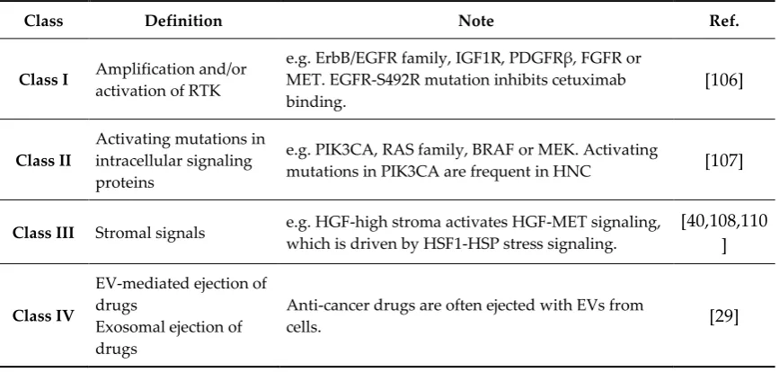
The mechanisms of drug resistance in cancer had previously been classified into three types, although the EV-oncosomal ejection of drugs can now be added as a fourth class (Table 2). Resistance Class I is the amplification and activation of alternative RTKs due to the known redundancy in RTK signaling pathways, e.g. ErbB/EGFR/Her family [106], IGF1R, PDGFRβ, FGFR or MET [32]. Indeed, the EGFR-S492R mutation inhibits the binding of cetuximab, an anti-EGFR antibody medication. Resistance Class II involves activating mutations in intracellular signaling proteins such as PIK3CA, RAS family, BRAF or MEK that enhance pro-tumorigenic signaling. For example, activating mutations in PIK3CA have been often detected in HNC [107]. Resistance Class III consists of stromal signals, including HGF-high stroma that activates HGF-MET signaling [108-110]. The HGF-MET signaling pathway is driven by the HSF1-HSP stress response system in triple-negative breast cancer (TNBC) [40]. Resistance Class IV is the exosome-mediated (EV-mediated) ejection of drugs discussed above.
Table 2. Classification of drug resistance in cancer
Class Definition Note Ref.
Class I Amplification and/or activation of RTK e.g. ErbB/EGMET. EGFR-S492R mutation inhibits cetuximab FR family, IGF1R, PDGFRβ, FGFR or
binding. [106]
Class II Activating mutations in intracellular signaling proteins
e.g. PIK3CA, RAS family, BRAF or MEK. Activating
mutations in PIK3CA are frequent in HNC [107]
Class III Stromal signals e.g. which is driven by HGF-high stroma activates HGFHSF1-HSP stress signaling. -MET signaling, [40,108,110 ]
Class IV
EV-mediated ejection of drugs
Exosomal ejection of drugs
Anti-cancer drugs are often ejected with EVs from
cells. [29]
Class I and class II drug resistance are generated by genetic alterations in tumor cells. Class III drug resistance is caused by stromal signals from the tumor microenvironment. Class IV drug resistance is
6. Conclusions
EV-mediated ejection of anti-cancer therapeutics is a novel mechanism of drug resistance that develops in cancer. Chemotherapeutics, as well as antibody drugs, can be released with EVs derived from the tumor cells. EV/drug-releasing phenotypes are often coupled with cellular transforming processes such as EMT and CSC/CIC. RASP is a marker of resistant phenotypes and a potential target to inhibit EV release from cancer cells.
Author Contributions: conceptualization, T.E.; writing—original draft preparation, T.E.; writing—review and editing, S.K.C., K.On, and K.Ok.; visualization, T.E.; supervision, S.K.C., K.Ok, T.E.; project administration, T.E.; funding acquisition, T.E.
Funding: T.E. was funded by JSPS Kakenhi, grant numbers 17K11642-TE, 19H04051-HO, 19H03817-MT, 18K09789-KN, 17K11643-CS, 17K11669-KO, 16K11722-JM, and 16K11863-KO and by Suzuki Kenzo Memorial
Foundation.
Acknowledgments: We thank Akira Sasaki for mentorable supports and Chiharu Sogawa, Yuka Okusha, Hotaka Kawai, Keisuke Nakano, and Heiichiro Udono for illuminating discussions.
Abbreviations
ABC ATP-binding cassette
ADCC antibody-dependent cellular cytotoxicity ATP Adenosine triphosphate
CAF Cancer-associated fibroblast CDK Cyclin-dependent kinase CIC Cancer-initiating cell CNV copy number variation CRC Colorectal cancer
CRPC Castration-resistant prostate cancer CSC Cancer stem cell
CTL Cytotoxic T-lymphocyte ECM Extracellular Matrix
EGF epidermal growth factor
EGFR Epidermal growth factor receptor
EGFRvIII Epidermal growth factor receptor variant III
EMT Epithelial to mesenchymal transition EV Extracellular vesicle
FcR Fragment-crystallizable receptor
GIST gastrointestinal stromal tumor
HGF Hepatocyte growth factor HNC Head and neck cancer
HSF Heat shock factor HSP Heat shock protein MDR Multidrug resistance MMP matrix metalloproteinase MSC Mesenchymal stem cell MZF1 Myeloid zinc finger 1 NK Natural killer
NSCLC Non-small cell lung carcinoma OMV outer membrane vesicles OSCC Oral squamous cell carcinoma PD-1 Programmed cell death-1 PD-L1 Programmed cell death-ligand 1
RASP Resistance-associated secretory phenotype RTK Receptor tyrosine kinase
TGFβ transforming growth factor β
References
1. Yanez-Mo, M.; Siljander, P.R.; Andreu, Z.; Zavec, A.B.; Borras, F.E.; Buzas, E.I.; Buzas, K.; Casal, E.; Cappello, F.; Carvalho, J., et al. Biological properties of extracellular vesicles and their physiological
functions. J Extracell Vesicles 2015, 4, 27066, doi:10.3402/jev.v4.27066.
2. Colombo, M.; Raposo, G.; Thery, C. Biogenesis, secretion, and intercellular interactions of exosomes and other extracellular vesicles. Annu Rev Cell Dev Biol 2014, 30, 255-289, doi:10.1146/annurev-cellbio-101512-122326.
3. Witwer, K.W.; Buzas, E.I.; Bemis, L.T.; Bora, A.; Lasser, C.; Lotvall, J.; Nolte-'t Hoen, E.N.; Piper, M.G.; Sivaraman, S.; Skog, J., et al. Standardization of sample collection, isolation and analysis methods in extracellular vesicle research. J Extracell Vesicles 2013, 2, doi:10.3402/jev.v2i0.20360.
4. Raposo, G.; Stoorvogel, W. Extracellular vesicles: exosomes, microvesicles, and friends. J Cell Biol 2013, 200, 373-383, doi:10.1083/jcb.201211138.
5. Fujita, Y.; Yoshioka, Y.; Ochiya, T. Extracellular vesicle transfer of cancer pathogenic components.
Cancer Sci 2016, 107, 385-390, doi:10.1111/cas.12896.
6. Lawson, C.; Vicencio, J.M.; Yellon, D.M.; Davidson, S.M. Microvesicles and exosomes: new players in metabolic and cardiovascular disease. J Endocrinol 2016, 228, R57-71, doi:10.1530/joe-15-0201.
7. Janowska-Wieczorek, A.; Wysoczynski, M.; Kijowski, J.; Marquez-Curtis, L.; Machalinski, B.; Ratajczak, J.; Ratajczak, M.Z. Microvesicles derived from activated platelets induce metastasis and angiogenesis in lung cancer. Int J Cancer 2005, 113, 752-760, doi:10.1002/ijc.20657.
8. Andreola, G.; Rivoltini, L.; Castelli, C.; Huber, V.; Perego, P.; Deho, P.; Squarcina, P.; Accornero, P.;
Lozupone, F.; Lugini, L., et al. Induction of lymphocyte apoptosis by tumor cell secretion of FasL -bearing microvesicles. J Exp Med 2002, 195, 1303-1316, doi:10.1084/jem.20011624.
9. Choi, D.; Spinelli, C.; Montermini, L.; Rak, J. Oncogenic Regulation of Extracellular Vesicle Proteome and Heterogeneity. Proteomics 2019, 19, e1800169, doi:10.1002/pmic.201800169.
10. Rak, J. Extracellular vesicles - biomarkers and effectors of the cellular interactome in cancer. Front Pharmacol 2013, 4, 21, doi:10.3389/fphar.2013.00021.
11. Al-Nedawi, K.; Meehan, B.; Micallef, J.; Lhotak, V.; May, L.; Guha, A.; Rak, J. Intercellular transfer of
the oncogenic receptor EGFRvIII by microvesicles derived from tumour cells. Nat Cell Biol 2008, 10,
619-624, doi:10.1038/ncb1725.
12. Vagner, T.; Spinelli, C.; Minciacchi, V.R.; Balaj, L.; Zandian, M.; Conley, A.; Zijlstra, A.; Freeman, M.R.;
Demichelis, F.; De, S., et al. Large extracellular vesicles carry most of the tumour DNA circulating in
prostate cancer patient plasma. J Extracell Vesicles 2018, 7, 1505403, doi:10.1080/20013078.2018.1505403. 13. Di Vizio, D.; Morello, M.; Dudley, A.C.; Schow, P.W.; Adam, R.M.; Morley, S.; Mulholland, D.; Rotinen, M.; Hager, M.H.; Insabato, L., et al. Large oncosomes in human prostate cancer tissues and in the circulation of mice with metastatic disease. Am J Pathol 2012, 181, 1573-1584, doi:10.1016/j.ajpath.2012.07.030.
14. Schmidt, J.R.; Kliemt, S.; Preissler, C.; Moeller, S.; von Bergen, M.; Hempel, U.; Kalkhof, S. Osteoblast-released Matrix Vesicles, Regulation of Activity and Composition by Sulfated and Non-sulfated Glycosaminoglycans. Mol Cell Proteomics 2016, 15, 558-572, doi:10.1074/mcp.M115.049718.
15. Chen, Q.; Bei, J.J.; Liu, C.; Feng, S.B.; Zhao, W.B.; Zhou, Z.; Yu, Z.P.; Du, X.J.; Hu, H.Y. HMGB1 Induces
Secretion of Matrix Vesicles by Macrophages to Enhance Ectopic Mineralization. PLoS One 2016, 11, e0156686, doi:10.1371/journal.pone.0156686.
Phospholipases of mineralization competent cells and matrix vesicles: roles in physiological and pathological mineralizations. Int J Mol Sci 2013, 14, 5036-5129, doi:10.3390/ijms14035036.
17. Huang, Y.; Zucker, B.; Zhang, S.; Elias, S.; Zhu, Y.; Chen, H.; Ding, T.; Li, Y.; Sun, Y.; Lou, J., et al. Migrasome formation is mediated by assembly of micron-scale tetraspanin macrodomains. Nat Cell Biol 2019, 21, 991-1002, doi:10.1038/s41556-019-0367-5.
18. Ma, L.; Li, Y.; Peng, J.; Wu, D.; Zhao, X.; Cui, Y.; Chen, L.; Yan, X.; Du, Y.; Yu, L. Discovery of the migrasome, an organelle mediating release of cytoplasmic contents during cell migration. Cell Res 2015, 25, 24-38, doi:10.1038/cr.2014.135.
19. Coelho, C.; Brown, L.; Maryam, M.; Vij, R.; Smith, D.F.Q.; Burnet, M.C.; Kyle, J.E.; Heyman, H.M.;
Ramirez, J.; Prados-Rosales, R., et al. Listeria monocytogenes virulence factors, including listeriolysin O, are secreted in biologically active extracellular vesicles. The Journal of biological chemistry 2019, 294, 1202-1217, doi:10.1074/jbc.RA118.006472.
20. Kim, O.Y.; Park, H.T.; Dinh, N.T.H.; Choi, S.J.; Lee, J.; Kim, J.H.; Lee, S.W.; Gho, Y.S. Bacterial outer membrane vesicles suppress tumor by interferon-gamma-mediated antitumor response. Nat Commun 2017, 8, 626, doi:10.1038/s41467-017-00729-8.
21. van Niel, G.; D'Angelo, G.; Raposo, G. Shedding light on the cell biology of extracellular vesicles. Nat Rev Mol Cell Biol 2018, 19, 213-228, doi:10.1038/nrm.2017.125.
22. Zhao, X.; Wu, X.; Qian, M.; Song, Y.; Wu, D.; Zhang, W. Knockdown of TGF-beta1 expression in human
umbilical cord mesenchymal stem cells reverts their exosome-mediated EMT promoting effect on lung cancer cells. Cancer Lett 2018, 428, 34-44, doi:10.1016/j.canlet.2018.04.026.
23. Fujiwara, T.; Eguchi, T.; Sogawa, C.; Ono, K.; Murakami, J.; Ibaragi, S.; Asaumi, J.-i.; Calderwood, S.K.;
Okamoto, K.; Kozaki, K.-i. Carcinogenic epithelial-mesenchymal transition initiated by oral cancer exosomes is inhibited by anti-EGFR antibody cetuximab. Oral Oncology 2018, 86, 251-257, doi:10.1016/j.oraloncology.2018.09.030.
24. Fuxe, J.; Karlsson, M.C. TGF-beta-induced epithelial-mesenchymal transition: a link between cancer
and inflammation. Semin Cancer Biol 2012, 22, 455-461, doi:10.1016/j.semcancer.2012.05.004.
25. Kalluri, R. EMT: when epithelial cells decide to become mesenchymal-like cells. J Clin Invest 2009, 119, 1417-1419, doi:10.1172/JCI39675.
26. Zavadil, J.; Bottinger, E.P. TGF-beta and epithelial-to-mesenchymal transitions. Oncogene 2005, 24,
5764-5774, doi:10.1038/sj.onc.1208927.
27. Nieto, M.A.; Huang, R.Y.; Jackson, R.A.; Thiery, J.P. Emt: 2016. Cell 2016, 166, 21-45, doi:10.1016/j.cell.2016.06.028.
28. Arai, K.; Eguchi, T.; Rahman, M.M.; Sakamoto, R.; Masuda, N.; Nakatsura, T.; Calderwood, S.K.; Kozaki, K.; Itoh, M. A Novel High-Throughput 3D Screening System for EMT Inhibitors: A Pilot Screening Discovered the EMT Inhibitory Activity of CDK2 Inhibitor SU9516. PLoS One 2016, 11, e0162394, doi:10.1371/journal.pone.0162394.
29. Fujiwara, T.; Eguchi, T.; Sogawa, C.; Ono, K.; Murakami, J.; Ibaragi, S.; Asaumi, J.; Okamoto, K.;
Calderwood, S.; Kozaki, K. Anti-EGFR antibody cetuximab is secreted by oral squamous cell carcinoma
and alters EGF-driven mesenchymal transition. Biochem Biophys Res Commun. 2018, 503, 1267-1272.
30. Okusha, Y.; Eguchi, T.; Sogawa, C.; Okui, T.; Nakano, K.; Okamoto, K.; Kozaki, K.I. The intranuclear PEX domain of MMP involves proliferation, migration, and metastasis of aggressive adenocarcinoma cells. J Cell Biochem 2018, 119, 7363-7376, doi:10.1002/jcb.27040.
-beta1-induced CK17 enhances cancer stem cell-like properties rather than EMT in promoting cervical cancer metastasis via the ERK1/2-MZF1 signaling pathway. Febs j 2017, 284, 3000-3017, doi:10.1111/febs.14162. 32. Shibue, T.; Weinberg, R.A. EMT, CSCs, and drug resistance: the mechanistic link and clinical
implications. Nat Rev Clin Oncol 2017, 14, 611-629, doi:10.1038/nrclinonc.2017.44.
33. Tisza, M.J.; Zhao, W.; Fuentes, J.S.; Prijic, S.; Chen, X.; Levental, I.; Chang, J.T. Motility and stem cell
properties induced by the epithelial-mesenchymal transition require destabilization of lipid rafts. Oncotarget 2016, 7, 51553-51568, doi:10.18632/oncotarget.9928.
34. Rak, J.; Guha, A. Extracellular vesicles--vehicles that spread cancer genes. Bioessays 2012, 34, 489-497, doi:10.1002/bies.201100169.
35. Di Vizio, D.; Kim, J.; Hager, M.H.; Morello, M.; Yang, W.; Lafargue, C.J.; True, L.D.; Rubin, M.A.; Adam, R.M.; Beroukhim, R., et al. Oncosome formation in prostate cancer: association with a region of frequent chromosomal deletion in metastatic disease. Cancer Res 2009, 69, 5601-5609, doi:10.1158/0008-5472.Can-08-3860.
36. D'Asti, E.; Garnier, D.; Lee, T.H.; Montermini, L.; Meehan, B.; Rak, J. Oncogenic extracellular vesicles in brain tumor progression. Front Physiol 2012, 3, 294, doi:10.3389/fphys.2012.00294.
37. Dovrat, S.; Caspi, M.; Zilberberg, A.; Lahav, L.; Firsow, A.; Gur, H.; Rosin-Arbesfeld, R. 14-3-3 and
beta-catenin are secreted on extracellular vesicles to activate the oncogenic Wnt pathway. Mol Oncol 2014, 8, 894-911, doi:10.1016/j.molonc.2014.03.011.
38. Ono, K.; Eguchi, T.; Sogawa, C.; Calderwood, S.K.; Futagawa, J.; Kasai, T.; Seno, M.; Okamoto, K.; Sasaki,
A.; Kozaki, K.I. HSP-enriched properties of extracellular vesicles involve survival of metastatic oral cancer cells. J Cell Biochem 2018, doi:10.1002/jcb.27039.
39. Murshid, A.; Eguchi, T.; Calderwood, S.K. Stress proteins in aging and life span. Int J Hyperthermia 2013, 29, 442-447, doi:10.3109/02656736.2013.798873.
40. Gong, J.; Weng, D.; Eguchi, T.; Murshid, A.; Sherman, M.Y.; Song, B.; Calderwood, S.K. Targeting the hsp70 gene delays mammary tumor initiation and inhibits tumor cell metastasis. Oncogene 2015, 34, 5460-5471, doi:10.1038/onc.2015.1.
41. Ciocca, D.R.; Arrigo, A.P.; Calderwood, S.K. Heat shock proteins and heat shock factor 1 in carcinogenesis and tumor development: an update. Arch Toxicol 2013, 87, 19-48, doi:10.1007/s00204-012-0918-z.
42. Eguchi, T.; Lang, B.J.; Murshid, A.; Prince, T.; Gong, J.; Calderwood, S.K. Regulatory roles for Hsp70 in cancer incidence and tumor progression. In Frontiers in Structural Biology, Galigniana, M.D., Ed. Bentham Science: 2018; Vol. 1, pp. 1-22.
43. Neckers, L.; Blagg, B.; Haystead, T.; Trepel, J.B.; Whitesell, L.; Picard, D. Methods to validate Hsp90 inhibitor specificity, to identify off-target effects, and to rethink approaches for further clinical development. Cell Stress Chaperones 2018, 23, 467-482, doi:10.1007/s12192-018-0877-2.
44. Eguchi, T.; Sogawa, C.; Okusha, Y.; Uchibe, K.; Iinuma, R.; Ono, K.; Nakano, K.; Murakami, J.; Itoh, M.; Arai, K., et al. Organoids with Cancer Stem Cell-like Properties Secrete Exosomes and HSP90 in a 3D NanoEnvironment. PLOS ONE 2018, 13, e0191109, doi:10.1371/journal.pone.0191109.
45. Taha, E.A.; Ono, K.; Eguchi, T. Roles of Extracellular HSPs as Biomarkers in Immune Surveillance and Immune Evasion. Int J Mol Sci 2019, 20, doi:10.3390/ijms20184588.
46. Dong, H.; Zou, M.; Bhatia, A.; Jayaprakash, P.; Hofman, F.; Ying, Q.; Chen, M.; Woodley, D.T.; Li, W.
47. Tsen, F.; Bhatia, A.; O'Brien, K.; Cheng, C.F.; Chen, M.; Hay, N.; Stiles, B.; Woodley, D.T.; Li, W.
Extracellular heat shock protein 90 signals through subdomain II and the NPVY motif of LRP-1 receptor to Akt1 and Akt2: a circuit essential for promoting skin cell migration in vitro and wound healing in vivo. Mol Cell Biol 2013, 33, 4947-4959, doi:10.1128/mcb.00559-13.
48. Najafi, M.; Goradel, N.H.; Farhood, B.; Salehi, E.; Solhjoo, S.; Toolee, H.; Kharazinejad, E.; Mortezaee,
K. Tumor microenvironment: Interactions and therapy. J Cell Physiol 2019, 234, 5700-5721, doi:10.1002/jcp.27425.
49. Hance, M.W.; Dole, K.; Gopal, U.; Bohonowych, J.E.; Jezierska-Drutel, A.; Neumann, C.A.; Liu, H.; Garraway, I.P.; Isaacs, J.S. Secreted Hsp90 is a novel regulator of the epithelial to mesenchymal transition (EMT) in prostate cancer. J Biol Chem 2012, 287, 37732-37744, doi:10.1074/jbc.M112.389015.
50. Nolan, K.D.; Franco, O.E.; Hance, M.W.; Hayward, S.W.; Isaacs, J.S. Tumor-secreted Hsp90 subverts
polycomb function to drive prostate tumor growth and invasion. J Biol Chem 2015, 290, 8271-8282, doi:10.1074/jbc.M115.637496.
51. Nagaraju, G.P.; Long, T.E.; Park, W.; Landry, J.C.; Taliaferro-Smith, L.; Farris, A.B.; Diaz, R.; El-Rayes,
B.F. Heat shock protein 90 promotes epithelial to mesenchymal transition, invasion, and migration in
colorectal cancer. Mol Carcinog 2015, 54, 1147-1158, doi:10.1002/mc.22185.
52. Nolan, K.D.; Kaur, J.; Isaacs, J.S. Secreted heat shock protein 90 promotes prostate cancer stem cell heterogeneity. Oncotarget 2017, 8, 19323-19341, doi:10.18632/oncotarget.14252.
53. Hsieh, C.L.; Liu, C.M.; Chen, H.A.; Yang, S.T.; Shigemura, K.; Kitagawa, K.; Yamamichi, F.; Fujisawa,
M.; Liu, Y.R.; Lee, W.H., et al. Reactive oxygen species-mediated switching expression of MMP-3 in stromal fibroblasts and cancer cells during prostate cancer progression. Sci Rep 2017, 7, 9065, doi:10.1038/s41598-017-08835-9.
54. Richards, K.E.; Zeleniak, A.E.; Fishel, M.L.; Wu, J.; Littlepage, L.E.; Hill, R. Cancer-associated fibroblast
exosomes regulate survival and proliferation of pancreatic cancer cells. Oncogene 2017, 36, 1770-1778, doi:10.1038/onc.2016.353.
55. Leca, J.; Martinez, S.; Lac, S.; Nigri, J.; Secq, V.; Rubis, M.; Bressy, C.; Serge, A.; Lavaut, M.N.; Dusetti, N., et al. Cancer-associated fibroblast-derived annexin A6+ extracellular vesicles support pancreatic cancer aggressiveness. J Clin Invest 2016, 126, 4140-4156, doi:10.1172/JCI87734.
56. Gascard, P.; Tlsty, T.D. Carcinoma-associated fibroblasts: orchestrating the composition of malignancy. Genes & Development 2016, 30, 1002-1019, doi:10.1101/gad.279737.
57. Cirri, P.; Chiarugi, P. Cancer associated fibroblasts: the dark side of the coin. Am J Cancer Res 2011, 1, 482-497.
58. Zhuang, J.; Lu, Q.; Shen, B.; Huang, X.; Shen, L.; Zheng, X.; Huang, R.; Yan, J.; Guo, H. TGFbeta1
secreted by cancer-associated fibroblasts induces epithelial-mesenchymal transition of bladder cancer cells through lncRNA-ZEB2NAT. Sci Rep 2015, 5, 11924, doi:10.1038/srep11924.
59. Weber, C.E.; Kothari, A.N.; Wai, P.Y.; Li, N.Y.; Driver, J.; Zapf, M.A.; Franzen, C.A.; Gupta, G.N.; Osipo,
C.; Zlobin, A., et al. Osteopontin mediates an MZF1-TGF-beta1-dependent transformation of
mesenchymal stem cells into cancer-associated fibroblasts in breast cancer. Oncogene 2015, 34, 4821-4833, doi:10.1038/onc.2014.410.
60. Hida, K.; Maishi, N.; Annan, D.A.; Hida, Y. Contribution of Tumor Endothelial Cells in Cancer Progression. Int J Mol Sci 2018, 19, doi:10.3390/ijms19051272.
Cells 2019, 8, doi:10.3390/cells8070761.
62. Hassona, Y.; Cirillo, N.; Heesom, K.; Parkinson, E.K.; Prime, S.S. Senescent cancer-associated fibroblasts secrete active MMP-2 that promotes keratinocyte dis-cohesion and invasion. Br J Cancer 2014, 111, 1230-1237, doi:10.1038/bjc.2014.438.
63. Qin, X.; Guo, H.; Wang, X.; Zhu, X.; Yan, M.; Wang, X.; Xu, Q.; Shi, J.; Lu, E.; Chen, W., et al. Exosomal miR-196a derived from cancer-associated fibroblasts confers cisplatin resistance in head and neck cancer through targeting CDKN1B and ING5. Genome Biol 2019, 20, 12, doi:10.1186/s13059-018-1604-0.
64. Ramteke, A.; Ting, H.; Agarwal, C.; Mateen, S.; Somasagara, R.; Hussain, A.; Graner, M.; Frederick, B.;
Agarwal, R.; Deep, G. Exosomes secreted under hypoxia enhance invasiveness and stemness of prostate cancer cells by targeting adherens junction molecules. Mol Carcinog 2015, 54, 554-565, doi:10.1002/mc.22124.
65. Principe, S.; Mejia-Guerrero, S.; Ignatchenko, V.; Sinha, A.; Ignatchenko, A.; Shi, W.; Pereira, K.; Su, S.; Huang, S.H.; O'Sullivan, B., et al. Proteomic Analysis of Cancer-Associated Fibroblasts Reveals a
Paracrine Role for MFAP5 in Human Oral Tongue Squamous Cell Carcinoma. J Proteome Res 2018, 17,
2045-2059, doi:10.1021/acs.jproteome.7b00925.
66. Cianciaruso, C.; Beltraminelli, T.; Duval, F.; Nassiri, S.; Hamelin, R.; Mozes, A.; Gallart-Ayala, H.; Ceada
Torres, G.; Torchia, B.; Ries, C.H., et al. Molecular Profiling and Functional Analysis of Macrophage -Derived Tumor Extracellular Vesicles. Cell Rep 2019, 27, 3062-3080 e3011, doi:10.1016/j.celrep.2019.05.008.
67. De Palma, M.; Nassiri, S.; Cianciaruso, C. Macrophage interference on chemotherapy. Nat Cell Biol 2019, 21, 411-412, doi:10.1038/s41556-019-0303-8.
68. Hida, K.; Maishi, N.; Akiyama, K.; Ohmura-Kakutani, H.; Torii, C.; Ohga, N.; Osawa, T.; Kikuchi, H.; Morimoto, H.; Morimoto, M., et al. Tumor endothelial cells with high aldehyde dehydrogenase activity show drug resistance. Cancer Sci 2017, 108, 2195-2203, doi:10.1111/cas.13388.
69. Hida, K.; Kikuchi, H.; Maishi, N.; Hida, Y. ATP-binding cassette transporters in tumor endothelial cells and resistance to metronomic chemotherapy. Cancer Lett 2017, doi:10.1016/j.canlet.2017.02.006.
70. Kanlikilicer, P.; Bayraktar, R.; Denizli, M.; Rashed, M.H.; Ivan, C.; Aslan, B.; Mitra, R.; Karagoz, K.; Bayraktar, E.; Zhang, X., et al. Exosomal miRNA confers chemo resistance via targeting Cav1/p-gp/M2-type macrophage axis in ovarian cancer. EBioMedicine 2018, 38, 100-112, doi:10.1016/j.ebiom.2018.11.004. 71. Steinbichler, T.B.; Dudas, J.; Skvortsov, S.; Ganswindt, U.; Riechelmann, H.; Skvortsova, II. Therapy
resistance mediated by exosomes. Mol Cancer 2019, 18, 58, doi:10.1186/s12943-019-0970-x.
72. Namba, Y.; Sogawa, C.; Okusha, Y.; Kawai, H.; Itagaki, M.; Ono, K.; Murakami, J.; Aoyama, E.; Ohyama, K.; Asaumi, J.I., et al. Depletion of Lipid Efflux Pump ABCG1 Triggers the Intracellular Accumulation of Extracellular Vesicles and Reduces Aggregation and Tumorigenesis of Metastatic Cancer Cells. Front Oncol 2018, 8, 376, doi:10.3389/fonc.2018.00376.
73. Neumann, J.; Rose-Sperling, D.; Hellmich, U.A. Diverse relations between ABC transporters and lipids: An overview. Biochim Biophys Acta Biomembr 2017, 1859, 605-618, doi:10.1016/j.bbamem.2016.09.023. 74. Noguchi, K.; Katayama, K.; Sugimoto, Y. Human ABC transporter ABCG2/BCRP expression in
chemoresistance: basic and clinical perspectives for molecular cancer therapeutics. Pharmgenomics Pers Med 2014, 7, 53-64, doi:10.2147/pgpm.S38295.
76. Xie, F.; Xu, M.; Lu, J.; Mao, L.; Wang, S. The role of exosomal PD-L1 in tumor progression and immunotherapy. Mol Cancer 2019, 18, 146, doi:10.1186/s12943-019-1074-3.
77. Lux, A.; Kahlert, C.; Grutzmann, R.; Pilarsky, C. c-Met and PD-L1 on Circulating Exosomes as Diagnostic and Prognostic Markers for Pancreatic Cancer. Int J Mol Sci 2019, 20, doi:10.3390/ijms20133305.
78. Li, C.; Li, C.; Zhi, C.; Liang, W.; Wang, X.; Chen, X.; Lv, T.; Shen, Q.; Song, Y.; Lin, D., et al. Clinical significance of PD-L1 expression in serum-derived exosomes in NSCLC patients. J Transl Med 2019, 17, 355, doi:10.1186/s12967-019-2101-2.
79. Kim, D.H.; Kim, H.; Choi, Y.J.; Kim, S.Y.; Lee, J.E.; Sung, K.J.; Sung, Y.H.; Pack, C.G.; Jung, M.K.; Han, B., et al. Exosomal PD-L1 promotes tumor growth through immune escape in non-small cell lung cancer. Exp Mol Med 2019, 51, 94, doi:10.1038/s12276-019-0295-2.
80. Theodoraki, M.N.; Yerneni, S.S.; Hoffmann, T.K.; Gooding, W.E.; Whiteside, T.L. Clinical Significance of PD-L1(+) Exosomes in Plasma of Head and Neck Cancer Patients. Clin Cancer Res 2018, 24, 896-905, doi:10.1158/1078-0432.Ccr-17-2664.
81. Chen, G.; Huang, A.C.; Zhang, W.; Zhang, G.; Wu, M.; Xu, W.; Yu, Z.; Yang, J.; Wang, B.; Sun, H., et al. Exosomal PD-L1 contributes to immunosuppression and is associated with anti-PD-1 response. Nature 2018, 560, 382-386, doi:10.1038/s41586-018-0392-8.
82. Safaei, R.; Larson, B.J.; Cheng, T.C.; Gibson, M.A.; Otani, S.; Naerdemann, W.; Howell, S.B. Abnormal lysosomal trafficking and enhanced exosomal export of cisplatin in drug-resistant human ovarian carcinoma cells. Mol Cancer Ther 2005, 4, 1595-1604, doi:10.1158/1535-7163.Mct-05-0102.
83. Federici, C.; Petrucci, F.; Caimi, S.; Cesolini, A.; Logozzi, M.; Borghi, M.; D'Ilio, S.; Lugini, L.; Violante,
N.; Azzarito, T., et al. Exosome release and low pH belong to a framework of resistance of human melanoma cells to cisplatin. PLoS One 2014, 9, e88193, doi:10.1371/journal.pone.0088193.
84. Xiao, X.; Yu, S.; Li, S.; Wu, J.; Ma, R.; Cao, H.; Zhu, Y.; Feng, J. Exosomes: decreased sensitivity of lung
cancer A549 cells to cisplatin. PloS one 2014, 9, e89534-e89534, doi:10.1371/journal.pone.0089534. 85. Crow, J.; Atay, S.; Banskota, S.; Artale, B.; Schmitt, S.; Godwin, A.K. Exosomes as mediators of platinum
resistance in ovarian cancer. Oncotarget 2017, 8, 11917-11936, doi:10.18632/oncotarget.14440.
86. Wang, M.; Qiu, R.; Yu, S.; Xu, X.; Li, G.; Gu, R.; Tan, C.; Zhu, W.; Shen, B. Paclitaxelresistant gastric cancer MGC803 cells promote epithelialtomesenchymal transition and chemoresistance in paclitaxelsensitive cells via exosomal delivery of miR1555p. Int J Oncol 2019, 54, 326-338, doi:10.3892/ijo.2018.4601.
87. Hu, Y.B.; Yan, C.; Mu, L.; Mi, Y.L.; Zhao, H.; Hu, H.; Li, X.L.; Tao, D.D.; Wu, Y.Q.; Gong, J.P., et al. Exosomal Wnt-induced dedifferentiation of colorectal cancer cells contributes to chemotherapy resistance. Oncogene 2019, 38, 1951-1965, doi:10.1038/s41388-018-0557-9.
88. Zhao, X.; Lei, Y.; Zheng, J.; Peng, J.; Li, Y.; Yu, L.; Chen, Y. Identification of markers for migrasome detection. Cell Discov 2019, 5, 27, doi:10.1038/s41421-019-0093-y.
89. Dorayappan, K.D.P.; Wanner, R.; Wallbillich, J.J.; Saini, U.; Zingarelli, R.; Suarez, A.A.; Cohn, D.E.; Selvendiran, K. Hypoxia-induced exosomes contribute to a more aggressive and chemoresistant ovarian cancer phenotype: a novel mechanism linking STAT3/Rab proteins. Oncogene 2018, 37, 3806-3821, doi:10.1038/s41388-018-0189-0.
91. Choi, S.-K.; Kam, H.; Kim, K.-Y.; Park, S.I.; Lee, Y.-S. Targeting Heat Shock Protein 27 in Cancer: A Druggable Target for Cancer Treatment? Cancers 2019, 11, doi:10.3390/cancers11081195.
92. Xiao, X.; Wang, W.; Li, Y.; Yang, D.; Li, X.; Shen, C.; Liu, Y.; Ke, X.; Guo, S.; Guo, Z. HSP90AA1-mediated autophagy promotes drug resistance in osteosarcoma. J Exp Clin Cancer Res 2018, 37, 201, doi:10.1186/s13046-018-0880-6.
93. Moses, M.A.; Kim, Y.S.; Rivera-Marquez, G.M.; Oshima, N.; Watson, M.J.; Beebe, K.E.; Wells, C.; Lee, S.; Zuehlke, A.D.; Shao, H., et al. Targeting the Hsp40/Hsp70 Chaperone Axis as a Novel Strategy to Treat Castration-Resistant Prostate Cancer. Cancer Res 2018, 78, 4022-4035, doi:10.1158/0008-5472.Can-17-3728.
94. Calderwood, S.K.; Gong, J. Heat Shock Proteins Promote Cancer: It's a Protection Racket. Trends Biochem Sci 2016, 41, 311-323, doi:10.1016/j.tibs.2016.01.003.
95. Calderwood, S.K.; Murshid, A. Molecular Chaperone Accumulation in Cancer and Decrease in
Alzheimer's Disease: The Potential Roles of HSF1. Front Neurosci 2017, 11, 192,
doi:10.3389/fnins.2017.00192.
96. Chou, S.D.; Murshid, A.; Eguchi, T.; Gong, J.; Calderwood, S.K. HSF1 regulation of beta-catenin in
mammary cancer cells through control of HuR/elavL1 expression. Oncogene 2015, 34, 2178-2188, doi:10.1038/onc.2014.177.
97. Chou, S.D.; Prince, T.; Gong, J.; Calderwood, S.K. mTOR is essential for the proteotoxic stress response,
HSF1 activation and heat shock protein synthesis. PLoS One 2012, 7, e39679,
doi:10.1371/journal.pone.0039679.
98. Calderwood, S.K.; Xie, Y.; Wang, X.; Khaleque, M.A.; Chou, S.D.; Murshid, A.; Prince, T.; Zhang, Y. Signal Transduction Pathways Leading to Heat Shock Transcription. Sign Transduct Insights 2010, 2, 13-24, doi:10.4137/STI.S3994.
99. Xie, Y.; Zhong, R.; Chen, C.; Calderwood, S.K. Heat shock factor 1 contains two functional domains that mediate transcriptional repression of the c-fos and c-fms genes. J Biol Chem 2003, 278, 4687-4698, doi:10.1074/jbc.M210189200.
100. Eguchi, T.; Calderwood, S.K.; Takigawa, M.; Kubota, S.; Kozaki, K.I. Intracellular MMP3 Promotes HSP Gene Expression in Collaboration With Chromobox Proteins. J Cell Biochem 2017, 118, 43-51, doi:10.1002/jcb.25607.
101. Eguchi, T.; Kubota, S.; Kawata, K.; Mukudai, Y.; Uehara, J.; Ohgawara, T.; Ibaragi, S.; Sasaki, A.; Kuboki, T.; Takigawa, M. Novel transcription-factor-like function of human matrix metalloproteinase 3
regulating the CTGF/CCN2 gene. Mol Cell Biol 2008, 28, 2391-2413, doi:10.1128/MCB.01288-07.
102. Eguchi, T.; Prince, T.L.; Tran, M.T.; Sogawa, C.; Lang, B.J.; Calderwood, S.K. MZF1 and SCAND1
Reciprocally Regulate CDC37 Gene Expression in Prostate Cancer. Cancers (Basel) 2019, 11, doi:10.3390/cancers11060792.
103. Eguchi, T.; Prince, T.; Wegiel, B.; Calderwood, S.K. Role and Regulation of Myeloid Zinc Finger Protein
1 in Cancer. J Cell Biochem 2015, 116, 2146-2154, doi:10.1002/jcb.25203.
104. Kim, M.S.; Haney, M.J.; Zhao, Y.; Mahajan, V.; Deygen, I.; Klyachko, N.L.; Inskoe, E.; Piroyan, A.; Sokolsky, M.; Okolie, O., et al. Development of exosome-encapsulated paclitaxel to overcome MDR in cancer cells. Nanomedicine 2016, 12, 655-664, doi:10.1016/j.nano.2015.10.012.
106. Shah, S.P.; Roth, A.; Goya, R.; Oloumi, A.; Ha, G.; Zhao, Y.; Turashvili, G.; Ding, J.; Tse, K.; Haffari, G., et al. The clonal and mutational evolution spectrum of primary triple-negative breast cancers. Nature 2012, 486, 395-399, doi:10.1038/nature10933.
107. Cancer Genome Atlas, N. Comprehensive genomic characterization of head and neck squamous cell carcinomas. Nature 2015, 517, 576-582, doi:10.1038/nature14129.
108. Tomihara, H.; Yamada, D.; Eguchi, H.; Iwagami, Y.; Noda, T.; Asaoka, T.; Wada, H.; Kawamoto, K.; Gotoh, K.; Takeda, Y., et al. MicroRNA-181b-5p, ETS1, and the c-Met pathway exacerbate the prognosis of pancreatic ductal adenocarcinoma after radiation therapy. Cancer Sci 2017, 108, 398-407, doi:10.1111/cas.13159.
109. Trusolino, L.; Bertotti, A.; Comoglio, P.M. MET signalling: principles and functions in development, organ regeneration and cancer. Nat Rev Mol Cell Biol 2010, 11, 834-848, doi:10.1038/nrm3012.