Copyright © 2000, American Society for Microbiology. All Rights Reserved.
Cloning and Expression of Immunoreactive Antigens from
Mycobacterium tuberculosis
RENEE LAY HONG LIM,
1* LI KIANG TAN,
1WAI FUN LAU,
1MAXEY CHING MING CHUNG,
1,2ROSEANNE DUNN,
1† HENG PHON TOO,
2ANDLILY CHAN
1Bioprocessing Technology Centre
1and Department of Biochemistry,
2The National University of Singapore, Singapore
Received 1 December 1999/Returned for modification 8 February 2000/Accepted 4 April 2000
Four immunoreactive proteins, B.4, B.6, B.10, and B.M, with molecular weights ranging from 16,000 to
58,000, were observed from immunoblots of
Mycobacterium tuberculosis
total lysates screened with sera from
individuals with active tuberculosis. These proteins were identified from microsequence analyses, and genes of
proteins with the highest homology were PCR amplified and cloned into the pQE30 vector for expression
studies. In addition, a 37.5-kDa protein, designated C17, was identified from a phage expression library of
M. tuberculosis
genomic DNA. Preliminary immunoblot assays indicated that these five resultant recombinant
proteins could detect antibodies in individuals with active pulmonary and extrapulmonary tuberculosis. The
overall ranges of sensitivities, specificities, positive predictive values, and negative predictive values for the
recombinant antigens were 20 to 58, 88 to 100, 69 to 100, and 56 to 71%, respectively. The B.6 antigen showed
preferential reactivity to antibodies in pulmonary compared to nonpulmonary tuberculosis serum specimens.
All of these recombinant antigens demonstrated potential for serodiagnosis of tuberculosis.
Tuberculosis (TB) is a major health problem in the
devel-oping world as well as a disease which is reemerging as a major
health threat in the developed world (6). World Health
Orga-nization (WHO) statistics indicate that one-third of the world’s
population is currently infected (34). TB is rated the second
most common infectious disease and has the highest mortality
rate of any infectious disease in the world. Currently there are
30 million cases of active TB worldwide, with approximately 8
million new cases and 3 million deaths reported annually. In
addition, 50 million people may already be infected with
mul-tidrug-resistant (MDR) strains of
Mycobacterium tuberculosis
.
A high prevalence of TB is also associated with human
immu-nodeficiency virus (HIV) infection and AIDS and is now
be-coming the leading cause of death among HIV-positive
indi-viduals, with a fatality rate of 80% (34).
Control of this disease revolves around good patient care
and management. In particular, early detection and treatment
of tuberculosis can limit transmission of the bacilli.
Conven-tional tests for the diagnosis of tuberculosis include chest
X-ray, direct sputum smear for acid-fast bacilli, culture test, and
the skin tuberculin PPD (purified protein derivative) test (29).
Among these, the culture method is time-consuming but
reli-able. PCR and nucleic acid-based methods for detecting
M.
tuberculosis
DNA sequences require complex equipment and
highly skilled staff, and they are expensive and unsuitable for
routine diagnostic testing in developing countries (5). Rapid
serological diagnostic tests such as the enzyme-linked
immu-nosorbent assay (ELISA) and membrane chromatography
tests, in contrast, are simple and inexpensive, and the latter can
be point-of-care devices (3). A major problem encountered in
serological techniques is the specificity and reactivity of
anti-gens used. A majority of
M. tuberculosis
antigens studied to
date have homology with analogous proteins of environmental
mycobacteria or other bacteria, resulting in unspecific
reactiv-ity to antibodies in patients with inactive TB or nontuberculous
infections (8, 23). Hence, positive test results produced by
these known antigens are generally unreliable, and
supplemen-tary tests are required to confirm tuberculosis infection. It was
shown that the use of recombinant
M. tuberculosis
antigens of
specific purity or particular epitopes may enhance the
speci-ficity and sensitivity of serological testing for TB when used in
a panel of recombinant antigens (1). Rapid diagnostic tests
that are specific and sensitive would be useful in both
sero-epidemiological and clinical studies pertaining to tuberculosis
control and prevention. In this report, we describe the
identi-fication, isolation, and characterization of five recombinant
antigens from
M. tuberculosis
for use as serodiagnostic markers
for tuberculosis.
MATERIALS AND METHODS
M. tuberculosistotal-protein extraction.M. tuberculosiscells (ATCC 27294) were cultured in MycoFlasks (Gibco, BRL) containing Lowenstein-Jensen me-dium at 37°C with 10% CO2in a humidified incubation chamber (Jouan IG/50
model). Confluent cells from six culture flasks were harvested by adding 3 ml of Middlebrook 7H9 medium (Difco Laboratories, Detroit, Mich.) into each flask and gently flushing the surface of the flask. Dislocated cells were placed into sterile plastic tubes (Falcon), and cells were pelleted by centrifugation at 1,100⫻ gfor 5 min. Cells were washed once in an equal volume of distilled H2O before
being resuspended in an equal volume of distilled H2O. The cell suspension was
then heated to 90°C for 2 h and frozen at⫺20°C overnight. Cells were then thawed on ice and pelleted by centrifugation at 20,000⫻gfor 10 min. Extraction of total protein was performed by adding 500l of 8 M urea solution to 0.5 g of cell pellet, vortexing the cell suspension at room temperature for 20 min, and heating it at 90°C for 2 min. Insoluble cellular debris was removed by centrifu-gation at 20,000⫻gfor 10 min, and the supernatant containing the extracted total protein was kept at⫺20°C until further use.
Western blot analysis.The total-protein extract ofM. tuberculosiswas
frac-tionated on a sodium dodecyl sulfate–7.5% polyacrylamide gel electrophoresis (SDS–7.5% PAGE) gel (20) and transferred onto a nitrocellulose membrane by Western blotting (33). Strips from the immunoblot were probed against pooled positive (tested positive by skin PPD and culture) and negative sera from nine individuals with active TB and seven healthy individuals, respectively. Incubation with pooled sera (1:100 in 1% skim milk-TBST [10 mM Tris, pH 7.5, 300 mM NaCl, 0.005% Tween 20]) was carried out with rocking for 1 h at room temper-ature. The blots were then washed four times in TBST before incubation with alkaline phosphatase-conjugated goat anti-human immunoglobulin (Ig) (Harlan Sera Lab, Loughborough, United Kingdom) (1:1000 in 1% skim milk-TBST) for another 1 h. The strips were again washed four times in TBST followed by
* Corresponding author. Mailing address: Bioprocessing
Technol-ogy Centre, National University of Singapore, 5th Floor, MD11, 10
Kent Ridge Crescent, Singapore 119260. Phone: 65-874-6222. Fax:
65-775 4933. E-mail: [email protected].
† Present address: Department of Cell and Molecular Biology,
Uni-versity of Technology, Sydney, NSW, Australia.
600
on August 17, 2020 by guest
http://cvi.asm.org/
incubation in 1 ml of bromochloroindolyl phosphate-nitroblue tetrazolium sub-strate (NBT-BCIP; Bio-Rad) for 4 min. The reaction was stopped by washing four times in distilled H2O.
N-terminal sequencing.Individual protein bands (which were shown to react
positively in the immunoscreening experiment) were excised from several pre-parative SDS–7.5% polyacrylamide gels and concentrated by reelectrophoresis (constant current of 18 mA at 8°C) on a long stacking gel (7 cm of 4% stacking gel, 5 cm of 10% resolving gel). The concentrated protein bands were blotted onto a polyvinylidene difluoride (PVDF) membrane (Bio-Rad), stained with Coomassie brilliant blue R-250 (Sigma, St. Louis, Mo.), and excised for N-terminal microsequencing. The protein bands were also blotted onto Hybond-C nitrocellulose membranes (Amersham Life Science, Little Chalfont, United Kingdom) for validation by immunoscreening using the same pooled sera sam-ples as described above.
Screening of a phage expression library.An expression library of Eco
RI-restricted genomic DNA of M. tuberculosiswas constructed in lambda ZAP Phage expression vector, according to the protocol by Stratagene (ZAP Express cDNA Synthesis kit manual, Stratagene Cloning Systems, 1998). The resultant library has 98% recombinants (2⫻106PFU/g arms) and insert sizes ranging
from 0.7 to 2 kb. A lawn of XL1-MRF⬘host cells infected with about 2⫻104
PFU of the phage stock was prepared on a 150-mm plate and incubated for 6 to 7 h at 42°C. The lawn was then overlaid with a Hybond-C nitrocellulose mem-brane (Amersham) presoaked in 1 mM isopropyl--D-thiogalactopyranoside
(IPTG) for induction of protein expression by further incubation at 37°C for 4 h. The plate and membrane were indexed for matching corresponding plate and membrane position. Approximately 0.4⫻106plaques were screened, and as a
negative control, a lawn of host cells infected with 2⫻104PFU of the
nonre-combinant lambda ZAP phage was used instead.
After transfer, membranes were washed twice in TBST buffer and blocked in 5% skim milk-TBST. Pooled sera from four individuals with active TB and from four healthy individuals were preabsorbed overnight, against negative-control membranes. Membranes from the expression library were incubated with the preabsorbed human sera for 2 h with rocking at room temperature, followed by three washes in TBST. A secondary antibody of alkaline phosphatase-conjugated goat anti-human Ig (diluted 1:1,000 1% skim milk-TBST) was added to the membranes and allowed to incubate for 1 h. After a final wash, colorimetric detection was performed using NBT-BCIP substrate as described previously. Positive plaques were cored out, and recombinant phage was eluted in SM buffer containing 2% chloroform (Stratagene manual). These were replated at about 100 to 200 PFU on 82-mm plates for secondary and tertiary screenings using the same preabsorbed pooled sera. Positive recombinant phage clones from tertiary screenings were subjected to pBlueScript SK plasmid excision using helper phage, and recombinant plasmids were DNA sequenced using the forward T3 primer (Stratagene manual).
Cloning and expression ofM. tuberculosisantigens.Synthetic oligonucleotides
were designed for PCR and cloning of the B.4, B.6, B.10, B.M, and C17 genes, including a known 38-kDa antigen (2).M. tuberculosisgenomic DNA was ex-tracted as previously described with a few modifications (7). The PCR mixture contained 0.1g ofM. tuberculosisgenomic DNA, 50 pmol of synthetic oligo-nucleotides (Genset, Singapore), 10l of GC-melt (from the Advantage-GC Genomic PCR kit [Clontech, Inc., Palo Alto, Calif.], 5l of 10⫻Expand High-Fidelity DNA Polymerase buffer (containing MgCl2), 0.2 mM deoxynucleoside
triphosphates (dNTPs), 0.75 U of Expand High-Fidelity DNA Polymerase (Boehringer GmbH, Mannheim, Germany), and distilled H2O to a total reaction
volume of 50l. PCR amplications were carried out in a DNA PTC-100 thermal cycler (MJ Research, Watertown, Mass.) under the following conditions: 94°C for 2 min; 9 cycles of 94°C for 15 s, 55°C for 30 s, and 72°C for 1 min; 19 cycles of 94°C for 15 s, 55°C for 30 s, and 72°C for 1 min plus 20 s per cycle; and a final extension step at 72°C for 7 min.
PCR products were initially cloned into pGEM-T Easy vector (Promega,
Madison, Wis.) and subsequently subcloned into pQE30 expression vector (Qia-gen, Inc., Valencia, Calif.). DH5␣and Epicurian Coli XL10-Gold (Stratagene)
E. colicells were used for cloning and maintenance of plasmid DNA vector and recombinant plasmid DNA, whereas M15E. colicells (Qiagen) were used for
expression studies. Transformation of plasmid DNA intoE. colicells was done by a heat shock method as described by Crouse et al. (13). Plasmid DNA extraction was performed using the Wizard Midi and Miniprep DNA purification systems (Promega). DNA sequencing was carried out on an Automated DNA Sequencer, model 373A (Perkin-Elmer Applied Biosystems, Foster City, Calif.) using the forward (5⬘-GAATTCATTAAAGAGGAGAAA-3⬘) and reverse (5⬘-GTTCTG AGGTCATTACTGG-3⬘) primers of the pQE30 vector and primers synthesized based on internal sequences obtained from DNA sequencing of the TB genes. Recombinant proteins were expressed as NH2-terminally polyhistidine-tagged
fusion proteins and purified from the M15E. colicell extracts to
near-homoge-neity by Ni-nitrilotriacetic acid (NTA) affinity chromatography (Qiagen, The QIAexpressionist, 3rd ed., July 1997). Quantitation of recombinant proteins was carried out using the DCProtein Assay Kit (Bio-Rad). SDS-PAGE gels for visualization were stained either with Coomassie brilliant blue R-250 or with silver stain (27).
SDS-PAGE and Western blotting ofM. tuberculosisrecombinant antigens.To
maintain consistency, Tris-HCl two-dimensional (2D) preparative ready gels (Bio-Rad) were used for Western blotting. A total of 10g of purified recom-binant antigen was subjected to SDS-PAGE and Western blotted onto Hy-bond-C nitrocellulose membranes (Amersham) using the Bio-Rad TransBlotter (according to the manufacturer’s protocol). After transfer, the membrane was blocked in 5% skim milk-TBST, air dried, and stored at 4°C until further use.
Immunoblot analysis of recombinant antigens.Each membrane containing
Western-blotted antigen was cut into strips of 3-mm width (a total of 23 strips from each blot), and each strip was used for screening with a serum specimen. One strip was used as an internal positive control probed with a positive serum specimen that is reactive to the recombinant protein antigens. A second strip was probed with the commercially available anti-RGS His probe (Qiagen). Screening was carried out by incubating each strip in trays with 1 ml of diluted serum specimen (1:100 in 1% skim milk-TBST) per well for 1 h with rocking at room temperature. The strips were then washed four times in TBST, followed by incubation with alkaline phosphatase-conjugated goat anti-human Ig (Harlan Sera Lab) for 1 h with rocking at room temperature. The strips were again washed four times in TBST and then allowed to develop in 1 ml of NBT-BCIP substrate (Bio-Rad) for 4 min. The reaction was stopped by washing four times in distilled H2O.
Western blot score.The reactivities of recombinant proteins to serum
speci-mens were interpreted based on the intensities of bands obtained on an X-Rite
400densitometer (X-Rite Inc., Grandville, Mich.). The cutoff values (expressed as densities) for each individual recombinant antigen were determined based on the range observed for normal serum specimens.
Serum specimens.A total of 139 human sera were used in this study, of which
119 serum specimens were purchased from BioClinical Partners, Inc., Franklin, Mass., whereas 20 were donated by healthy laboratory workers. The control groups consisted of two panels, (i) a panel of 50 serum specimens from healthy individuals, of whom 20 (laboratory workers) had been Mycobacterium bovis
BCG vaccinated previously and 30 (BioClinical) had unknown BCG status, and (ii) a panel of 19 serum specimens (BioClinical) from individuals with non-TB respiratory disease (lung cancer).
The test group consisted of 48 serum specimens (BioClinical) from bacterio-logically confirmed active TB patients and 22 serum specimens (BioClinical) from patients with inactive TB. The active-TB serum specimens comprised 28 from pulmonary-TB and 20 from extrapulmonary-TB patients. Serum specimens from the inactive-TB panel were from patients with positive PPD skin tests but negative acid-fast stains of sputum and bacterial culture. All sera were aliquoted and stored at⫺70°C before use.
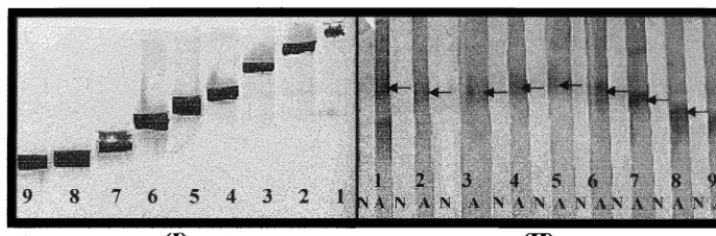
FIG. 1. (I) Gel-purified and concentratedM. tuberculosisprotein antigens (1, 2, 3, 4, 5, 6, 7, 8, and 9) blotted onto PVDF membranes were excised for N-terminal sequencing. (II) These protein antigens were blotted onto nitrocellulose membranes and immunoscreened against pooled normal (N) and active-TB (A) sera, respectively. Positive bands (arrows) were observed with A but not with N.
on August 17, 2020 by guest
http://cvi.asm.org/
Databases and software.Nucleotide and protein sequence analysis was carried out using the basic BLAST 2.0 search program from the National Center for Biotechnology Information (NBCI) and theM. tuberculosisBLAST server at the Sanger Centre (Cambridge, United Kingdom).
Statistical analysis.Sensitivities, specificities, and positive and negative
pre-dictive values were calculated using the Win Episcope 1.0 (Borland International Inc.).
RESULTS
Identification and isolation of
M. tuberculosis
antigens.
Im-munoblot analysis of
M. tuberculosis
total proteins revealed
protein bands which reacted with the pooled active sera but not
with the pooled normal sera. When the respective bands were
concentrated on a long stacking gel, excised, and Western
blotted, these bands were reactive with the pooled active sera
but not with pooled normal sera, thus confirming the
authen-ticity of these excised proteins as those initially observed in the
primary screening (Fig. 1). These proteins were identified by
homology searches against protein sequence databases, which
gave a high percentage of homology to
Mycobacterium
proteins
(Table 1). PCR primers were designed to isolate and clone the
genes coding for four proteins (B.4, B.6, B.10, and B.M) which
gave the highest matches based on a BLAST homology search
against the SwissProt database.
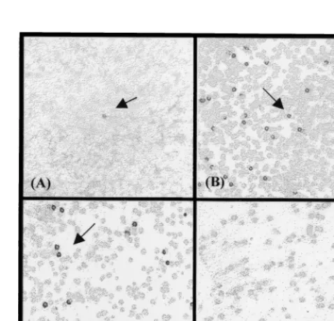
Concurrently, primary screening of the phage expression
library gave eight reactive phage recombinants, of which six
were further confirmed positive by secondary and tertiary
screenings (Fig. 2). These clones were subjected to plasmid
DNA excision, and restriction enzyme digestions with
Eco
RI
indicated that all the clones contained a 2-kb insert. DNA
sequencing revealed that all the clones were identical, having a
1.161-kb open reading frame (in frame with the vector’s ATG
initiation codon) which coded for a proline-rich protein. A
summary of all five TB antigens, with the respective gene sizes,
theoretical molecular masses, and pI values is shown in Table 2.
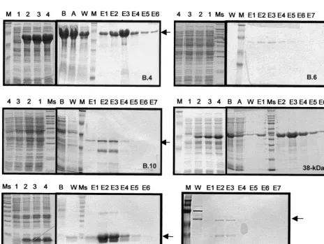
Expression and purification of recombinant antigens.
Ex-pression was detected by probing immunoblots containing
these antigens using the commercial anti-RGS His antibody.
The levels of expression observed were high for the B.4, B.M,
and 38-kDa proteins, moderate for the B.6 and B.10 proteins,
and low for the C17 protein (Fig. 3). All of these recombinant
proteins, except for C17, were present in the insoluble fraction
of an SDS-PAGE analysis (data not shown), indicating that
these proteins formed inclusion bodies and were insoluble. As
such, these recombinant proteins were purified by Ni-NTA
affinity chromatography in 8 M urea, and the SDS-PAGE
pro-file of the purified antigens subsequently used for immunoblots
is shown in Fig. 4. The approximate yields of recombinant
antigens purified through Ni-NTA were 36 mg/liter for B.4, 0.5
mg/liter for B.6, 0.2 mg/liter for B.10, 15 mg/liter for B.M,
⬍
0.1
mg/liter for C17, and 10 mg/liter for the 38-kDa protein.
Reactivities of the recombinant antigens to TB serum
spec-imens.
The different reactivities of recombinant TB antigens
on immunoblot strips probed with serum specimens and
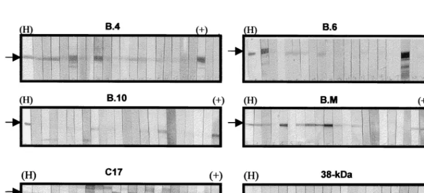
anti-RGS His are shown in Fig. 5. The respective cutoff values for
determining reactivity to the different recombinant antigens
were obtained based on the mean densities observed in sera
from the control group of healthy individuals (
n
⫽
50). The
cutoff values were densities of
⬎
0.04 for the B.4 and B.6 bands,
ⱖ
0.04 for the B.10 and B.M bands,
ⱖ
0.15 for the C17 band,
and
⬎
0.15 for the 38-kDa band.
Based on the Western blot assay, the reactivities of these
antigens to a panel of active-TB serum specimens are shown in
Table 3. Percentages of reactivities and positive and negative
predictive values for each antigen were calculated based on
sera from infected individuals (
n
⫽
48; pulmonary and
ex-trapulmonary TB) and sera from the control group of healthy
individuals (
n
⫽
50), with a
P
value of
⬍
0.05. The specificities
for the B.4, B.6, B.10, B.M, C17, and 38-kDa recombinant
antigens are 94, 88, 100, 96, 90, and 98%, respectively. All of
the recombinant TB antigens showed substantial reactivity to
FIG. 2. Screening of the lambda ZAP Phage expression library ofEco RI-restricted genomic DNA of M. tuberculosis, using pooled human sera from individuals with active TB. (A) Primary screening; (B and C) secondary screen-ing; (D) negative control consisting of plaques of nonrecombinant phages. Ar-rows indicate plaques containing positive recombinant phage.
TABLE 1. Results of homology searches against the GenBank protein sequence databases
aRelative molecular
size (kDa) N-terminal sequence Match (NBCI)b
⬃58
SKLIEYDELALEAME
db:
2SKLIEYDETARRAME16; 55.7 kDa; GroEL1/protein Cpn60 (18, 30); pID⫽
g44601;
X60350 (80% match)
⬃48
AEVDAYKFDPDAVD
db:
161AEFDAYRRDPMA172; probable exported protease, has signal sequence, very similarto three proteases/peptidases from
Streptomyces
; pID
⫽
e235164; MTCY427.04c (51%
match)
⬃34
MEIDILAVAAP
db:
117IEVDLLDLDAP127; 33 kDa; mycocerosic acid synthase; pID⫽
g149978; M95808
(56.9% match)
⬃14
ATTLPVQRHDARL
db:ATTLPVQRHPRSL; 14/16 kDa (21); pID
⫽
g244562; M76712 (69.0% match)
aProteins showing the highest homology to theM. tuberculosisproteins excised for N-terminal sequencing are shown. bdb, database; pID, protein identification.
on August 17, 2020 by guest
http://cvi.asm.org/
active-TB specimens, both pulmonary and extrapulmonary.
The B.4 antigen was reactive with 58.3% of the active-TB
panel, compared to 37.5% detected by the known 38-kDa
antigen. In addition, the B.4 antigen showed reactivity to
27.3% of the inactive-TB specimens compared to other TB
antigens, which exhibited lower percentages of reactivity to
these specimens (Table 3). The B.6 antigen was found to
ex-hibit specific reactivity to pulmonary-TB specimens (46.4%)
compared to extrapulmonary specimens (5%) (Fig. 6). All the
other antigens were able to detect antibodies in both
pulmo-nary- and extrapulmopulmo-nary-TB specimens.
DISCUSSION
A number of
M. tuberculosis
antigens have been identified
and characterized by various methods employing polyclonal
antibodies from rabbits or monoclonal antibodies (MAbs)
from hybridomas generated from immunized mice. Such
anti-bodies were used widely for identification and purification of
protein antigens by affinity chromatography (22),
immuno-screening of clones from DNA libraries of
M. tuberculosis
(24,
37), and analyses of total-cell lysates or secretory proteins from
culture medium by both one- and two-dimensional gel
electro-phoresis (16, 32). Immunogenicity in animals (e.g., mice or
rabbits), however, may not reflect relevance to human immune
responses. Thus, attempts were made to search for candidate
serodiagnostic antigens by directly testing mycobacterial
pro-teins with tuberculous-patient sera on immunoblots of
one-FIG. 3. Expression and affinity chromatography purification profiles of theM. tuberculosisantigens expressed in 100 ml of M15/E. colicultures. Lanes: M and Ms,
protein molecular weight markers (M, Kaleidoscope standards; Ms, Sigma Broad range); 1 through 4, aliquots taken at 0, 1, 2, and 3 h, respectively, after induction with 1 mM IPTG; B, total cell lysate before passing through Ni-NTA column; A, total lysate after passing through column; W, wash fractions in 8 M urea buffer (pH 6.5 to 5.9); E1 to E7, eluted fractions in 8 M urea buffer (pH 4.5). The bulk of the recombinant proteins were observed to be eluted in fractions in 8 M urea buffer (pH 4.5). The bulk of the recombinant proteins were observed to be eluted in fractions E2 and E3 (arrows). All the gels were stained with Coomassie brilliant blue, except for C17, which was silver stained.
TABLE 2. TB antigens genes which were cloned and
expressed in pQE30
aAntigen Size of gene(kb)
Theoreticalb:
Molecular mass
(kDa) pI
B.4
1.617
55.8
5.12
B.6
1.560
55.0
5.03
B.10
0.903
32.9
4.95
B.M
0.432
16.1
5.00
C17
1.161
37.5
9.43
aThe resultant recombinant proteins will be approximately 1.4 to 1.5 kDa
larger than the theoretical molecular mass shown, due to the histidine tag at the N terminus.
bObtained using the software “Compute pI/Mwt” from the ExPASy home
page, Swiss Institute of Bioinformatics, Geneva, Switzerland.
on August 17, 2020 by guest
http://cvi.asm.org/
dimensional and two-dimensional separations of antigenic
ex-tracts or culture filtrates of
M. tuberculosis
H37Rv (4, 26).
In this study, we used antibodies present in the sera of
infected individuals to screen total-cell lysates and a phage
expression library of
M. tuberculosis
DNA. To date, there is no
single immunodominant species-specific antigen for detection
of tuberculosis. We have chosen to use pooled sera from
sev-eral infected individuals to allow identification of sevsev-eral
im-munoreactive antigens reactive to antibodies present in each
serum. In addition, the
M. tuberculosis
genome database
com-pleted by the Sanger Centre (12) allowed for the rapid
iden-tification of these immunoreactive antigens by homology
searches against available protein and gene databases, which
also facilitated the identification of these gene sequences for
cloning.
We have successfully identified and characterized five
anti-gens using a Western blot total-cell-lysate approach; of these,
the B.6, B.10, and C17 antigens are novel and showed high
degrees of nucleotide identity to unpublished
M. tuberculosis
H37Rv genes. Based on DNA sequencing results, the B.6
an-tigen gene was found to have 99% nucleotide identity to a gene
coding for a protein with homology to exported proteases or
peptidases. The B.10 antigen exhibited 99 and 98% nucleotide
identities to the
M. bovis
acyl coenzyme A (CoA) synthase
(accession no. U75685) and mycocerosic acid synthase
(acces-sion no. M95808) genes, respectively. The C17 antigen
exhib-ited 99 to 100% nucleotide identity to a gene coding for
PE-PGRS (polymorphic GC-rich repetitive sequence) proteins, a
member of PE (proline-glutamic acid) families of clustered
genes coding for glycine-rich proteins which may have
immu-nological and pathogenic implications (12).
The B.4 antigen exhibited 99.8% amino acid and 98.9%
nucleotide sequence homology to the Cpn-60 protein reported
by Kong et al. (18). The diagnostic potential of a 65-kDa
protein (also a Cpn-60 family of heat shock proteins) by both
serological and PCR methods has been demonstrated (28).
The B.M antigen has 99% nucleotide identity to the reported
M. tuberculosis
14-kDa antigen gene (accession no. M76712)
and the gene for the 19-kDa major membrane protein purified
from the virulent Erdman strain of
M. tuberculosis
(21). The
serological value of this 19-kDa antigen was shown by 85%
reactivity to a panel of 56 sera from individuals with active
pulmonary TB (9).
Antigens of diagnostic importance for
M. tuberculosis
iden-tified to date include the 65-, 45-, 30/31-, 19-, and 12-kDa
proteins and the 38-kDa lipoprotein (9, 11, 14, 36). Among
FIG. 4. RecombinantM. tuberculosisantigens (arrows) purified by Ni-NTA affinity chromatography as observed on an SDS-PAGE gel (silver stained) (A) and on a Western blot of a duplicate gel which was probed with anti-RGS His antibody followed by detection with alkaline phosphatase-conjugated goat anti-mouse Ig and NBT-BCIP substrate (B). Some truncated products of the recombinant proteins were observed which were detected by the antibody. Lanes: M, Kaleidoscope prestained standards; 1, B.4; 2, B.6; 3, B.10; 4, B.M; 5, 38-kDa protein; 6, C17.
FIG. 5. Immunoscreening of recombinant TB antigens by a Western blot assay. Arrows indicate the positions of recombinant antigens on the immunoblots. Each strip was probed with different serum specimens. A strip was probed with anti-RGS His (H) to indicate the position of protein on the blots. The positive control (⫹) is represented by a strip probed with a known serum specimen which is reactive to the specific recombinant antigen.
on August 17, 2020 by guest
http://cvi.asm.org/
these, the 38-kDa protein was shown to be the most specific
and sensitive for detecting antibodies against
M. tuberculosis
and is specific for TB complex species (35). As such, we have
chosen to clone and express this antigen to be used in
immu-noscreening against the serum panels for comparison with our
recombinant TB antigens. This 38-kDa antigen was expressed
as an insoluble protein in the pQE30/
E. coli
system; similarly,
Singh et al. reported the expression of this 38-kDa antigen as
an insoluble unfused protein in
E. coli
(31).
We have chosen the Qiagen expression system for cloning
and expression of the TB antigens. Each expressed
recombi-nant protein contained a nonimmunogenic 6
⫻
His tag at the N
terminus which could be immunodetected by anti-RGS His. A
substantially low expression level was observed for the C17
protein. This may be due to the codon usage of this protein,
which is rich in proline (46.6%), as it is reported that the
expression level of a gene decreases with an increase in the use
of rare codons (17). In addition, the Kyte-Doolittle hydropathy
plot revealed that it is very hydrophilic, which explains the
soluble nature of this protein (19). As most of the recombinant
antigens were insoluble, we have chosen a Western blot assay
for preliminary screening against serum specimens.
Immunoblot assays using human sera were described
previ-ously for analysis of HPLC-purified 45/47-kDa antigen
com-plex (15). Rovatti et al. reported a semiquantitative Western
blot serological test to identify PPD-positive individuals, using
a discriminate score for the
M. bovis
BCG antigen complex
A60 against MAbs (25). In our immunoblot assay system, we
used affinity-purified recombinant proteins to detect antibodies
in serum specimens and have included the known 38-kDa
lipoprotein as a control for our purification and Western blot
assay system. Zhou et al. reported the use of this antigen in a
rapid membrane-based assay that gave a specificity of 92%,
very close to our in-house immunoblot assay of the 38-kDa
protein, which gave 98% specificity (38). The 38-kDa antigen
was included in the assay system as a further control for
spec-ificity and sensitivity. This was further compared to a
commer-cially available diagnostic test kit which uses two antigens, one
of which is the 38-kDa antigen. All 13 of the 18 active-TB
serum specimens that tested positive with the 38-kDa protein
in our Western blot assay also tested positive with the kit. The
percentage of specificity of the 38-kDa antigen in this assay is
comparable to that with the kit (98 and 100%, respectively).
Data analysis from our assay system indicated that the B.4,
B.6, B.10, B.M, C17, and 38-kDa recombinant antigens reacted
with antibodies in serum specimens of TB-infected individuals.
The reactivities were sufficiently differentiated from those of
serum specimens from healthy individuals and from individuals
with inactive TB or lung cancer. Screening of the same serum
panels using two commercially available TB diagnostic kits of
high specificity indicated the presence of immunologically
spe-cific antibodies reacting to these TB antigens (submitted for
publication). Reports have also shown that other recombinant
antigens did exhibit low levels of cross-reactivities to sera from
healthy individuals, which may be due to cross-reactive epitopes
or analogues to other bacterial proteins (8). To circumvent this
problem, the use of MAbs with recombinant antigens in
com-petition with patient sera as a test assay was reported to give a
high degree of specificity (10). Alternatively, we have to further
optimize the immunoblot assay to decrease cross-reactivities.
In conclusion, we have demonstrated the identification and
cloning of five TB antigen genes for expression in an
E. coli
system and shown the potential of each recombinant antigen as
a serodiagnostic marker for detection of TB infections. We are
in the process of validating the diagnostic utility of these
an-tigens in various test formats.
ACKNOWLEDGMENTS
We are grateful to Christopher Froggatt, Kean Chong Loh, Cindy
Luo, and Adrian Lee at the Fermentation and Purification Laboratory
of BTC for generously helping us to produce and purify the
recombi-nant antigens for screenings.
REFERENCES
1.Amicosante, M., S. Barnini, V. Corsini, G. Paone, C. A. Read, Jr., P. L.
Tartoni, M. Singh, C. Albera, A. Bisetti, S. Senesi, M. Campa, and C. Saltini.
1995. Evaluation of a novel tuberculosis complex-specific 34-kDa protein in the serological diagnosis of tuberculosis. Eur. Respir. J.8:2008–2014.
2.Andersen, A. B., and E. B. Hansen.1989. Structure and mapping of antigenic
domains of protein antigen b, a 38,000-molecular-weight protein of Myco-bacterium tuberculosis. Infect. Immun.57:2481–2488.
3.Banica, D., G. Algeorge, A. Moisoiu, A. Petre, and M. Toanca.1994. The
possibilities for improving the serological diagnosis of active tuberculosis by using new mycobacterial antigens and immunoblot and ELISA technique. Pneumoftiziologia43:173–177.
4.Bassey, E. O. E., D. Catty, D. S. Kumararatne, and C. Raykundalia.1996.
Candidate antigens for improved serodiagnosis of tuberculosis. Tubercle Lung Dis.77:136–145.
5.Beige, J., J. Lokies, T. Schaberg, U. Finckh, M. Fischer, H. Mauch, H. Lode,
B. Kohler, and A. Rolfs.1995. Clinical evaluation of aMycobacterium
tuber-culosisPCR assay. J. Clin. Microbiol.33:90–95.
6.Bloom, B. R., and C. J. Murray.1992. Tuberculosis: commentary on a
reemergent killer. Science257:1055–1064.
7.Bose, M., A. Chander, and R. H. Das.1993. A rapid and gentle method for
the isolation of genomic DNA from mycobacteria. Nucleic Acids Res.21:
2529–2530.
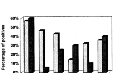
FIG. 6. Graph showing percentages of reactivity for the recombinant TB antigens against the pulmonary-TB (n⫽28) and extrapulmonary-TB (n⫽20) serum specimens. The B.6 antigen detected antibodies in 46.4% of the pulmo-nary-TB compared to only 5% of the extrapulmopulmo-nary-TB serum specimens. The rest of the TB antigens did not exhibit such significant differentiation of reactivity between the two serum panels.
TABLE 3. Reactivities and positive and negative predictive values
of recombinant TB antigens against different sera panels
Antigena
Reactivity (%) of sera from:
PPVb
(%) NPV
c
(%) Healthy
controls (n⫽50)
Active-TB patients (n⫽48)
Inactive-TB patients (n⫽22)
Lung cancer patients (n⫽19)
B.4
6.0
58.3
27.3
10.5
90.3 70.2
B.6
12.0
29.2
9.1
5.3
70.0 56.4
B.10
0
35.4
0
5.3
100.0 61.7
B.M
4.0
20.8
4.6
0
83.3 55.8
C17
10.0
22.9
0
0
68.8 54.9
38-kDa antigen
2.0
37.5
9.1
10.5
96.6 71.0
aA known 38-kDa antigen (GenBank accession no. M30046 [2]) ofM.
tuber-culosiswas included in the screening.
bPPV, positive predictive value,P⬎0.05. cNPV, negative predictive value,P⬎0.05.
on August 17, 2020 by guest
http://cvi.asm.org/
8.Bothamley, G. H.1995. Serological diagnosis of tuberculosis. Eur. Respir. J. Suppl.20:676S–688S.
9.Bothamley, G., H. Batra, V. Ramesh, A. Chandramui, and J. Ivanyi.1992.
Serodiagnosis value of the 19-kilodalton antigen ofMycobacterium tubercu-losisin Indian patients. Eur. J. Clin. Microbiol. Infect. Dis.11:912–915.
10. Bothamley, G. H., and R. M. Rudd.1994. Clinical evaluation of a serological
assay using a monoclonal antibody (TB72) to the 38-kDa antigen ofM. tuberculosis. Eur. Respir. J.7:240–246.
11. Coates, A. R., H. Nicolai, M. J. Pallen, A. Guy, S. D. Chaparas, and D. A.
Mitchison.1989. The 45-kDa molecule ofMycobacterium tuberculosis
iden-tified by immunoblotting and antibodies as antigenic in patients with tuber-culosis. Br. J. Exp. Pathol.70:215–225.
12. Cole, S. T., R. Brosch, J. Parkhill, and T. Garnier.1998. Deciphering the
biology ofMycobacterium tuberculosisfrom the complete genome sequence. Nature393:537–544.
13. Crouse, G. F., A. Frishauf, and H. Lehrach.1983. An integrated and
sim-plified approach to cloning into plasmids and single-stranded phages. Meth-ods Enzymol.101:78–79.
14. Deshpande, E. R., M. B. Khan, and R. G. Navalkar.1993. Immunological
evaluation of a 12-kDa protein ofMycobacterium tuberculosisby enzyme-linked immunosorbent assay. Tubercle Lung Dis.74:382–387.
15. Diagbouga, S., F. Fumoux, A. Zoubga, P. T. Sanou, and G. Marchal.1996.
Immunoblot analysis for serodiagnosis of tuberculosis using a 45/47-kilodal-ton antigen complex ofMycobacterium tuberculosis. Eur. Respir. J.9:288– 292.
16. Espitia, C., R. Espinosa, R. Saavedra, R. Mancilla, F. Romain, A.
Laqueyre-rie, and C. Moreno. 1995. Antigenic and structural similarities between
Mycobacterium tuberculosis50- to 55-kilodalton andMycobacterium bovis
BCG 45- to 47-kilodalton antigens. Infect. Immun.63:580–584.
17. Grantham, R., C. Gautier, M. Gouy, M. Jacobzone, and R. Mercier.Codon
catalog usage is a genome strategy modulated for gene expressivity. Nucleic Acids Res.9:43r–74r.
18. Kong, T. H., A. R. Coates, P. D. Butcher, C. J. Hickman, and T. M. Shinnick.
1993.Mycobacterium tuberculosisexpresses two chaperonin-60 homologs. Proc. Natl. Acad. Sci. USA90:2608–2612.
19. Kyte, J., and R. F. Doolittle.1982. A simple method for displaying the
hydropathic character of a protein. J. Mol. Biol.157:105–132.
20. Laemmli, U. K.1970. Cleavage of structural proteins during the assembly of
the head of bacteriophage T4. Nature (London)227:680–685.
21. Lee, B. Y., S. A. Hefta, and P. J. Brennan.1992. Characterization of the
major membrane protein of virulentMycobacterium tuberculosis. Infect. Im-mun.60:2066–2074.
22. Ljungqvist, L., A. B. Andersen, P. Andersen, K. Haslov, A. Worsaae, J.
Bennedsen, and I. Heron.1990. Affinity purification, biological
characteriza-tion and serological evaluacharacteriza-tion of defined antigens from Mycobacterium tuberculosis. Trop. Med. Parasitol.41:333–335.
23. Lyashchenko, K., R. Colangeli, M. Houde, H. A. Jahdali, D. Menzies, and
M. L. Gennaro.1998. Heterogeneous antibody responses in tuberculosis.
Infect. Immun.66:3936–3940.
24. Parra, C. A., L. P. Londono, P. D. Portillo, and M. E. Patarroyo.1991.
Isolation, characterization, and molecular cloning of a specific Mycobacte-rium tuberculosisantigen gene: identification of a species-specific sequence. Infect. Immun.59:3411–3417.
25. Rovatti, E., M. P. Corradi, M. Amicosante, P. L. Tartoni, W. Panini, A.
Ancora, A. M. Cenci, L. Zucchi, L. Monno, G. Angarano, and C. Saltini.
1995. Evaluation of a Western blot serum test for the diagnosis of Mycobac-terium tuberculosisinfection. J. Commun. Dis.27:208–214.
26. Samanich, K. M., J. T. Belisle, M. G. Sonnenberg, M. A. Keen, S.
Zolla-Pazner, and S. Laal.1998. Delineation of human antibody responses to
culture filtrate antigens ofMycobacterium tuberculosis. J. Infect. Dis.178:
1534–1538.
27. Shevchenko, A., M. Wilm, O. Vorm, and M. Mann.1996. Mass spectrometric
sequencing of proteins from silver-stained polyacrylamide gels. Anal. Chem.
68:850–858.
28. Shinnick, T. M.1987. The 65-kilodalton antigen ofMycobacterium
tubercu-losis. J. Bacteriol.169:1080–1088.
29. Shinnick, T. M., and R. C. Good.1995. Diagnostic mycobacteriology
labo-ratory practices. Clin. Infect. Dis.21:291–299.
30. Shinnick, T. M., M. H. Vodkin, and J. C. Williams.1988. TheMycobacterium
tuberculosis65-kilodalton antigen is a heat shock protein which corresponds to common antigen and to theEscherichia coliGroEL protein. Infect. Im-mun.56:446–451.
31. Singh, M., A. B. Andersen, J. E. G. McCarthy, M. Rohde, H. Schutte, E.
Sanders, and K. N. Timmis.1992. TheMycobacterium tuberculosis38-kDa
antigen: overproduction inEscherichia coli, purification and characteriza-tion. Gene117:53–60.
32. Sonnenberg, M. G., and J. T. Belisle.1997. Definition ofMycobacterium
tuberculosisculture filtrate proteins by two-dimensional polyacrylamide gel electrophoresis, N-terminal amino acid sequencing, and electrospray mass spectrometry. Infect. Immun.65:4515–4524.
33. Towbin, H., T. Staehelin, and J. Gordon.1979. Electrophoretic transfer of
proteins from polyacrylamide gels to nitrocellulose sheets: procedure and some applications. Proc. Natl. Acad. Sci. USA76:4350–4354.
34. World Health Organization.1999. The World Health Report. World Health
Organization, Geneva, Switzerland.
35. Young, D., L. Kent, A. Rees, J. Lamb, and J. Ivanyi.1986. Immunological
activity of a 38-kilodalton protein purified fromMycobacterium tuberculosis. Infect. Immun.54:177–183.
36. Young, D. B.1988. Structure of mycobacterial antigens. Br. Med. J.44:562–
583.
37. Young, R. A., B. R. Bloom, C. M. Grosskinsky, J. Ivanji, D. Thomas, and
R. W. Davies.1985. Dissection ofMycobacterium tuberculosisantigens using
recombinant DNA. Proc. Natl. Acad. Sci. USA82:2583–2587.
38. Zhou, A. T., W. L. Ma, P. Y. Zhang, and R. A. Cole.1996. Detection of
pulmonary and extrapulmonary tuberculosis patients with the 38-kilodalton antigen fromMycobacterium tuberculosisin a rapid membrane-based assay. Clin. Diagn. Lab. Immunol.3:337–341.