Case – Recurrent incarcerated inguinal bladder hernia with small bowel obstruction
Jeff McLellan, BComm1; Zuhaib M. Mir, MD2; Michael Di Lena, MD3; Sulaiman Nanji, MD, PhD FRCSC2; Darren Beiko, MD, MBA, FRCSC3
1Northern Ontario School of Medicine, Sudbury, ON, Canada; 2Department of Surgery, Division of
General Surgery, Queen’s University, Kingston, ON, Canada; 3Department of Urology, Queen’s University, ON, Canada
Cite as: Can Urol Assoc J 2019 February 26; Epub ahead of print. http://dx.doi.org/10.5489/cuaj.5740
Published online February 26, 2019
***
Introduction
Inguinal hernias are a common surgical complaint, with a lifetime incidence requiring repair of 27% in men and 3% in women.1 Recurrence of an inguinal hernia requiring repair depends on a number of factors including repair type, surgeon experience, and patient factors, but has been estimated at 1-4%.2,3 The bladder is involved in 1-4% of inguinal hernias in the general population, and as high as 10% of cases in men over age 50.4 Risk factors for urinary bladder involvement in an inguinal hernia include male gender, age over 50, and obesity.5 The mainstay of treatment for an inguinal bladder hernia is herniorrhaphy.
Most small bladder hernias are discovered intraoperatively due to the lack of urinary symptoms prior to herniorrhaphy. In fact, less than 7% are diagnosed
preoperatively4. Small urinary bladder hernias are often asymptomatic, but clinical manifestations can include urinary frequency, urgency, nocturia, hematuria, and double-phase micturition.6 Extensive herniation of the bladder, commonly referred to as a scrotal cystocele, is much less frequent. These are more likely to present with urinary symptoms, and urgent intervention is usually required as bladder infarction and/or obstruction may result.6 Inguinal bladder hernias with associated small bowel obstruction are rare and rates have not been characterized in the literature.
Case report
Our patient was an 80-year old male, who presented to the emergency department with a two-day history of vomiting, diarrhea, expressive aphasia, and confusion. Co-morbidities prior to presentation included Alzheimer’s disease, prostate adenocarcinoma (T2a, Gleason 6/10, treated with curative radiotherapy), hypertension, gout, previous atrial tachycardia, previously-resected vestibular schwannoma, and interestingly, a previous left inguinal hernia repaired with Shouldice technique. He was seen urgently by the Neurology service, however, acute cerebrovascular pathology was subsequently ruled out. The focus then shifted to the patient’s gastrointestinal symptoms, and initial
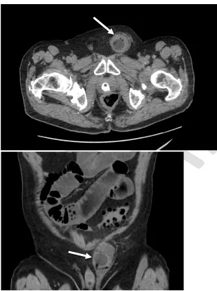
bloodwork revealed a mild leukocytosis (11.2 x10⁹/L), elevated creatinine (165 µmol/L), but an otherwise unremarkable hematologic and biochemical profile. Blood cultures and urine culture were drawn and ultimately negative, ruling out underlying sepsis. Initial CT of the abdomen and pelvis demonstrated a recurrent left-sided inguinal hernia containing a loop of dilated small bowel with resultant proximal small bowel dilatation. The hernia also contained a portion of non-opacified bladder with the foley catheter balloon/tip within the herniated segment (figure 1), causing the bladder to remain distended.
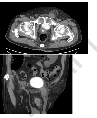
The foley catheter balloon was deflated and repositioned. In addition, manual reduction of the hernia was attempted in the emergency department under procedural sedation. Repeat CT images showed successful reduction of the small bowel from the hernia, but persistent incarceration of the urinary bladder within the left inguinal hernia (figure 2). There was no contrast extravasation seen from the bladder, suggesting that its wall was intact.
Nonetheless, given the persistent incarceration of the bladder, as well as the need to assess viability and ischemia via cystoscopy, the patient was consented and booked for urgent operative repair of the hernia. An open plug and patch repair was performed. Intra-operatively, the hernia was noted to be indirect, with no evidence of residual small bowel herniation. Externally, the bladder tissue did not appear to be necrotic or perforated. An intra-operative cystoscopy was also performed, which demonstrated mucosal edema at the dome of the bladder without any necrosis or perforation. A foley catheter was left in-situ and the patient tolerated the procedure well.
Discussion
Inguinal hernias are a common surgical complaint, with bladder involvement in approximately 1-4% of cases. While there are many reported cases of inguinal bladder hernias, we believe this to be the first case where the foley catheter balloon was inflated in the herniated portion of the bladder, thereby helping identify the underlying pathology. There is one prior case where a suprapubic catheter balloon was seen in the hernia, however that was a case involving complete bladder herniation.6
Small inguinal bladder hernias tend to be asymptomatic from a urinary perspective and therefore, most often discovered intraoperatively which can lead to a more challenging repair and the possibility of bladder injury during herniorrhaphy. Imaging modalities such as CT and cystograms should be considered pre-operatively to clearly delineate the anatomy if there is suspicion for bladder involvement, as well as to guide surgical repair.
When recognized preoperatively, the mainstay of treatment is surgical repair through an open inguinal approach, reducing the bladder to its original location in the pelvis. Partial cystectomy is rarely indicated, except in the case of bladder wall necrosis, herniated bladder diverticulum, tight hernia neck, or bladder tumour within the herniated segment.7,8 As such, we recommend routine intra-operative cystoscopy as an easy strategy to avoid missing such pathologies.
In this case, there was also an associated small bowel obstruction. While the small bowel was able to be reduced without operative intervention, incarceration of the bladder segment was an indication for urgent surgery. A review of the literature returns 18 cases of incarcerated inguinal bladder hernias, only two of which had associated small bowel obstructions.
Conclusion
To the best of our knowledge, this is the first reported case of an incarcerated inguinal bladder hernia where the foley catheter balloon was inflated in the herniated portion of the bladder. The incarcerated small bowel was reduced pre-operatively, however,
References
1. Primatesta P, Goldacre MJ. Inguinal hernia repair: Incidence of elective and
emergency surgery, readmission and mortality. Int J Epidemiol 1996;25(4):835-39. 2. Magnusson J, Gustafsson U, Nygren J, et al. Rates of and methods used at
reoperation for recurrence after primary inguinal hernia repair with Prolene Hernia System and Lichtenstein. Hernia 2018;22(3):439-44.
3. Ramjist J, Dossa F, Stukel T, et al. Reoperation for inguinal hernia recurrence in Ontario: A population-based study. Hernia 2018.
4. Moufid K, Touiti D, Mohamed L. Inguinal bladder hernia: Four case analyses. Rev Urol 2013;15(1):32-6.
5. Oruç MT, Akbulut Z, Özozan Ö, et al. Urological findings in inguinal hernias: A case report and review of the literature. Hernia 2004;8(1):76-79.
6. Bisharat M, O’Donnell ME, Thompson T, et al. Complications of inguinoscrotal bladder hernias: A case series. Hernia 2009;13(1):81-84.
7. Wagner AA, Arcand P, Bamberger MH. Acute renal failure resulting from huge inguinal bladder hernia. Urology 2004;64(1):156–57.