Original Article
Physiological and induced apoptosis in sea urchin larvae
undergoing metamorphosis
MARIA C. ROCCHERI*
,1, CINZIA TIPA
1, ROSA BONAVENTURA
2and VALERIA MATRANGA
2 1Dipartimento di Biologia Cellulare e dello Sviluppo “A Monroy”, Università di Palermo, Italy and2 Istituto di Biomedicina ed Immunologia Molecolare "A. Monroy", C.N.R., Palermo, Italy
ABSTRACT Paracentrotus lividus embryos at the early pluteus stage undergo spontaneous apoptosis. Using a TUNEL (TdT-mediated dUTP Nick-End Labelling) assay on whole mount embryos, we showed that there was a different distribution of the apoptotic cells in different optical sections. Not more than 20% of cells in plutei were spontaneously apoptotic, as confirmed by the counts of dissociated ectoderm and intestine cells. Observation of larva stages closer to metamor-phosis confirmed that apoptosis is a physiological event for the development of the adult. In particular, larvae at different developmental stages showed apoptotic cells in the oral and aboral arms, intestine, ciliary band and both apical and oral ganglia. Moreover, we found that the number of apoptotic cells decreased in later larva stages, possibly because in the organism approaching metamorphosis, a smaller number of cells needs to be eliminated. Furthermore, combined phorbol ester (TPA) and heat shock treatment enhanced apoptosis by increasing the number of cells involved in the phenomenon.
KEY WORDS: apoptosis, sea urchin, development, metamorphosis
0214-6282/2002/$25.00 © UBC Press
Printed in Spain www.ijdb.ehu.es
*Address correspondence to: Dr. Maria C. Roccheri. Dipartimento di Biologia Cellulare e dello Sviluppo “A Monroy”, Università di Palermo, Viale delle Scienze, Parco d’Orleans, 90128 Palermo, Italy. Fax: +39-091-657-7430. e-mail: [email protected]
Abbreviations used in this paper: DAB, diaminobenzide; mfs, Millipore filtered
seawater; PBS, phopsphate buffered saline; PI, propidium iodide; SSC, saline sodium citrate; TdT, terminal deoxynucleotidyl transferase; TPA, 12-O-tetradecanoylphorbol-12-acetate; TUNEL, TdT-mediated dUTP Nick-End Labelling.
Introduction
Programmed cell death or apoptosis is a physiological process of cell elimination that plays a critical role in development and in tissue homeostasis. This evolutionary conserved form of cell suicide is defined by morphological characteristics that include cell shrinkage, chromatin condensation, membrane blebbing, internucleosomal DNA fragmentation (Ellis et al., 1991; Clarke and Clarke, 1996; Nagata, 1997; Green, 1998; Messmer and Pfeilscifter, 2000; Stennicke and Salvesen, 2000; Wang, 2001). Programmed cell death is particularly important during development, morpho-genesis and metamorphosis (Sanders and Wride, 1995; Nishikawa and Hayashi, 1995; Jacobson et al., 1997; Hirata and Hall, 2000). Studies in Xenopus laevis have focused on the cell death that occurs during the early developmental stages, showing activation of apoptosis in gastrulation following appropriate external stimuli (Stack and Newport, 1997; Hensey and Gautier, 1997).
It has been previously shown that sea urchin embryos posses the machinery for apoptosis when induced by different agents (Roccheri et al., 1997; Sato and Yazaki, 1999; Voronina and Wessel, 2001). The aim of this study was to understand if, in the sea urchin embryo/ larva, the programmed cell death is a physiological process that naturally happens in view of remodelling and cellular homeostasis occurring at metamorphosis, as observed in the case of tadpole tail and intestine (Shi et al., 2001).
Previous studies on cell death in the Paracentrotus lividus sea urchin embryos showed that at the stages of gastrula and pluteus a treatment with TPA followed by an increase of the temperature to 31°C had a clear apoptotic effect, as judged by DNA analysis and supported by the other known apoptotic signs. Apoptosis was also found in some cells of non-treated plutei (Roccheri et al., 1997).
Results
Qualitative and Quantitative Estimates of Apoptosis in Pluteus Stage Embryos
In previous studies we have shown that no signs of apoptosis were detected in normally developing gastrula embryos, by using DNA electrophoresis, hematoxylin-eosin stainings and TdT assay, yet spontaneous apoptosis occurs at the pluteus stage especially in arm and intestine districts (Roccheri et al., 1997). This finding has been interpreted as due to the disappearance or regression of the above mentioned structures during metamorphosis. However, since the number of cells undergoing apoptosis in these structures seemed to be very high for an embryo with a life expectation of 3-4 weeks to reach metamorphosis, the possibility that apoptosis was due to starvation could not be excluded. At first it was important to understand how many cells undergo spontaneous apoptosis;
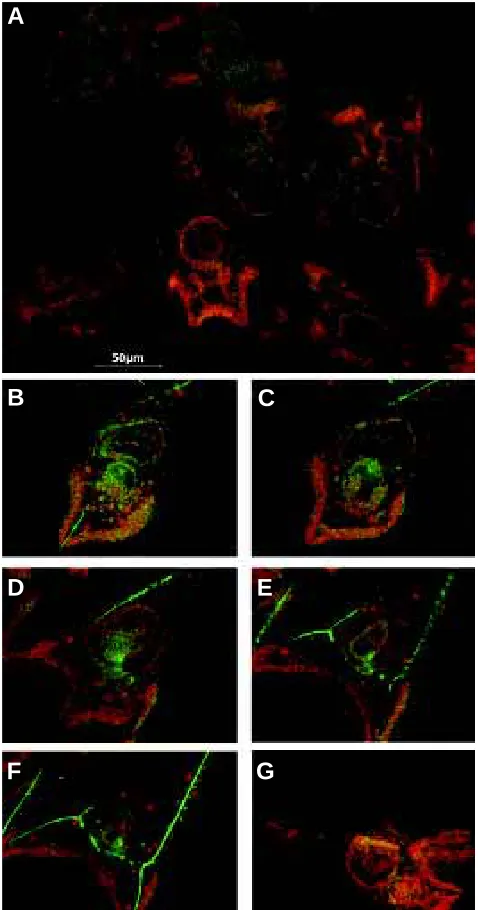
Fig. 1.Whole mounts of pluteus embryos observed by confocal laser microscopy after TdT assay. Panoramic view of some pluteus embryos (A). Serial optical sections of a pluteus embryo (B-F).Section of a pluteus as a positive control (G). The apoptotic nuclei appear orange and the non-apoptotic ones green. Scale bar, 50 µm.
Fig. 2.Dissociated cells from pluteus embryos observed by fluores-cence microscopy after TdT assay. Ectoderm cells (A,C). Endoderm cells (B,D). Negative (C) and positive (D) controls. The nuclei appear with a bright fluorescence when positive for apoptotis and with a faint flourescence when negative, due to PI counterstaining. Scale bar, 50 µm.
therefore, we used a TdT assay on whole mount embryos to localize apoptotic cells in any given serial section, visualized by confocal microscopy. As a consequence of this reaction, the nuclei were stained in orange when positive for apoptosis; counterstained nuclei showed green fluorescence (see Materials and Methods). As shown in Fig. 1A, which depicts a whole mount image of plutei observed by fluorescence, it seems that most of the cells contained in oral and aboral arms and intestine are apoptotic. A more accurate analysis by the examination of some serial optical sec-tions of a pluteus embryo, under a confocal laser microscope (Fig. 1 B-F), allowed us to observe a different distribution of the apoptotic cells in the different fields of focus. Optical sections were selected starting from the oral arms (Fig. 1B) and going down to the aboral arms (Fig. 1F). It is clear that not all the cells are apoptotic in a single optical section, as was suggested by the whole mount embryo total image, probably due to the cumulative effect of the fluorescence caused by the overlapping of different focal levels (Fig. 1A). As positive control to TdT assay we used embryos pre-treated with DNase I in order to induce total and not specific apoptosis (Fig. 1G). It should be noticed that the embryo skeleton is highly green fluorescent probably because primary mesenchyme cells, which synthetize the sea urchin skeleton, completely surround the spi-cules (Urry et al., 2000). In addition the possibility exists that the observed fluorescence is partially due to self-fluorescence of spicules.
Then, in order to quantify the apoptotic cells in these districts, we dissociated the pluteus embryos and separated the ectoderm cells from intestine cells (Roccheri et al., 1979), and thereafter carried out in situ TdT reaction (Fig. 2). In this case, the observations were
A
B
D
F
C
E
G
A
C
B
nomena differ according to each species, they all involve: eversion of the adult rudiment which develops within the larva, re-adsorption of the larval body into the juvenile and later differentiation of adult forms (for more details on sea urchin metamorphosis see Yokota et al., 2002). For the analysis of physiological and induced apoptosis in later stages of development, early plutei, 8-arms larvae, compe-tent larvae and juveniles, were tested with the colorimetric TUNEL
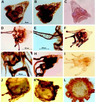
(TdT-mediated dUTP Nick-End Labelling) assay. It should be pointed out that no starvation was taking place, since live control larvae of the same batch reached metamorphosis in due time. As shown in Fig. 3, apoptotic cells are found in restricted areas of the oral and aboral arms and intestine in the early pluteus (Fig. 3A). Apoptotic cells are found in the same districts as shown above and also in the ciliary band, apical and oral ganglia in normal 8-arms larvae (Fig. 3 D,F), as confirmed in the enlargements of arms and ganglia regions respectively (Fig. 3 E,G). In addition, more exten-sive apoptotic areas were observed in induced plutei and larvae (Fig. 3 B,H,K). At later larval stages, corresponding to competent larva and juvenile stage, we found that the number of apoptotic cells decreases (Fig. 3 J,L), possibly because in the organism approaching metamorphosis, a smaller number of cells needs to be eliminated. The negative controls were obtained by omitting the TdT enzyme in the reaction (Fig. 3 C,I).
To further confirm that apoptotic cells decrease in number as larvae reach metamorphosis, larvae at two different stages ap-proaching metamorphosis were tested by florescence TdT assay
Fig. 3.Whole mounts of control or induced to apoptosis pluteus embryos and larvae observed after TUNEL assay.Pluteus embryo (A). Treated pluteus embryo (TPA+31°C) (B). Pluteus embryo negative control (C). 8-arm larva (D,F). Enlargements of 8-arms larva (E,G). Treated larva (TPA+31°C) (H). Larva negative control (I). Competent larva (J). Treated juvenile (TPA+31°C) (K). Juvenile (L). Scale bars: 50 µm (A,B,C); 200 µm (D,F,H,I,J,K,L); 600
µm (E); 400 µm (G). carried out using a fluorescence microscope with
a longpass filter (see Materials and Methods), which emphasised the bright fluorescence of nu-clei when positive for apoptosis and faint fluores-cence when negative due to propidium iodide (PI) counterstaining. Figure 2 shows some representa-tive fields of ectoderm (Fig. 2A) and intestine cells (Fig. 2B) in comparison to a negative control, obtained with ectoderm cells by omitting the TdT enzyme in the reaction (Fig. 2C), and a positive control of intestinal cells, induced by a DNase I pre-treatment (Fig. 2D). The quantitative analysis of the apoptotic cell frequency found in plutei, sum-marized in Table 1, indicated that about 12% of ectoderm cells and 8% of intestinal ones under-went spontaneous apoptosis. The results come from three different experiments. It should be men-tioned that in preliminary experiments, apoptotic cells were not observed in dissociated gastrula-stage embryos. This gastrula-stage has been shown to have no physiological apoptosis (Roccheri et al., 1997), indicating that the dissociation procedure does not induce apoptosis.
Physiological and Induced Apoptosis in Plutei, Larvae and Juveniles
Then we extended our observation to larval stages closer to metamorphosis to confirm if sea urchin apoptosis is a physiologic event for the development of the adult. After the sea urchin embryo has developed to the pluteus stage, on the left side of the larval intestine the adult rudiment is formed. Its growth will continue to give rise to the juvenile sea urchin through a process of metamor-phosis in response to proper feeding of the larva and to environmental cues (Cameron and Hinergardner, 1974; Burke, 1983; Yazaki and Harashima, 1994). The process of metamorphosis has been described in detail for many sea urchin species and, although the developmental
phe-TABLE 1
APOPTOTIC CELL FREQUENCY IN CELLS DISSOCIATED FROM PLUTEUS EMBRYOS
Ectoderm Intestin
apoptotic/scored cells apoptotic/scored cells Experiment
1 115/950 90/1070
2 120/1010 95/1100
3 134/1120 70/980
Mean frequency 12.10% 8.09%
A
B
C
D
E
F
G
H
I
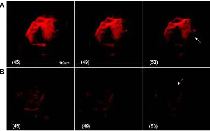
and analysed by confocal microscopy. Results in Fig. 4 show the analysis of 3 confocal serial sections (45-49-53, of the 107 total sections) of a 8-arms larva with small rudiment (A) and a competent larva with big rudiment (B). These results show that the fluorescent signal is found in the same larval districts (ciliary band and arm tips), but in the competent larva the number of fluorescent cells is lower than that found in 8-arms larva. This finding suggests that apoptosis decreases as larvae approach metamorphosis.
Discussion
Everthough echinoderm metamorphosis has been amply de-scribed in literature, little is known about the mechanisms and cellular process of reabsorption of larval structures. Echinoderm metamorphosis involves, in most cases, the transformation of a bilateral symmetric larva into a radial symmetric and benthonic adult. This radical event has attracted the attention of many zoologists. In the past Cameron (Cameron and Holland, 1985), studying the metamorphosis of the sea urchin Lytechinus pictus, noted autolysis of cells of the larval epidermis. In the sand dollar Dendraster excentricus, Chia and Burke (1978) found that nuclei of some epidermal cells became picnotic and fragmented upon metamorphosis. Their finding can be interpreted now as the first evidence of apoptotic signs. As new techniques and probes for examining apoptosis become more accessible and widely applied, it should be possible to clarify the role of specific pathways and involved genes.
Recently, investigators have examined the expression of regu-latory and structural genes during echinoderm metamorphosis in several species. Results from different groups suggest that in echinoderms instead of patterning the embryos, as in the case of arthropods and chordates, the hox genes primary function is patterning the post-embryonic body plan (Lowe and Wray, 1997; Arenas-Mena et al., 1998; Wray, 2000).
longer needed (Jacobson et al., 1997).
It should be stressed here that oral and aboral arms and intestine are considered to be very proliferative districts which involve pro-grammed cell death, similarly to the regression of tadpole tail muscles and intestinal epithelium, (Ishizuya-Oka and Ueda, 1996; Sachs et al., 1997; Shi et al., 2001). Furthermore, ciliary bands, oral and aboral arms, intestine and ganglia all disappear after metamor-phosis when adult remodelling takes place (Yokota et al., 2002).
In agreement with studies reported here our previous investiga-tions on Paracentrotus lividus sea urchin embryos at the blastula, gastrula and early pluteus stages have demonstrated the presence of an apoptotic machinery capable of executing programmed cell death upon TPA and heat combined treatment (Roccheri et al., 1997). More recently, similar results were found in oocytes, eggs and early embryos of the species Lytechinus variegatus experimentally induced to apoptosis with staurosporine (Voronina and Wessel, 2001). Furthermore it should be recalled here that Sato and Yazaki (1999) have provided some evidence of apoptosis, during the metamorphosis of the Japanese sea urchin species Anthocidaris crassispina, while apoptosis-like cells were recently found in early embryos of the species Hemicentrotus pulcherrimus (Mizoguchi et al., 2000).
In the future, it will be extremely interesting to compare the pathways of apoptosis utilized during development or in response to stress, in order to investigate if developmental and defensive apoptotic processes have a common origin. On the other hand, it has been hypothesized that apoptosis may have initially evolved as a defence mechanism but later, perhaps with the evolution of multicellularity, it was adopted for use during development (Vaux and Korsmeyer 1999; Ameisen, 2002)
Therefore, the developing sea urchin will represent an ideal model with which to study both physiological and induced apoptosis, in order to understand whether cellular death occurring during development regulates modelling.
Fig. 4. Whole mounts of larvae observed by confocal laser microscopy after TdT assay. Three optical serial sections (45-49-53, of the 107 total sections) of an 8-arms larva with small rudiment (A) and a competent larva with big rudiment (B). Arrows point to larvae rudiments. Scale bar, 100 µm.
In this study we investigated the possi-bility that apoptosis could occur as a physi-ological event in view of metamorphosis of the sea urchin. First, we found, using a TUNEL assay on whole mount embryos, apoptotic cells in specific and discrete dis-tricts of plutei, having a different spatial distribution in the embryo at a given time. Furthermore, not more than 20% of all the cells undergo spontaneous apoptosis, as confirmed by the counts of dissociated cells. Here we also documented the evidence that apoptosis occurs in the physiological development of the sea urchin embryos and larvae which undergo metamorphosis. Since we found that approaching metamor-phosis fewer cells were becoming apoptotic, we postulate that the elimination of useless cells is the result of a programmed cell death, needed for the development of the adult. Our hypothesis is in agreement with studies that claim that in the course of animal development various structures are formed and then removed by programmed cell death at later stages when they are no
A
Materials and Methods
Embryo and Larva Cultures
Adult sea urchins of the Mediterranean species Paracentrotus lividus were collected along Sicily’s western coast. Eggs were fertilized and reared at 18°C to the desired stages in Millipore filtered seawater (mfs) containing antibiotics, in a beaker with a rotating propeller, at the concentration of 5000/ml (Giudice and Mutolo, 1967).
After the pluteus stage embryos were fed Chaetocerus gracilis every 2 or 3 days, as described by Falugi (Falugi et al., 1993). Metamorphosis was obtained over a period of 30 days, beginning from the 25th after fertilization. Embryos and larvae to be used for detection of apoptosis were harvested at different developmental stages from the same batch of embryos. Live control embryos from the same batch always reached metamorphosis.
Induction of Apoptosis
We followed the procedure already reported for the induction of apoptosis in Paracentrotus lividus sea urchin embryos (Roccheri et al., 1997). Briefly, living embryos or larvae were treated with 25 nM TPA (12-O-tetradecanoylphorbol-12-acetate) for 2 hours at 20°C and then for 1 hour at 31°C (TPA+31°C). Thereafter, the specimen were washed twice, sus-pended in fresh mfs and utilized for following analyses.
Ectoderm and Endoderm Pluteus Cell Dissociation and Fixation
Pluteus embryos were incubated in 1 M glycine-2 mM EDTA for 10 min, washed 3 times in Ca2+ and Mg2+ free seawater, then the ectoderm and endoderm cells were separated as previously described by Roccheri et al. (1979). Mesenchyme cells, in little amounts, are usually associated with endoderm cell population. Cell pellets, corresponding to about 1x 10⁷ cells, were re-suspended in 1 ml of paraformaldehyde-glutaraldehyde fixative (5µl 25%glutaraldehyde in 8 ml 4%paraformaldehyde) for 1 hour at room temperature. Cells were washed twice in PBS (0.15 M Na2HPO4, 0.04 M NaH2PO4, 0.15 M NaCl) for 5 minutes and permeabilized in 0.05% saponin for 30 min at room temperature. After two washes with PBS, cells were resuspended in PBS and 100µl of cell suspension were spread onto poly-l-lysine-coated glass slides by cytocentrifugation. Slides were then utilized for the TdT assay (Amersham) described below.
Embryos and Larvae Fixation
Embryos and larvae pellets were suspended in 0.1% formalin, followed by 2 washes in mfs, permeabilized in 100% methanol, 2 washes in mfs, 2 washes in PBS, then utilized for the TdT assay (Amersham) or the Dead-End Colorimetric Apoptosis detection system (TUNEL) (Promega).
Apoptosis Detection Systems
TdT Assay. Fixed embryos, or dissociated cells, were incubated in 50 µl of a solution containing TdT buffer, Cy3TM –dCTP (0.3 nmoles), “cold” dCTP (0.033 nmoles), TdT enzyme (10 Units) for 60 min at 37°C in a humidified chamber (Amersham reagents). Whole mount embryos and cells incubated as above reported omitting the TdT enzyme served as negative controls. Pre-treatment with DNAaseI (10 µg/ml) for 10 min provided positive controls (Gorczyca et al., 1993). Incubation in 2X SSC (300mMNaCl-30mMNaCitrate) for 30 min at room temperature and 3 times 5 min each PBS washings stopped the reaction. In some cases, nuclei were counterstained with propidium iodide (PI) (2 µg/ml) in antifade solution (1 mg/ml p-phenylendiamine-dihydrocloride in 1 part PBS and 9 parts 87% glycerol; pH 8.0). Whole mount embryos were observed under an Olympus confocal laser Ar/Kr scanning microscope, by a 665 nm long band pass and 510-550 nm Cy3 band pass filters, with simultaneous observation. Positive reaction gave orange fluorescence; counterstaining gave green fluorescence.
Dissociated cells were counted and scored for apoptosis under a Nikon fluorescence microscope equipped with a HBO 100-W mercury lamp and suitable filters. Photographs were recorded on Kodak colour film.
TUNEL Assay. This assay was preferentially used for large sized larvae.
We used a DeadEndTM Colorimetric Apoptosis Detection System, a modified TUNEL (TdT-mediated dUTP Nick-End Labelling) assay (Promega), which labels in situ fragmented DNA of apoptotic cells. Control and treated plutei and larvae were fixed as described above; then immersed in 100 µl of equilibration buffer (provided by manufacturer) for 10 min at room temperature. Labelling was carried out in 25 µl of a biotinylated nucleotide labelling mix (purchased from Promega) for 30 min, and the reaction was terminated with 2X SSC for 15 min at room temperature. Horseradish-peroxidase labelled streptavidin was then bound to these biotinylated nucleotides; detection was achieved by the peroxi-dase substrate, hydrogen peroxide and the stable chromogen, diaminobenzide (DAB). Using this procedure, apoptotic nuclei are stained dark brown.
Acknowledgements
We are deeply indebted to Giovanni Morici for his assistance in preparing drawnings and figures. This work was partially supported by funds of the Italian M.U.R.S.T., CNR Contract N° 99.02499 CTO4 and by an EU program to V.M., Contract N°EVK3-CT-1999-00005. R. Bonaventura has been financially supported within the context of the above mentioned EU grant.
References
AMEISEN, J.C. (2002). On the origin, evolution, and nature of programmed cell death: a timeline of four billion years. Cell Death Differ. 9: 367-393.
ARENAS-MENA, C., MARTINEZ, P., CAMERON, R.A. and DAVIDSON, E.H. (1998). Expression of the Hox gene complex in the indirect development of a sea urchin. Proc. Natl. Acad. Sci. USA 95: 13062-13067.
BURKE, R.D. (1983). The induction of metamorphosis in marine invertebrate larvae: stimulus and response. Can. J. Zool. 61: 1701-1719.
CAMERON, R.A. and HINERGARDNER, R.T. (1974). Initiation of metamorphosis in laboratory cultured sea urchins. Biol. Bull. 146: 335-342.
CAMERON, R.A. and HOLLAND, N.D. (1985). Demonstration of the granular layer and the fate of the hyaline layer during the development of a sea urchin (Lytechinus variegatus). Cell Tissue Res. 239: 455-458.
CHIA, F. and BURKE, R.D. (1978). Echinoderm metamorphosis: fate of larval structures. In Settlement and metamorphosis of marine invertebrate larvae. (Eds Chia, F. and Rice, M.E.) Elsevier, New York, pp. 219-234.
CLARKE, A.R. and CLARKE, S. (1996). Nineteenth Century research of naturally occurring cell death and related phenomena. Anat. Embryol. 193: 81-99.
ELLIS, R.E., YUAN, J.Y. and HORVITZ, H.R. (1991). Mechanism and functions of cell death. Ann. Rev. Cell Biol. 7: 663-698.
FALUGI, C., MARCHI, B., TRIELLI, F., CORRE, M.C. and FENAUX, L. (1993). Cholinergic molecules are involved in the regulation of induction and growth during sea urchin metamorphosis. Anim. Biol 2: 140-145.
GIUDICE, G. and MUTOLO, V. (1967). Synthesis of ribosomal RNA during sea urchin development. Biochem. Biophys. Acta 138: 276-285.
GORCZYCA, W., GONG, J. and DARZYNKIEWICZ, Z. (1993). Detection of DNA strand breaks in individual apoptotic cells by the in situ terminal deoxynucleotidil transferase and nick translation assays. Cancer Research 53: 1945-1951.
GREEN, D.R. (1998). Apoptotic Pathway: the roads to ruin. Cell 94: 695-698.
HENSEY, C. and GAUTIER, J. (1997). A developmental timer that regulates apoptosis at the onset of gastrulation. Mech. Dev. 69: 183-195.
HIRATA, M. and HALL, B.K. (2000). Temporospatial patterns of apoptosis in chick embryos during the morphogenetic period of development. Int. J. Dev. Biol. 44: 757-768.
ISHIZUYA-OKA, A. and UEDA, S. (1996). Apoptosis and cell proliferation in the Xenopus small intestine during metamorphosis. Cell Tissue Res. 286: 467-476.
JACOBSON, M.D., WEIL, M. and RAFF, M.C. (1997). Programmed cell death in animal development. Cell 88: 347-354.
MESSMER, U.K. and PFEILSCIFTER, J. (2000). New insights into the mechanism for clearance of apoptotic cells. BioEssays 22: 878-881.
MIZOGUCHI, H., KUDO, D., SHIMIZU, Y., HIROTA, K., KAWAI, S. and HINO, A. (2000). Distribution of apoptosis-like cells in sea urchin early embryogenesis. Zygote 8: S 76.
NAGATA, S. (1997). Apoptosis by death factor. Cell 88: 355-365.
NISHIKAWA, A. and HAYASHI, H. (1995). Spatial, temporal and hormonal regulation of programmed muscle cell death during metamorphosis of the frog Xenopus laevis. Differentiation 59: 207-214.
ROCCHERI, M.C., DI BERNARDO, M.G. and GIUDICE, G. (1979). Archenteron cells are responsible for the increase in ribosomal RNA synthesis in sea urchin gastrulae. Cell Biol. Intern. Reports 3: 733-737.
ROCCHERI, M.C., BARBATA, G., CARDINALE, F., TIPA, C., BOSCO, L., OLIVA, O.A., CASCINO, D. and GIUDICE, G. (1997). Apoptosis in sea urchin embryos. Biochem. Biophys. Res. Commun. 240: 359-366.
SACHS, L.M., ABDALLAH, B., HASSAN, A., LEVI, G., DE LUZE, A., REED, J.C. and DEMENEIX. B.A. (1997). Apoptosis in Xenopus tadpole tail muscles involves Bax-dependent pathways. FASEB J. 11: 801-808.
SANDERS, E.J. and WRIDE, M.A. (1995). Programmed cell death in development. In Review of Cytology. (Eds Jeon, K.W. and Jarvik, J.) Academic Press, London, vol. 163. pp. 105-173.
SATO, Y. and YAZAKI, I. (1999). A cellular analysis of sea urchin metamorphosis induced by L-glutamine. In Echinoderm Research 1998, (Eds Candia-Carnevali, M.D. and Bonasoro, F. Balkema, Rotterdam, pp. 221-226.
SHI, Y.B., FU, L., HSIA, S.C., TOMITA, A. and BUCHHOLZ, D. (2001). Thyroid hormone regulation of apoptotic tissue remodelling during anuran metamorpho-sis. Cell Res. 11 (4): 245-252.
STACK, J.H. and NEWPORT, J.W. (1997). Developmentally regulated activation of apoptosis early in Xenopus gastrulation results in cyclin A degradation during interphase of the cell cycle. Development 124: 3185-3195.
STENNICKE, H.R. and SALVESEN, G.S. (2000). Caspases-controlling intracellular signals by protease zymogen activation. Biochim. Biophys. Acta 1477: 299-306.
URRY, L.A., HAMILTON, P.C., KILLIAN, C.E., WILT, F.H. (2000). Expression of spicule matrix proteins in the sea urchin embryo during normal and experimentally altered spiculogenesis. Dev. Biol. 225: 201-213
VAUX, D.L. and KORSMEYER, S. (1999). Cell Death in Development. Cell 96: 245-254.
VORONINA, E. and WESSEL, G.M. (2001). Apoptosis in sea urchin oocytes, eggs, early embryos. Mol. Reprod. Dev. 60: 553-561.
WANG, J.Y.J. (2001). DNA damage and apoptosis. Cell Death and Differ. 8: 1047-1048.
WRAY, G.A. (2000). Gene expression during echinoderm metamorphosis. Zigote 8: S48-S49.
YAZAKI, I. and HARASHIMA, H. (1994). Induction of metamorphosis in sea urchin, Pseudocentrotus depressus, using L-Glutamine. Zoolog. Sci. 11: 253-260.
YOKOTA, Y., MATRANGA, V. and SMOLENICKA, Z. (2002). The Sea Urchin: from basic biology to aquaculture. Swet & Zeitlinger, Lisse, the Netherlands.