Electronic Thesis and Dissertation Repository
8-18-2016 12:00 AM
Clinical and Cost-Effectiveness of a Locking Versus Non-Locking
Clinical and Cost-Effectiveness of a Locking Versus Non-Locking
Fixation Plate in Medial Opening Wedge High Tibial Osteotomy
Fixation Plate in Medial Opening Wedge High Tibial Osteotomy
Codie PrimeauThe University of Western Ontario Supervisor
Dr. Trevor Birmingham
The University of Western Ontario Graduate Program in Kinesiology
A thesis submitted in partial fulfillment of the requirements for the degree in Master of Science © Codie Primeau 2016
Follow this and additional works at: https://ir.lib.uwo.ca/etd
Part of the Orthopedics Commons
Recommended Citation Recommended Citation
Primeau, Codie, "Clinical and Cost-Effectiveness of a Locking Versus Non-Locking Fixation Plate in Medial Opening Wedge High Tibial Osteotomy" (2016). Electronic Thesis and Dissertation Repository. 3986.
https://ir.lib.uwo.ca/etd/3986
This Dissertation/Thesis is brought to you for free and open access by Scholarship@Western. It has been accepted for inclusion in Electronic Thesis and Dissertation Repository by an authorized administrator of
i
We investigated the clinical and cost-effectiveness of using a locking versus non-locking fixation plate in medial opening wedge high tibial osteotomy (HTO) for patients with medial compartment knee osteoarthritis. Medical charts were retrospectively reviewed up to 12 months following HTO for 144 patients who received a locking plate and 105 patients who received a non-locking plate. Surgeon notes provided the time to return to full weight-bearing. Participants had completed the Knee injury and Osteoarthritis Outcome Score (KOOS) preoperatively, six and 12 months postoperatively. Hospital and provincial administrative databases provided direct and indirect cost data. Improvements in KOOS scores were similar between groups. The locking plate was more expensive and therefore its use was not cost-effective from the healthcare payer perspective. However, the locking plate enabled statistically shorter time to return to full weight-bearing, translating to a faster return to work, and therefore its use was cost-effective from the societal perspective.
Keywords
ii
Co-
Authorship Statement
iii
Acknowledgments
I would like to thank my supervisors, Drs. Trevor Birmingham, J. Robert Giffin and Jacquelyn Marsh, for their continuous guidance and support through this graduate
experience and for contributing to my growth as an independent researcher. I am extremely grateful that you have all gone above and beyond to provide me with the best opportunities to make my graduate studies exceptionally enriching and for preparing me for future endeavors.
I would also like to thank Dr. Rebecca Moyer for her encouragement in the lab and for always being available to provide advice and guidance, along with Mr. Ian Jones for
sharing his lab expertise and assistance. Thank you both for providing an extremely positive lab atmosphere and for helping me develop countless skills that I will carry with me in the future.
To my fellow WOBL and FKSMC students, thank you for all of the encouragement and laughs through my graduate studies. I am extremely grateful that we were able to share this experience and you all made it enjoyable for me. I would like to extend a special thank you to Ryan Pinto for always being there to lend a helping hand and for being a great friend.
Cheryl Pollard, Cathy Cuthbert and Marsha Yerema, thank you all for all of your assistance, support, good company and humor in the clinic.
To the Bone & Joint Institute for providing me with the opportunity to expand my MSK research knowledge and apply this knowledge in a transdisciplinary manner through the Collaborative Program in Musculoskeletal Health Research.
iv
Table of Contents
Abstract ... i
Co-Authorship Statement ... ii
Acknowledgments ... iii
Table of Contents ... iv
List of Tables ... viii
List of Figures ... ix
List of Appendices ... xi
List of Abbreviations ... xii
Chapter 1 ... 1
1 Introduction ... 1
1.1 Background & Rationale ... 1
1.2 Study Objectives and Hypotheses ... 5
1.3 Review of Literature ... 6
1.3.1 Osteoarthritis ... 6
1.3.1.1 Epidemiology ... 6
1.3.2 Knee Osteoarthritis ... 7
1.3.2.1 Knee Anatomy ... 7
1.3.2.2 Pathophysiology ... 8
1.3.2.3 Etiology ... 8
1.3.2.4 Risk Factors for Knee OA ... 9
1.3.2.5 Diagnosis and Clinical Manifestations ... 18
1.3.3 Management of medial compartment knee OA ... 19
1.3.4 Medial Opening Wedge High Tibial Osteotomy ... 20
v
1.3.4.3 Benefits of Medial Opening Wedge HTO ... 24
1.3.4.4 Medial Opening Wedge HTO Success ... 25
1.3.4.5 Weight-bearing and Return to Work after HTO ... 25
1.3.4.6 Locking Plate vs. Non-Locking Plate Designs ... 26
1.3.5 The Arthrex ContourLock HTO Plate® ... 28
1.3.6 Health Economics ... 31
1.3.6.1 Economic Evaluation ... 31
1.3.6.2 Economic Burden of Osteoarthritis ... 31
Chapter 2 ... 34
2 Methods ... 34
2.1 Study Design ... 34
2.2 Eligibility Criteria ... 34
2.3 Intervention ... 35
2.3.1 Operative Procedure ... 35
2.3.2 Post-operative Care ... 35
2.4 Radiographic Assessment ... 36
2.5 Outcome Measures ... 37
2.5.1 Return to full weight-bearing ... 37
2.5.2 Knee injury and Osteoarthritis Outcome Score ... 38
2.5.3 Cost ... 39
2.5.3.1 Surgical Costs ... 39
2.5.3.2 Healthcare Resource Use ... 39
2.6 Data Analysis ... 41
2.6.1 Objective 1 ... 41
vi
2.6.3.1 Economic Analysis ... 42
2.6.3.2 Incremental Cost-Effectiveness Ratio ... 42
2.6.3.3 Net Benefit Regression ... 42
2.6.3.4 Uncertainty ... 43
2.6.3.5 Sensitivity Analysis ... 44
2.6.4 Missing Data ... 44
Chapter 3 ... 45
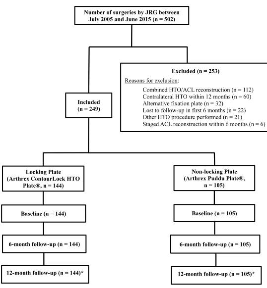
3 Results ... 45
3.1 Patient Flow ... 45
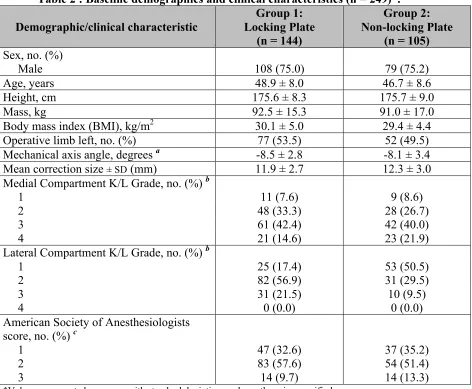
3.2 Demographics and Clinical Characteristics ... 47
3.3 Surgical Characteristics ... 49
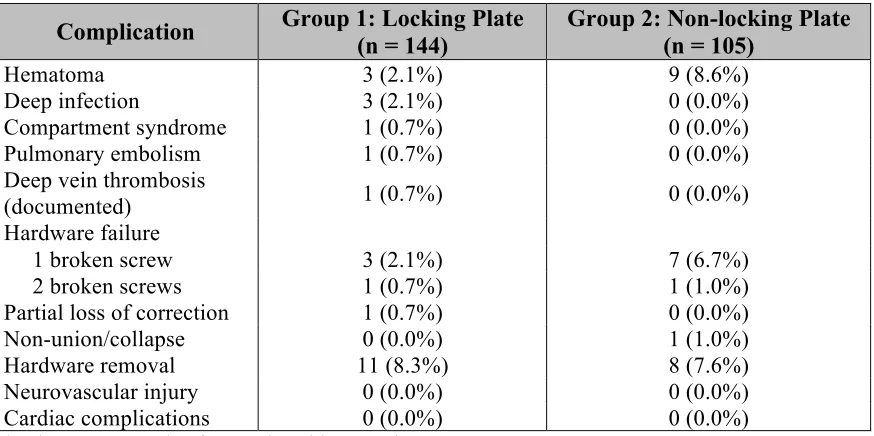
3.4 Surgical and Post-operative Complications ... 50
3.5 Outcome Measures ... 51
3.5.1 Objective 1 ... 51
3.5.2 Objective 2 ... 51
3.5.3 Objective 3 ... 55
3.5.3.1 ICER ... 55
3.5.3.2 Net Benefit Regression ... 55
3.5.3.3 Sensitivity Analysis ... 59
Chapter 4 ... 63
4 Discussion ... 63
4.1 Strengths & Limitations ... 68
Chapter 5 ... 72
5 Conclusion ... 72
vii
Appendices ... 95
viii
Table 1: Ideal patient criteria for a medial opening wedge HTO. ... 22
Table 2 : Baseline demographics and clinical characteristics (n = 249). ... 48
Table 3: Adverse event rates within first 12 months after HTO (n = 249) ... 50
Table 4: Time to return to weight-bearing and patient reported outcome measures (KOOS) for all patients (n = 244). ... 52
Table 5: Cost and effect outcomes. ... 57
Table 6: Net benefit regression results. ... 57
Table 7: Sensitivity analyses cost and effect outcomes. ... 60
ix
List of Figures
Figure 1: Schematic representation of the interacting systemic (biochemical) and local (biomechanical) risk factors that are associated with the development of knee OA and progression of the disease. Also presented are the radiographic and clinical criteria used to
diagnose knee OA according to Kellgren and Lawrence, 1957 and Altman et al., 1986. ... 11
Figure 2: A) The mechanical axis angle (MAA) of the lower limb is measured as the angle
between the line connecting the center of the hip and knee joints and the line connecting the
center of the knee and ankle joints. B) The weight-bearing line (WBL) is drawn from the center of the hip joint to the center of the ankle joint. ... 13
Figure 3: The “vicious cycle” of medial compartment knee osteoarthritis. Varus alignment leads to excess loading on the medial compartment, promotes articular cartilage breakdown,
narrowing of medial joint space and further malalignment of the joint. ... 15
Figure 4: The external knee adduction moment (KAM) about the knee is the product of the perpendicular distance between the knee joint center and the ground reaction force (GRF) vector in the front plane, forming the lever arm, and the magnitude of the GRF vector. Figure
adapted from Perry J Gait Analysis: Normal and Pathological Function 1992108. ... 17
Figure 5: Patient radiographs (A) before and (B) 12 months after HTO surgery. The yellow lines provide an estimate of the weight-bearing line (WBL) to display the shift to a more
neutral position. ... 21
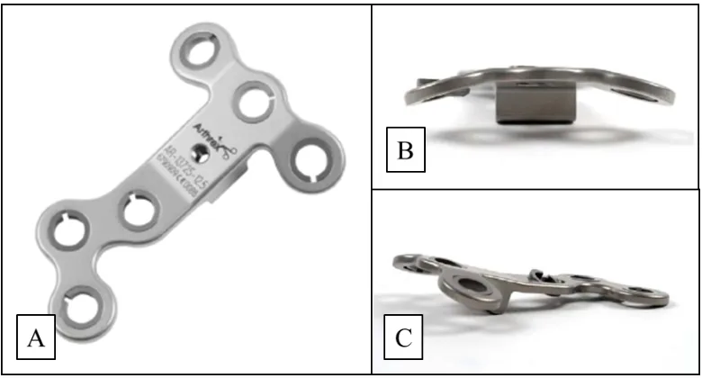
Figure 6: A) The Arthrex ContourLock HTO Plate® design (Arthrex, Naples, FL) possesses
a wide frame which lengthens the distance between fixation screws and provides additional stability. B) & C) Top and side views of the Arthrex ContourLock HTO Plate® illustrate the anatomically curved body of the implant. ... 29
Figure 7: Participant flow through the study. Asterisk represents a smaller sample size for the
cost-effectiveness analysis from the societal perspective (n = 106 locking plate, n = 58
x
use (in weeks with 95% confidence intervals) for patients undergoing a medial opening wedge high tibial osteotomy with a locking or non-locking internal fixation plate. Stars
indicate a significant difference. ... 53
Figure 9: Mean Knee injury and Osteoarthritis Outcome Score (KOOS) total and subdomain change scores from baseline to six months postoperative (with 95% confidence intervals) for patients undergoing a medial opening wedge high tibial osteotomy with an locking or
non-locking internal fixation plate. ... 54
Figure 10: Cost-effectiveness acceptability curves (CEAC) displaying the probability that the locking plate is cost-effective compared to the non-locking plate from A) the healthcare payer’s perspective and B) the societal perspective, over a range of willingness to pay values for an additional one-point improvement in the Knee injury and Osteoarthritis Outcome
Score total change score (baseline to 12 months). ... 58
Figure 11: Sensitivity analysis cost-effectiveness acceptability curves (CEAC) displaying the probability that the locking plate is cost-effective compared to the non-locking plate from the societal perspective over a range of willingness to pay values for an additional one-point improvement in the Knee injury and Osteoarthritis Outcome Score total change score (baseline to 12 months). A) Adjusting the dollar value for retirement or home making time,
xi
xii
List of Abbreviations
ADL Activities of Daily Living
ACL Anterior Cruciate Ligament
AP Anteroposterior
ASA American Society of Anesthesiologists
BMI Body Mass Index
CEAC Cost-effectiveness Acceptability Curve
CI Confidence Interval
HTO High Tibial Osteotomy
ICER Incremental Cost-effectiveness Ratio
INB Incremental Net Benefit
KL Kellgren-Lawrence (grading of OA severity)
KOOS Knee injury and Osteoarthritis Outcome Score
MAA Mechanical Axis Angle
NBR Net Benefit Regression
OA Osteoarthritis
PCL Posterior Cruciate Ligament
SD Standard Deviation
TKA Total Knee Arthroplasty
UKA Unicompartmental Knee Arthroplasty
WBL Weight-bearing Line
Chapter 1
1
Introduction
1.1
Background & Rationale
Osteoarthritis (OA) is a common degenerative health condition and a leading cause of
pain, disability and reduced quality of life in adult populations worldwide1. As a chronic
condition, the symptoms of the disease can persist for decades, resulting in substantial economic burden to healthcare systems. In Canada, arthritis accounted for $6.4 billion of direct and indirect healthcare costs in the year 2000, OA being responsible for the
majority of these costs2. These costs are expected to continue to grow with the aging
population and the rising rate of obesity3. Therefore, the identification of cost-effective
treatments for OA is of utmost concern for public health strategists2.
Osteoarthritis commonly involves the knee, with an estimated 250 million people
currently affected globally4. Although there is no known cure for knee OA, there are
several identified risk factors for knee OA progression that form the targets of various
interventions5–8. Varus alignment of the lower limb is a particularly strong risk factor for
the progression of knee OA due to its effect on loading the medial tibiofemoral
compartment9–13. Surgical and non-surgical treatments aimed at altering loads on the
medial tibiofemoral compartment are therefore common14–17.
Medial opening wedge high tibial osteotomy (HTO) is a surgical realignment procedure for patients with varus malalignment and OA of the medial compartment of the
tibiofemoral joint15,18,19. The goals of HTO are to correct lower limb malalignment,
redistribute loads laterally across the knee to lessen the compressive force on the diseased medial compartment, and thereby improve pain and function. The medial opening wedge technique requires cutting into the medial proximal tibia, wedging the bone open to a predetermined correction size to correct the malalignment, and securing the osteotomy
Adequate fixation of the osteotomy, typically achieved using an internal fixation plate, is
vital for bone healing and recovery during rehabilitation22,23, that involves progressive
bearing using ambulatory aids (i.e. crutches, canes, etc.). The patient’s weight-bearing status is progressed based on postoperative assessments of radiographic bone healing and pain. The suggested duration of return to full weight-bearing after medial opening wedge HTO ranges from 2 to 12 weeks, and highly depends on the type of
fixation used24–30.
HTO fixation plates are similar to those used for fracture fixation and can generally be categorized as non-locking and locking. The mechanical principles are quite different for non-locking and locking plates, providing distinct mechanical environments for bone healing. Non-locking plates rely on plate compression and high friction at the
bone-plate interface to provide fracture site stability31. At higher loads, however, non-locking
screws that are drilled into the bone can begin to loosen. This reduces bone-plate friction, may render the plate unstable and increases the risk of complications such as hardware
failure, delayed union, non-union and loss of correction32. Locking plate designs address
mechanical issues with threaded fixed-angle screws or interference washers that control the axial rotation between the screw and the plate, and eliminate screw-plate-bone
motion33. The mechanism does not rely on high friction at the bone-plate interface, but
rather maintains stability at the angular-stable screw-plate interface31. Locking plates also
convert shear stresses to compressive stresses, improving fixation since bone has a stronger resistance to compressive stress compared to shear. The mechanical advantages of locking plates provide stronger implant stability and resistance to higher load-bearing, and are therefore suggested to be advantageous for healing after medial opening wedge HTO23,34.
In vitro biomechanical studies have suggest that locking plates do provide greater
mechanical stability in response to compression and torsion35,36. Clinical studies suggest
locking plates maintain the osteotomy correction size better than non-locking plates37,38,
achieve bone healing37 and earlier return to full weight-bearing after surgery37,39,40. Although results are mixed, some studies also suggest that the rates of delayed and
non-union22,38, loss of correction and hardware failure have decreased since the introduction
of locking plates 41.
The Arthrex ContourLock HTO Plate® (ContourLock) is a relatively new locking plate designed to enable a precise fit proximally and distally on the tibia. The locking plate is proposed to provide advantages when compared to the commonly used non-locking Arthrex Puddu Plate® (Puddu). In-vitro biomechanical studies suggest the locking plate provides greater stability under high physiological stress loading and cyclical testing when compared to other locking and non-locking plates due to the wider distance
between its fixed-angle screws42,43. Although the greater stability is proposed to permit
faster recovery after surgery, there is currently no study evaluating clinical outcomes after HTO using the plate.
The cost associated with using different HTO fixation plates is another important consideration. Costs of HTO include direct (healthcare resources consumed and out-of-pocket expenses) and indirect (time and productivity losses) costs. If locking plates can limit the number of postoperative complications that require revision surgery (i.e. non-union), locking plates could provide direct cost savings. Additionally, if locking plates enable quicker return to weight-bearing, patients could also return to work sooner, thus financially benefiting society with indirect cost savings from productivity. Alternatively, the cost of locking plates are substantially greater than non-locking plates because of their more complex design and number of screws (typically six or more, compared to four) used to achieve fixation. If clinical results are similar regardless of plate design then the extra costs of locking plates may not be warranted. Furthermore, the relative
bulkiness of locking plates may cause irritation to the patients and require the plate to be
surgically removed40,44 which can increase direct costs associated with the procedure.
1.2
Study Objectives and Hypotheses
1. To compare the time to return to full weight-bearing following medial opening
wedge HTO in patients receiving a locking versus non-locking fixation plate. Hypothesis: Patients receiving the locking plate will return to full weight-bearing sooner postoperatively.
2. To compare the change in patient-reported outcomes (Knee injury and
Osteoarthritis Outcome Score – KOOS) following a medial opening wedge HTO in patients receiving a locking versus non-locking fixation plate.
Hypothesis: Patients receiving the locking plate will experience greater
improvements in patient-reported outcomes from baseline to 6 months after the surgery.
3. To estimate the cost-effectiveness of a locking plate compared to a non-locking
plate, from the healthcare payer (Ministry of Health) and societal perspectives, using change in KOOS total score at 12 months postoperative as the measure of effectiveness.
1.3
Review of Literature
1.3.1 Osteoarthritis
Osteoarthritis (OA) is the most common form of arthritis and is rapidly becoming one of
the most disabling health conditions worldwide4,45–47. It is a chronic musculoskeletal
disease that can affect single or multiple joints, and is characterized by localized joint
pain, functional limitations and diminished quality of life1,48. In Canada, there are more
than 4.4 million people (1 in 8) living with OA and this number is projected to double by
the year of 2040 due to the aging population and the obesity epidemic49.
1.3.1.1
Epidemiology
According to the 2013 Global Burden of Disease (GBD) study, the prevalence of OA has grown 72% from the year 1990 with approximately 240 million people burdened by the
disease worldwide 4. The chronic nature of OA also makes it one of the fastest growing
health conditions in terms of disability46. It accounted for more than 17 million years
living with disability (YLDs) globally in 2010, a 64% increase from the year of 199050.
The World Health Organization (WHO) projects OA to become the fourth leading cause
of disability worldwide by the year 202051.
The disease is not isolated to a specific population group, but affects people of various
ethnic backgrounds and in different geographical locations worldwide52. Although OA
can be seen in people as young as 15 years of age, the majority of people affected by the
disease are older individuals3,53 with women being affected approximately twice as often
1.3.2 Knee Osteoarthritis
OA develops more frequently in the knee than any other weight-bearing joint in the body54–56 with over 10% of the adult population currently affected by symptomatic knee
OA49. Due to the high loading demands on the joint, knee OA specifically is considered
one of the leading causes of physical disability worldwide57,58. The lifetime risk of
developing knee OA is estimated to be 45% (47% in women, 40% in men) with increased odds seen in those who possess predisposing risk factors for the disease such as obesity
and malalignment, among others59.
1.3.2.1
Knee Anatomy
The knee is a complex synovial hinge joint between the patella, the distal femur and the
proximal tibia60. The bone surfaces are lined with hyaline (articular) cartilage which aids
in dissipating forces within the joint and limiting friction between bones. The cartilage tissue is not innervated by pain receptors, nor is it well vascularized which limits the
tissue’s ability to repair itself61. Aside from the surfaces concealed by articular cartilage,
1.3.2.2
Pathophysiology
Knee OA is a degenerative condition that affects the various tissues of the joint and is
considered a whole-joint disease62. It is characterized by the disruption of the natural
cartilage remodeling process, ultimately leading to the fibrillation and softening of the
articular cartilage and intensified degeneration of the tissue63. As cartilage continues to
breakdown, subchondral bone becomes exposed within the joint resulting in bone-on-bone articulation. Continuous friction between exposed bone-on-bone leads to the development of osteophytes and subchondral cysts at the articulating bone extremities due to excessive
bone remodeling48,64. In later stages of the disease, the subchondral bone tissue will begin
to thicken and become sclerotic. Additionally, it is common to see inflammation of the
synovial lining of the joint46 as well of the overproduction of several proteolytic enzymes
and cytokines that have been shown to promote cartilage degradation and breakdown of the extracellular matrix of the joint. These intra-articular changes and inflammatory responses ultimately lead to loss of joint space, destabilization of the joint, abnormal joint loading and a number of clinical symptoms for the affected individual.
1.3.2.3
Etiology
Similar to OA in other joints of the body, knee OA is a complex condition. The initial onset of the disease can be idiopathic in nature, developing naturally over time as a result of various interacting risk factors (known as primary OA), or can develop following excessive or repetitive trauma to the knee joint such as ligament tears, cartilage impact,
etc. (known as secondary OA)47. There are still quite a few uncertainties that surround the
etiology of the disease. Knee OA is unpredictable in its method of initiation and its progression, with some individuals exhibiting mild degeneration of the joint sustained over an extended period of time, while others progress in disease severity very rapidly. The medial compartment is the most commonly affected area of the joint when compared
to other compartments of the knee8,65–67. The disease does not affect the entire joint
1.3.2.4
Risk Factors for Knee OA
As a disease whose onset and progression is quite variable, it is important to better understand the multiple risk factors that predispose individuals to developing knee OA and accelerate the progression of the disease. These risk factors can act systemically or can be considered local by acting directly on the joint itself (Figure 1).
Systemic risk factors have a biochemical influence on the knee joint. These factors can cause direct damage to the joint tissues or limit the tissue’s ability to repair itself after being damaged, both of which make the affected individual susceptible to further injury. Systemic risk factors that have been associated with development of knee OA and its
progression include age46,47, genetics46,47,62, gender6,46,62,68, overweight/obesity (BMI ≥
25)69,70, nutritional deficiencies71,72, inactivity72,73 and elevated bone mineral density74,75.
Local risk factors influence the joint mechanically. These factors are associated with exposure to joint injury or excessive joint loading that leads to degeneration of tissues. Local risk factors that have been associated with development of knee OA and its
progression include knee malalignment10,55,76,77, congenital deformities of the joint78,
previous injuries to the tissue components of the joint6,62,79,80, overweight/obesity (BMI ≥
25)81–83, occupation62,79,84, muscle weakness85,86, elevated peak knee adduction moment9– 11,87,88, elevated knee adduction impulse89, and varus thrust90.
Although risk factors have been shown to independently promote knee OA disease progression, the risks are intensified as individuals are exposed to multiple risk factors simultaneously. For example, mechanical varus alignment of the lower limb has been
shown to increase the risk of medial compartment OA progression by a fourfold10,76 as a
result of increased mechanical axial loading on the joint past a normal physiological range to maintain proper cartilage function. In overweight and obese individuals, the sheer excess weight increases this level of mechanical loading of the medial compartment leading to further articular cartilage breakdown. Various studies have suggested there is
Figure 1: Schematic representation of the interacting systemic (biochemical) and local (biomechanical) risk factors that are associated with the development of knee OA and progression of the disease. Also presented are the radiographic and clinical
1.3.2.4.1
Varus Alignment
Lower limb alignment is typically determined using full-limb standing hip-knee-ankle (HKA) anteroposterior (AP) radiographs. The gold standard measure of lower limb alignment is the mechanical axis angle (MAA), defined as the angle formed between the line connecting the femoral head center of the hip and the knee joint center and the line
connecting the knee joint center and the ankle joint center91,92 (Figure 2). It has been
Figure 2: A) The mechanical axis angle (MAA) of the lower limb is measured as the angle between the line connecting the center of the hip and knee joints and the line
connecting the center of the knee and ankle joints. B) The weight-bearing line
(WBL) is drawn from the center of the hip joint to the center of the ankle joint.
Based on the MAA measure, lower limb alignment can be assessed to identify patients with varus (“bow-legged”) or valgus (“knock-kneed”) alignment. Individuals with an inward angulation of the distal segment of the lower limb (tibia and fibula) and a negative MAA are considered varus aligned, while those with an outward angulation and a
positive MAA are considered valgus aligned. Many epidemiological studies suggest that
lower limb malalignment is an important risk factor for knee OA progression5,55,76,95,96
and that the direction of this malalignment will affect which compartment of the knee joint is most affected. Medial compartment knee OA is most often seen in individuals with varus alignment, whereas lateral compartment knee OA is more commonly seen in
individuals with valgus alignment55,76,97,98.
The role alignment plays in the degenerative process of medial knee OA is related to
increased loading of the knee joint10,96,99. The distribution of loading within the joint is
related to the lower limb weight-bearing line (WBL), a line drawn from the center of the femoral head to the center of the ankle (Figure 2). Individuals who are neutrally aligned will bear 75% of the overall knee load in the medial compartment while standing on one
leg91 with a WBL passing through the medial compartment of the joint. As alignment
steers away from neutral and the WBL is shifted medially, the load distribution within the knee joint will undergo aberrant changes. Individuals with varus alignment will
experience an increase in medial compartment loading9,91,100. This in turn, will lead to a
heightened degree of articular cartilage degeneration. In fact, a longitudinal study found that for every additional 1 degree of varus, patients will lose 17.7µl of femoral articular
cartilage on average annually, with similar losses seen in the tibial cartilage volume97.
Figure 3: The “vicious cycle” of medial compartment knee osteoarthritis. Varus alignment leads to excess loading on the medial compartment, promotes articular cartilage breakdown, narrowing of medial joint space and further malalignment of
the joint. Increased varus
alignment
Increased loading of medial compartment
Medial articular cartilage degeneration Medial joint space
1.3.2.4.2
Gait and Knee Osteoarthritis
For the general population, one of the most common daily activities performed is
walking, with thousands of steps taken per day101. Three-dimensional (3D) motion
analysis of gait kinematics and kinetics has been used extensively in the literature to better understand biomechanical factors associated with knee OA. Specifically, authors have investigated the role of the external knee adduction moment (KAM) (Figure 4) on
increased loading of the medial compartment10,102,103. During stance phase of gait,
individuals will generate a ground reaction force (GRF) vector that projects upwards and medial to the knee joint’s center of rotation. The perpendicular line that connects the GRF to the knee joint center is known as the lever arm and the product of this lever arm and the GRF vector generates what is known as the external KAM. The external KAM creates a torque force that causes the tibia to adduct in relation to the femur, which results in greater compressive loading to the medial compartment of the joint. As the GRF is projected more medially, the lever arm grows longer and thus, increases the magnitude of the external KAM suggesting that the increase in external KAM is also related to
alignment. Halder at al. suggest that the magnitude of medial compartment loading
increases 5% for every 1 degree increase in varus while walking104.
Several studies have shown that the external KAM is strongly associated with
characteristics of knee OA such as knee pain in previously asymptomatic knees88 and
measures of OA disease severity87,105. It has also been proven to be a reliable, valid and
clinically meaningful proxy measure of medial compartment loading during gait9,106,107
and more importantly, a predictor for OA disease progression10,89. Thus, treatment
Figure 4: The external knee adduction moment (KAM) about the knee is the product of the perpendicular distance between the knee joint center and the ground
reaction force (GRF) vector in the front plane, forming the lever arm, and the
magnitude of the GRF vector. Figure adapted from Perry J Gait Analysis: Normal
1.3.2.5
Diagnosis and Clinical Manifestations
Knee OA can develop in one or more of the three knee joint compartments: the medial tibiofemoral joint, the lateral tibiofemoral joint or the patellofemoral joint. Clinicians will use both clinical and radiographic assessment to diagnose patients with knee OA. Often,
clinical assessment follows the guidelines set by Altman et al.109, while radiographic
assessment follows the criteria set by Kellgren and Lawrence110 (Figure 1). According to
Altman et al., the required clinical criteria to diagnose a patient with knee OA includes knee pain, as well as one of the following; crepitus (popping sound/sensation or
cracking) of the joint, over 50 years of age, or morning stiffness that lasts no longer than 30 minutes. For radiographic assessment, Kellgren and Lawrence developed a four point joint degeneration rating scale (1 = mild OA, 4 = severe OA) to assess OA disease severity in the knee joint by evaluating the presence or absence of osteophytic bone, sclerosis of subchondral bone and whether marked joint space narrowing is present on anteroposterior radiographs. Lateral and skyline radiographic views can also be helpful in
confirming compartment disease severity111.
Patients diagnosed with knee OA can exhibit a number of clinical symptoms which include recurrent joint pain (frequently activity-induced and persistent), stiffness, and
swelling, reduced function, reduced range of motion, crepitus and deformity109,112. Often,
these symptoms will limit individuals and restrict participation in their daily activities113
1.3.3 Management of medial compartment knee OA
Currently, there is no established cure for OA. However, a number of treatment modalities are available to aid patients in the management of associated pain and symptoms. Clinical guidelines outline non-surgical and surgical treatment interventions
for patients with symptomatic knee OA1,114. Available non-surgical interventions include
physiotherapy, pharmacotherapy, foot orthoses, bracing, lifestyle modifications and activity management. These modalities typically target symptom management, but do not alter joint anatomy and benefits are not considered to be permanent. Surgical
interventions include high tibial osteotomy (HTO), unicompartmental knee arthroplasty (UKA) and total knee arthroplasty (TKA). These modalities physically alter joint bony structures with permanent anatomical changes which modify risk factors that promote knee OA disease progression such as knee malalignment. Ultimately, selection of management method is decided upon mutually between the clinician and the patient based on the patient’s personal characteristics, physical functional limitations, symptom severity and the current level of disease severity.
For patients in earlier stages of the disease, clinicians typically attempt non-surgical interventions before opting for surgery. However, patients who are at more progressed stages of the disease often display substantial mobility restrictions, severe pain, decreases in quality of life and severe degenerative changes in a single or multiple compartments of the knee warranting a referral to an orthopaedic surgeon. The surgeon may recommend a UKA or a TKA to replace the articular components of the joint or the surgeon may recommend a HTO to correct malalignment of the lower limb, a risk factor for progression of OA, while preserving the components of the joint. All three surgical procedures have shown evidence of long-term benefits for pain management, improved quality of life and mobility, but differ in terms of recovery time, invasiveness, potential adverse events, limitations in activity participation following surgery and costs associated
with the procedures115–119. Thus, selection of the appropriate surgical intervention must
1.3.4 Medial Opening Wedge High Tibial Osteotomy
Medial opening-wedge high tibial osteotomy is a surgical treatment for patients with
varus alignment of the lower limb and medial compartment knee OA15,19,20,120. The
procedure corrects knee malalignment by shifting the weight-bearing load of the joint laterally to a more neutral position (usually slight valgus) and away from the affected
portion of the knee (Figure 5) 121. The redistribution of load decreases the magnitude of
both static (standing) and dynamic (during walking) loading in the medial compartment with the goal of relieving patient symptoms and slowing the progression of the
Figure 5: Patient radiographs (A) before and (B) 12 months after HTO surgery. The yellow lines provide an estimate of the weight-bearing line (WBL) to display the
shift to a more neutral position.
1.3.4.1
Preoperative Assessment
Similar to consideration in other surgical procedures, surgeons take a thorough patient history that identifies previous lower limb injuries or comorbidities that would
contraindicate the patient from undergoing surgery and help ensure patient compliance during the post-operative period. A physical examination is then conducted to identify ligamentous instabilities of the joint and to provide additional information to aid the surgeon in establishing an appropriate treatment plan. Previous authors suggest the surgery is ideally performed on healthy young, active patients, where the level of joint degeneration is isolated to the medial compartment with associated varus alignment as
determined by the mechanical axis angle19,111,121. It is usually recommended to patients
whose activities of daily living are typically more physically demanding. Inactive patients with tricompartmental disease, have complaints of rest/ night pain and those who are
above 60 years of age may be better suited for a TKA121. Appropriate patient selection is
considered crucial to maximize the likelihood that the procedure will be successful120.
Ideal patient criteria for medial opening wedge HTO is presented in Table 1.
Table 1: Ideal patient criteria for a medial opening wedge HTO. Varus deformity of the lower limb
Pronounced degeneration in the medial compartment of the tibiofemoral joint
Moderate to high activity levels Younger than 60 years of age A certain degree of pain tolerance
Radiographic assessment is considered an important component for preoperative
planning20,120 with full-limb standing anteroposterior (AP) radiographs111,123. Radiographs
are used to determine the patient’s MAA and anatomical axes of the femur and tibia, as well as identifying the degree of arthritic joint degeneration in both the medial and lateral compartments of the joint. Ultimately, the AP radiographs are used to calculate the suggested degree of surgical correction for the procedure. Using the technique described by Dudgale et al., the desired correction has the WBL shifting laterally to a maximum position of 62.5% of the medial-to-lateral tibial plateau width, otherwise known as the
“Fujisawa point” 124.
1.3.4.2
Surgical Procedure
The classic technique for a medial opening wedge HTO has previously been described by
Amendola and Fowler20,120. First, a guide pin is drilled medially into the proximal tibia at
an angle approximately 3cm below the medial joint line. An oscillating saw is then used to make surgical cuts medially, anteriorly and posteriorly into the proximal tibia where both flexible and rigid osteotomes are used to complete the osteotomy and open the wedge to a predetermined correction size. Once achieved, the proximal and distal portions of the bone are fixed with an internal fixation plate using both cancellous and cortical screws. Bone graft or a synthetic substitute is typically used to fill in the wedge
space for corrections larger than 7.5mm to assist with the bone healing process20,125. The
surgery is done under fluoroscopic control to ensure that the desired correction is
accurately achieved and to avoid breaching the lateral tibial cortex when making the cut.
Often, surgeons will also perform knee arthroscopy preceding the HTO to investigate OA severity in both the medial and lateral compartments of the tibiofemoral joint. The degree of degeneration in the lateral compartment is important to consider as shifting the loading from the affected medial compartment to a lateral compartment that is equally as
must decide whether proceeding with the HTO is in the best interest for the patient. Arthroscopy also allows a full visual inspection of the joint entirely to examine the overall knee condition and to surgically treat unstable chondral or meniscal tissue if the
surgeon feels that it is indicated120.
Following surgery, patients are monitored during their rehabilitation and given early joint exercises. These exercises are geared towards retraining patient gait, improving their range of motion, managing pain and improving overall function. Patients undergo a progressive returning to weight-bearing protocol based on evidence of radiographic bone healing and subsidized pain.
1.3.4.3
Benefits of Medial Opening Wedge HTO
The most common surgical methods of HTO reported in the literature are the medial opening wedge HTO and the lateral closing wedge HTO. Between these techniques,
medial opening wedge HTO has grown in popularity over the last few years126. The
method easily allows simultaneous bi-planar correction of the frontal and sagittal planes to correct limb alignment. The ability to increase the wedge opening gradually to the desired correction also allows for a more precise adjustment in both the frontal and sagittal planes and makes it easier to achieve smaller corrections (< 5 degrees) than in a
lateral closing wedge HTO20. Moreover, the lateral closing wedge HTO requires two cuts
to be made in the bone which can make it difficult to achieve the proper correction size
1.3.4.4
Medial Opening Wedge HTO Success
Many studies have shown that medial opening wedge HTO is beneficial to the patient both clinically and biomechanically. Measures of pain, symptoms, function and quality of life have all been shown to improve significantly at one to five year follow-up
assessments after the procedure27,28,122,128–130. It has also been shown to reduce the level
of loading on the medial compartment of the knee joint by significantly decreasing the
degree of malalignment122,131 and reducing the external knee adduction moment during
ambulation28,95,122,128,131 as well as other relevant kinematic and kinetic measures such as
varus thrust90 that promote disease progression. Overall, medial opening wedge HTO is
suggested to be a successful procedure with survival rates reported as high as 98% after
five years132, 90% after ten years133 and 71% after 15 years132 following the HTO.
Despite the many benefits of HTO, there are a number of complications associated with the surgery. Reports of surgical complications vary between authors, ranging from 1% to
45% of cases24,126,134–139. The most frequently reported are lateral cortex hinge fractures,
hardware failure often resulting in loss of correction, delayed and non-union of the bone (insufficient healing of the fracture site after a given time lapse) and hardware failure. However, many authors suggest that the rate of complication is dependent on the type of
internal fixation used for the procedure37,38,126,134,140,141.
1.3.4.5
Weight-bearing and Return to Work after HTO
Postoperative care following medial opening wedge HTO typically involves a 2 week period of toe-touch or feather weight -bearing with limb stabilization from a tracker brace, followed by a progressive increase in weight-bearing to the surgeon’s discretion based on radiographic healing of the bone and knee pain. The typical progression would have patients graduate to toe-touch or feather-touch weight-bearing, followed by
The length of time for protected weight-bearing ranges between two and 12 weeks24–30,142
.
A return to full weight-bearing without gait aid (i.e. crutches, cane, walker, etc.) after
HTO surgery is dependent on the ability of the bone to consolidate enough to safely bear
weight on the limb. This is a major concern for surgeons, as allowing patients to early
weight-bear after HTO has the potential to increase complication rates if the plate does
not provide enough stability. As a result, studies have shown that the return to
weight-bearing process can be related to the type of fixation hardware used for the HTO37.
Optimized stable implant designs are therefore essential to warrant a safe earlier return to
weight-bear.
Another important factor to consider associated with the time to return to weight-bearing is the time to return to work following the HTO. Time lost from employment and leisure
accounts for an estimated 80% of the overall annual costs for OA in Canada143. It is
important for healthcare providers to target OA treatment interventions that minimize these productivity losses to society and help reduce the overall OA burden worldwide. Previous studies have reported that the time to return to work following medial opening
wedge HTO ranges between three to six months142,144–146. It is important that more stable
plate designs are developed to allow patients to return to weight-bearing earlier, translating to a faster return to work which will benefit society as a whole.
1.3.4.6
Locking Plate vs. Non-Locking Plate Designs
Early studies suggest that an optimal balance between micro-motion and implant stability
is needed to promote osteotomy healing147,148 by ensuring that the plate is not too stiff
(suppresses micro-motion and healing149
) but is stable enough to evade non-union of the
osteotomy site. Over the years, technological advancements have allowed manufacturers
to design fixation plates that provide the required components to optimize HTO success.
Fixation plates used in HTO can generally be divided into non-locking and locking plate
osteotomy with resistance to various types of loading33
. The force generated from the
axial load is countered by the normal force of the plate (i.e. the product of the friction
force between the plate and bone, and the force generated by screw torque) which forms a
shear stress at the bone-plate interface. As the screw torque or the friction coefficient
decreases however, bone-plate motion increases. Excessive motion results in mechanical
environment that discourages primary or secondary bone healing. Furthermore, frictional
forces that are overcome by axial loading rely on the axial stiffness of the screw most
distal to the plate to maintain stability. The lack of axial control in non-locking screws
forces it to be maintained by the bone at the bone-plate interface. Here, the bone is the
load-determining factor in maintaining stability under compressive loads. The high
mechanical shear stresses generated therefore leaves the bone vulnerable to failure under
compressive load or susceptible to absorption of the bone that results in screw loosening.
Since, locking plate designs have been developed to address the mechanical pitfalls of
conventional non-locking plates. Locking plates control the axial orientation of the
screws to the plate, which improves bone-plate-screw stability33
. Locking screw-plate
constructs act as fixed-angle devices that provide stability maintenance without relying
on bone-plate friction and can convert shear stresses to compressive stresses when subject
to loading. This improves the stability of the fixation as bone has a high resistance to
compressive stress and a low tolerance for shear stress. The strength of the fixation also
combines the strength of all bone-screw interfaces, as opposed to relying on the axial
stiffness of a single screw (i.e. in non-locking plates), which further increases the stability
of the implant150
. Additionally, threaded locking screws or interference washers provide
angular and axial stability that optimize the rigidity of the fixation and optimized strain
under loading conditions. The latter provides a favourable biological environment for
secondary bone healing with callus formation, important for fractures located in the
metaphysis (Schutz, 2003) such as the case in the proximal tibia after HTO. The lack of
frictional forces between the bone and plate also allows blood supply under the plate to
be preserved and is suggested to promote faster bone healing151
Although all fixation plate designs have the same goal of maintaining correction size and
promoting bone healing, the mechanical and biological benefits that locking plates
provide are suggested to optimize rehabilitation outcomes after HTO surgery 27,127,152
.
Many studies have evaluated different plate designs through biomechanics and clinical
outcomes23,29,34–38,42,43,140,141,153,154. General conclusions suggest that locking plates provide
better stability for patients with a higher resistance to mechanical stresses35 and optimized
micro-motion at the osteotomy site23. Locking plates are also suggested to allow patients
to return to full weight-bearing and achieve consolidation of the osteotomy faster37,
improve clinical outcomes faster37,141, and reduce the cases of delayed and non-union22,38,
hardware failure41, loss of correction41,155 and post-surgical lateral cortex fractures38 than
using a non-locking plate.
1.3.5 The Arthrex ContourLock HTO Plate®
The Arthrex ContourLock HTO Plate® is a new titanium fixation device designed with a
locking construct (Figure 6), an anatomically curved body, a wider frame than previously
introduced locking plates and screws that diverge proximally. To date, no studies have
compared clinical outcomes between the Arthrex ContourLock HTO Plate® and other
more conventionally used plate designs. However, a few studies have examined the
biomechanical differences between the Arthrex ContourLock HTO Plate® and other
Figure 6: A) The Arthrex ContourLock HTO Plate® design (Arthrex, Naples, FL) possesses a wide frame which lengthens the distance between fixation screws and
provides additional stability. B) & C) Top and side views of the Arthrex
Studies have shown that the Arthrex ContourLock HTO Plate® provides great stability
under static loading, similar to other implant designs (Tomofix sm, Tomofix std,
iBalance, Peek Power), while it provides superior stability under dynamic cyclic loading
conditions, likely as a result of a wider frame and larger distance between fixation
screws42,43
. Although these studies suggest that the maximum forces at moment of failure
are considered too low to warrant full dynamic loading (full weight-bearing) immediately
after the surgery for all the plate designs studied, the maximum force at failure for the
Arthrex ContourLock HTO Plate® is almost twice as high as other designs, suggesting
that full dynamic loading may be achievable for patients much earlier and would require
less healing of the osteotomy site to safely begin weight-bearing. Furthermore, a study
using finite element modeling showed that at higher compressive loadings, the Arthrex
ContourLock HTO Plate® experiences low hardware stresses and small wedge
micromotion which can be beneficial for fracture site healing156
.
Although there is currently only a small body of evidence to support the use of the
Arthrex ContourLock HTO Plate® design, the aforementioned biomechanical studies
suggest that the Arthrex ContourLock HTO Plate® is a great implant choice for patients
who require a strong, stable locking construct for an early return to weight-bearing
following the HTO surgery. A faster return to full weight-bearing could translate to faster
improvements in clinical outcomes relating to pain, symptoms and quality of life for the
patient, as well as faster returns to daily activity and sport. In turn, patients could return
to work much earlier, which would provide socioeconomically benefit by reducing losses
1.3.6 Health Economics
Healthcare costs are rising at an alarming rate. The current economic climate requires a high level of accountability from budgetary decision-makers for expended healthcare
dollars157. As a result, decision-makers are seeking treatment interventions that provide
the best quality of care for patients while minimizing dollars spent for the intervention. Programs are now requesting evidence-based research to support the economic efficiency
of treatments to better judge the value for their money158.
1.3.6.1 Economic Evaluation
Economic evaluation provides a framework to compare clinical and cost data
simultaneously between competing interventions to assess value for money159. In Canada,
along with many other countries, economic analyses are a requirement for manufacturers wishing to make their products available as treatment options with Ontario’s Health
Insurance Plan160. They are also a useful evaluative tool for decision-makers operating on
a given budget in order to make choices concerning the deployment of finances for maximum health benefit.
1.3.6.2
Economic Burden of Osteoarthritis
Symptoms of OA typically do not resolve, and are associated with chronic pain that can persist for decades, resulting in a substantial number of health-care visits over a lifetime, which poses a large economic burden on healthcare systems. In industrialized countries such as Canada, the US, UK, Australia and France, OA accounts for anywhere between 1
and 2.5% of the country’s gross national product161. In Canada alone, there is an average
annual cost of $12,200 ($CAN) per patient with OA143 with the annual economic burden
of OA estimated to increase to $405 billion dollars by 202049. As the burden of OA
1.3.6.2.1
Cost-Effectiveness of Medial Opening Wedge HTO
Medial opening wedge HTO is a procedure that has been shown to benefit patients with varus alignment and medial compartment knee OA both clinically and biomechanically however, economic evaluation of the HTO procedure is an evolving area of research. Studies are beginning to examine the economic impact of medial opening wedge HTO in comparison to alternative treatment methods.
The first study to evaluate the utility of HTO concluded that UKA is a more
cost-effective treatment method for medial compartment knee OA than HTO162 (Brown,
2010). A study that soon followed found results favoring the KineSpring® Knee Implant
System (an implantable load absorber)163. However, the authors from this study claim to
report an ICER when the values reported are in fact average cost-effectiveness ratios
(ACERs). This incorrect use of terminology can lead to misinterpretation of results164 and
inaccurate conclusions.
More recently, two studies have investigated the cost-utility of HTO compared to both UKA and total knee arthroplasty (TKA) for younger patients with medial compartment
knee OA118,119. According to the results of these studies, HTO is the most cost-effective
treatment for patients below 60 years of age and the authors strongly support the use of HTO as a first line treatment method for this younger patient population.
The aforementioned studies provide some evidence to suggest that HTO is a cost-effective treatment intervention for patients who are varus aligned and with medial compartment knee OA. However, no studies have been conducted to compare economic impact of using different internal hardware devices when performing a medial opening wedge HTO surgery. Locking plates have been suggested to provide functional and patient-important benefits when compared to non-locking plates for medial opening
wedge HTO35,36,43 and are thought to reduce the number of post-operative complications
return to full weight-bearing sooner, and therefore productivity losses may be lessened by allowing patients to return to work sooner. In both cases, costs associated with the
surgery can be minimized. However, locking plates and screws are generally more expensive than non-locking designs, and the bulkiness of locking plates can be irritating
to the patient requiring surgical plate removal29,40,44 further increasing the costs
Chapter 2
2
Methods
2.1
Study Design
We conducted a retrospective analysis using prospectively collected data from patients who had undergone medial opening wedge HTO at the Fowler Kennedy Sport Medicine Clinic between July 2005 and June 2015, performed by one fellowship-trained
orthopaedic surgeon (JRG). All surgeries were completed using either a locking (Arthrex ContourLock HTO Plate®) or a non-locking (Arthrex Puddu Plate®) internal fixation plate. Patients at earlier time points of the study received the non-locking plate.
Availability of the locking plate in 2009 resulted in a shift in clinical practice where most patients received the locking plate. The study was approved by the University of Western Ontario’s Research Ethics Board for Health Sciences Research Involving Human
Subjects. All patients had provided informed consent prior to study enrollment to have their data entered into a research database.
2.2
Eligibility Criteria
We included patients who underwent a medial opening wedge HTO for mechanical varus alignment and had been diagnosed with knee OA according to the American College of
Rheumatology classification criteria109 affecting primarily the medial compartment of the
tibiofemoral joint. We did not exclude patients with evidence of lateral compartment knee OA as long as the patient’s symptoms and radiographic severity of OA was more pronounced in the medial compartment. We excluded patients who had a combined HTO and anterior cruciate ligament (ACL) reconstruction, as well as those who received a bilateral HTO. We also excluded patients that underwent a revision ACL reconstruction surgery on the same limb or an HTO on the contralateral limb within 12 months
2.3
Intervention
2.3.1 Operative Procedure
Preoperative hip-knee-ankle full-limb standing anteroposterior radiographic views were used to calculate the desired correction size for the osteotomy using the method described
by Dugdale et al.124 This technique suggests a shift in the weight-bearing line to 62.5% of
the medial-to-lateral tibial plateau width. Other considerations for preoperative
templating were the condition of the articular cartilage in the lateral compartment and the degree of correction required to achieve neutral alignment.
The HTO was performed using a medial opening wedge technique similar to the
procedure described by Fowler et al.20 Fluoroscopy was used to insert a guide pin and
osteotomes, both flexible and rigid, were used to perform the osteotomy. Once the tibia was opened to the desired width, fluoroscopy was again used to confirm correction size and limb alignment. If necessary, adjustments were made to the posterior tibial slope to provide address sagittal instability. One of two plate designs was used as an internal implant: a 4-hole Arthrex Puddu Plate® non-locking plate (Arthrex, Naples, FL, USA) or an Arthrex ContourLock HTO Plate® locking plate (Arthrex, Naples, FL, USA). Cortical and cancellous bone screws were used to fixate the osteotomy both proximally and distally and confirmed using fluoroscopy. For corrections larger than 7.5mm, cancellous bone allograft was used to fill in the osteotomy gap.
2.3.2 Post-operative Care
Following surgery, the operative limb was placed in a hinged knee brace. At this time, patients were instructed to feather-touch weight-bear (WB) with the assistance of
progress the weight-bearing status of the patient during rehabilition was decided by the surgeon using radiographic evidence of osteotomy healing (i.e. the extent of
consolidation on x-ray), perceived stability of the fixation device and the level of pain or discomfort reported by the patient during ambulation.
Patients were also given a progressive rehabilitation protocol to allow them to
re-establish full range of motion, strength and function, in addition to reducing swelling, and avoiding joint contracture and muscle atrophy from disuse. Patients began this program at three weeks postoperative with lighter exercises and progressed in exercise difficulty until they exhibited a normal gait pattern at the discretion of the physiotherapist. All patients followed the same rehabilitation protocol with slight modifications if deemed necessary.
All patients returned to clinic for a follow-up visit with the surgeon at two and six weeks and three, six and 12 months after surgery. Patients who experienced intraoperative or post-operative complications (i.e. infection, delayed union, etc.) were reviewed as needed.
2.4
Radiographic
Assessment
A full-limb standing digital radiograph of the lower limb was obtained for each patient at baseline, three, six and 12 months following surgery. Patients stood with patellae
centered over their femoral condyles with feet pointed straight ahead. The position controls for effects of foot rotation on alignment measures that could result in inaccurate
frontal plane images165. Additional imaging was taken for patients who displayed delayed
bone healing and/or suspected complication to monitor consolidation more closely.
Baseline radiographs were assessed using a customized computer software program
(HTO Pro; Wolf Orthopaedic Biomechanics Laboratory, London, Ontario, Canada)93.
angle (MAA) and to assess OA severity in both the medial and lateral tibiofemoral compartments. The MAA was defined as the angle generated between the mechanical
axis of the femur and the mechanical axis of the tibia91,166. In other words, it is the angle
formed by the lines connecting the center of the hip to center of the knee, and the center of the knee to the center of the ankle. The center of the hip was characterized as the center of a circular outline positioned over the femoral head. The center of the knee was characterized as the midpoint of a line drawn between the peaks of the tibial spines, extrapolated inferiorly to the surface of the intercondylar eminence. The center of the ankle was characterized as the midpoint between the fibula and tibia at the height of the tibial plafond. A negative MAA value indicated varus alignment. Previous studies conducted in our lab have shown excellent reliability for the MAA when using the
HTOPro program (ICC2,1 = 0.97)93. Joint degeneration in the medial and lateral
compartments of the tibiofemoral joint was measured using the Kellgren-Lawrence rating
scale110. Although the reliability of reporting the Kellgren-Lawrence grade using HTOPro
has not yet been reported, evaluators were given original atlases of individual
radiographic features for Kellgren-Lawrence grading of knee OA. These guidelines have proven to be reliable in measuring the severity of knee OA.
2.5
Outcome Measures
2.5.1 Return to full weight-bearing
We defined time to return to full weight-bearing as the time to discontinuation of gait aid use (i.e. crutches, cane, or walker), as documented in the medical record. A single
reviewer (CAP) reviewed patient clinic follow-up reports from each follow up visit time point up to 12 months following the HTO. Identified weight-bearing status terms
patient was ambulating without the use of crutches (or other gait aid). A weight-bearing status timeline was created for each patient to determine the total time it took for each patient to return to full weight-bearing without use of a gait aid. The total time to return to weight-bearing was expressed in weeks.
If the time to return to full weight-bearing was specified by the surgeon, the time from surgery to that appointment date was attributed as the total time to return to weight-bearing for the patient. For patients whose reports did not explicitly provide a value in weeks, we made assumptions to determine total return to weight-bearing time. Patients were given a time to return to full weight-bearing one week past the date of the follow-up visit if they were instructed to wean off of their crutches and were fully weight-bearing by their next appointment. Similarly, patients instructed to slowly wean off crutches were attributed a time to return to weight-bearing two weeks later than the date of the follow-up visit. A patient who was described as having already been off crutches was attributed a return to weight-bearing time one week earlier than said appointment as a conservative measure. If the surgeon specified exactly how many weeks earlier that they were off crutches, that value was assigned to the patient.
In cases where the patient missed an appointment visit and the time to return to weight-bearing was unclear, a conservative measure of worst possible outcome was attributed to that patient. For example, if a patient was still on crutches at 10 weeks, was not seen at 3 months, but was off crutches at a 4 month appointment visit, the given outcome value was one week earlier than the 4 month appointment date (i.e. 16 weeks).
2.5.2 Knee injury and Osteoarthritis Outcome Score
The Knee injury and Osteoarthritis Outcome Score (KOOS) is a 42-item
related to the knee (4 items). The tool uses a five-point ordinal scale for each item and generates a standardized mean value score to represent each of the five domains ranging from 0 (worst outcome) to 100 (best outcome). The KOOS has been shown to exhibit excellent test-retest reliability in each domain (range 0.75-0.93), face validity, construct validity and responsiveness to change for individuals with knee OA and ligamentous
injuries167,168. A change of ten points in a given KOOS domain is considered to be
clinically meaningful169.
2.5.3 Cost
2.5.3.1
Surgical Costs
All direct costs associated with the HTO procedure were reported using the average
procedure cost from the Ontario Case Costing Initiative170 in addition to costs associated
with any additional surgeries (e.g. revisions, hardware removals, irrigation and
debridement for infection). These costs included operating room (OR) costs, equipment used, and other medical tests performed during the procedure, as well as the total length of stay in the hospital (outpatient or inpatient) following surgery. Surgeon and
anaesthesiologist billing fees were obtained through the Ontario Ministry of Health
Schedule of Benefits171. The individual costs for the locking (Arthrex ContourLock HTO
Plate®) and non-locking (Arthrex Puddu Plate®) plates and their associated fixation screws were obtained from our hospital’s cost report data.
2.5.3.2
Healthcare Resource Use
performed and additional procedures performed for postoperative complications were recorded. Costs were attributed using the Ontario Ministry of Health Schedule of
Benefits171.
Additionally, we recorded patient time to return to work. The time to return to work was defined as the time loss of employment, retirement or homemaking following surgery. The total time was determined in one of two ways. First, if patients returned to the clinic for a follow up visit during the study period, they were asked to indicate their
employment status at the time of surgery, time off work from paid employment (or retirement, homemaking activities, etc.) as a result of the HTO, change in employment status (i.e. modified or restricted duties), and level of activity of employment. If patients did not return to the clinic during the study period, we reviewed the surgeon dictated clinic follow-up reports up to 12 months following the HTO to identify the patient’s occupation and references of date to return to employment. The total time was reported as either below 3 months (with specification of total time), 3-4 months, 5-6 months, 7-8 months, more than 8 months (with specification of total time) or “I did not return to work”.
The 2015 average Canadian wage reported by Statistics Canada was used to account for
time off employment172. We assigned the current value of minimum wage in Ontario to
account for time off for patients who were retired, or who lost time from home making activities.