Containing Protective Epitopes
Shannon E. Niedermeyer,a,cThomas A. Penfound,a,cClaudia Hohn,a,cYi Li,a,cRamin Homayouni,dJingnan Zhao,dJames B. Dalea,b,c Departments of Medicineaand Microbiology, Immunology, and Biochemistry,bUniversity of Tennessee Health Science Center, and the Department of Veterans Affairs Medical Center,cMemphis, Tennessee, USA; Bioinformatics Program, University of Memphis, Memphis, Tennessee, USAd
Group A streptococci (GAS) (Streptococcus pyogenes) are common causes of infections in humans for which there is no licensed vaccine. Decades of work has focused on the role of the surface M protein in eliciting type-specific protective immunity. Recent studies have identified additional surface proteins of GAS that contain opsonic epitopes. In the present study, we describe a sero-type M65 GAS originally isolated during an epidemiologic study in Bamako, Mali, which simultaneously expressed M, M-related protein (Mrp), and streptococcal protective antigen (Spa) on the bacterial surface. Theemm,mrp, andspagenes were sequenced from PCR amplicons derived from the M65 chromosome. Rabbit antisera raised against synthetic peptides copying the N-termi-nal regions of M, Mrp, and Spa were highly specific for each peptide, reacted with the surface of M65 GAS, and promoted bacteri-cidal activity against the organism. A mixture of antisera against all three peptides was most effective in the bacteribacteri-cidal assays. Immunofluorescence microscopy revealed that the M, Mrp, and Spa antisera bound to the bacterial surface in the presence of human plasma proteins and resulted in the deposition of complement. Five additionalspagenes were identified in the Mrp-posi-tive GAS serotypes, and their sequences were determined. Our results indicate that there are multiple antigens on the surface of GAS that evoke antibodies that promote bacterial killing. A more complete understanding of the relative contributions of M, Mrp, and Spa in eliciting protective immunity may aid in the development of GAS vaccines with enhanced coverage and efficacy.
G
roup A streptococci (GAS) (Streptococcus pyogenes) are com-mon causes of infections in humans that range from uncom-plicated pharyngitis and pyoderma to more serious infections, including sepsis, necrotizing fasciitis, pneumonia, and toxic shock syndrome. The nonsuppurative complications include poststrep-tococcal glomerulonephritis and acute rheumatic fever. The main driving force behind the development of safe and effective vac-cines designed to prevent GAS infections has been the morbidity and mortality associated with invasive infections and rheumatic heart disease (1), which together account for an estimated excess mortality of 500,000 annually (2). Although there are several GAS candidate vaccines in development (3), most efforts have focused on the surface M protein, which contains epitopes that evoke op-sonic (bactericidal) antibodies that protect against infection (4).The seminal work of Lancefield (5) defined the central role of the M protein in protective immunity and resulted in the classical view of serotype-specific protection following natural infection. Since these early studies, additional cell wall proteins have been described that confer virulence and also contain protective epitopes (6–8). M18 and M36 GAS were shown to express the M protein and streptococcal protective antigen (Spa), both of which evoked antibodies that promoted bactericidal activity. M4 GAS expressed M4 and M-related protein (Mrp), and opsonic antibod-ies were directed against both surface antigens. To our knowledge, the expression of all three protective antigens by a single serotype has not been described. In the present study, we identified a sero-type M65 GAS recovered during an epidemiologic study of phar-yngitis in Bamako, Mali (9), which simultaneously expressed M, Mrp, and Spa on its surface. We show that antibodies against each surface protein opsonized the organism and promoted bacteri-cidal activity in whole human blood. We also identified and se-quenced five additionalspagenes from different serotypes of GAS, all of which have been predicted to be Mrp positive (10),
suggest-ing that the expression of these three distinct protective proteins by GAS may not be limited to the M65 serotype.
MATERIALS AND METHODS
Synthesis of M, Mrp, and Spa peptides and immunization of rabbits.
The M65emmand M65mrpsequences were determined from the PCR products that were generated using genomic DNA from the Malian isolate of M65 GAS as the template, as previously described (6). The M65spa
sequence was determined using the methods described below. The pep-tides synthesized (Bio-Synthesis, Lewisville, TX) were those that copied the N-terminal regions of the deduced amino acid sequences of each pro-tein using peptide-antigen algorithms that predict antigenic epitopes (Peptide Antigen Database; Protein Lounge). Each peptide contained a C-terminal cysteine to facilitate coupling to keyhole limpet hemocyanin (KLH), and the peptides were designated sM65, sM65Mrp, and sM65Spa for M, Mrp, and Spa, respectively. The sequence of each peptide was Y17SKLLNENDILRDKQDDYL35C (sM65), L13PGKEANKVFEERKALE
KQA32C (sM65Mrp), and K16DSSELIKLITDRNRNRNKM35C (sM65Spa). New Zealand White rabbits were immunized with 100g of each peptide adsorbed to an equal amount of alum at 0, 4, and 8 weeks (11). Serum samples were obtained prior to the first injection and 2 weeks after the final booster injection.
ELISA.Enzyme-linked immunosorbent assay (ELISA) was performed
using the synthetic peptides or whole bacteria as solid-phase antigens, as previously described (6). Nonspecific immunoglobulin binding to the
Received27 June 2014 Returned for modification9 July 2014
Accepted27 July 2014
Published ahead of print30 July 2014
Editor:D. L. Burns
Address correspondence to James B. Dale, [email protected].
Copyright © 2014, American Society for Microbiology. All Rights Reserved.
doi:10.1128/CVI.00448-14
on August 17, 2020 by guest
http://cvi.asm.org/
surface of whole streptococci was blocked by the addition of a mixture of 3% pig and 2% goat serum prior to the addition of the test antiserum.
Bactericidal assays. The bactericidal activities of antisera against
sM65, sM65Mrp, and sM65Spa were assessed individually and in various combinations, using previously described assays (12), with some modifi-cations. Briefly, each reaction mixture consisted of 50l of M65 GAS grown to early log phase (inoculum of⬃25 CFU), test serum, and 350l of fresh nonimmune human blood. For the preparation of the test sera, 33
l of each of the three antisera was added in various combinations, and sterile phosphate-buffered saline (PBS) was added to bring the final vol-ume added to the rotation mixture to 100l. After end-over-end rotation for 3 h at 37°C, an aliquot was removed, and pour plates were made with melted sheep’s blood agar. The surviving colonies were enumerated, and the percent killing was expressed as the fraction of the growth of the organism in immune serum compared to that in preimmune serum. The results were expressed as the average killing⫾the standard deviation (SD) observed from four experiments, and statistical analyses were performed using a one-way analysis of variance (ANOVA) of the independent sam-ples, followed by Tukey’s honestly significant difference (HSD) test.
Immunofluorescence assays.To detect the binding of M, Mrp, and
Spa antibodies to the surface of M65 GAS, bacteria were grown at 37°C in Todd-Hewitt broth (THB) plus 0.5% yeast extract to an optical density (OD) at 546 nm of⬃0.200. Fifteen microliters of 10 mg/ml hyaluronidase (Sigma, St. Louis, MO) was added, and the bacteria were allowed to incu-bate for approximately 30 min at 37°C. The bacteria were then washed with cold PBS plus 0.05% Tween (PBS-Tween) and resuspended in rabbit antiserum diluted 1:20 in nonimmune human plasma. The bacteria, plasma, and antiserum mixture was rotated at 37°C for 15 min and then washed with PBS-Tween. Fluorescein-conjugated goat anti-rabbit IgG (IgG fraction; MP Biomedicals, Solon, OH) was diluted 1:100 in PBS-Tween and used to resuspend the bacteria. The mixture was again rotated at 37°C for 15 min, washed twice, and resuspended in sterile water. Twenty microliters of the final mixture was dropped on a slide, air-dried, and then mounted in Gelvatol (EMD Chemicals, San Diego, CA) with a coverslip. Fluorescent images were captured using a Zeiss fluorescence microscope (Carl Zeiss Microscopy, Thornwood, NY).
Complement binding to the bacterial surface in the presence and ab-sence of M, Mrp, and Spa antibodies was also detected by fluorescence microscopy. The bacteria were grown and washed as described above and resuspended in nonimmune human plasma that had been heat-inacti-vated at 56°C for 25 min. The bacteria-and-plasma mixture was incubated at 37°C for 15 min, antiserum diluted 1:20 in PBS was added, and the mixture was incubated at 37°C for 15 min. The bacteria were pelleted, fresh human plasma was added as a source of complement, and the mix-ture was incubated again at 37°C for 15 min. The bacteria were washed twice with cold PBS-Tween and resuspended in fluorescein-conjugated goat anti-human complement C3 (MP Biomedicals, Solon, OH) diluted 1:50 in cold PBS-Tween, incubated for 15 min at 37°C, washed twice, and resuspended in sterile water. The slides were prepared for fluorescence microscopy as described above.
Identification and sequencing ofspagenes.Our previous studies
in-dicated that thespa genes of M18 and M36 GAS had identical leader sequences, highly conserved 3=sequences, and shared locations in the chromosome between RelA and NRDI (8). In the current study, the PCR primers used to amplify the M18 and M36spagenes were unsuccessful, presumably because they contained different 3=sequences and/or occu-pied different chromosomal locations. From an ongoing and unrelated genome sequencing project of multiple GAS serotypes, we identified a putative open reading frame in the M15 chromosome that showed ho-mology with M18Spa and M36Spa by BLAST analysis. Thus, we synthe-sized new PCR primer pairs complementing the putative leader sequence and the 3=end of the M15spagene (Sigma, St. Louis, MO), TGTCTTTT TCGTCTTTTAGGAATAGGA and AAAAGGAGAATAAACAATGCCT AAAAC. These primers amplified putativespagenes using chromosomal templates from M15, M65, M67, M74, and M78. The DNA sequences
were determined directly from the purified PCR products at the Univer-sity of Tennessee Health Science Center Molecular Resource Center. A phylogenetic tree showing the sequence relatedness of the N-terminal 50 amino acids of the Spa proteins was constructed using the Jukes-Cantor tree builder within Geneious (version 6.0.6; Biomatters, Auckland, New Zealand).
RESULTS
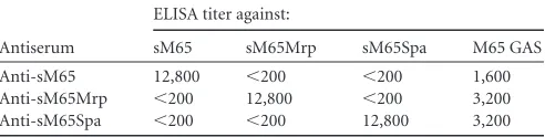
Specificities of M, Mrp, and Spa antibodies and binding to M65 streptococci.Rabbit antisera raised against the synthetic peptides copying the N-terminal regions of M65, M65Mrp, and M65Spa were assayed by ELISA using each of the synthetic peptides as solid-phase antigens (Table 1). Each antiserum contained anti-bodies with a titer of 12,800 against the respective immunogen. There was no reactivity with the heterologous antigens, indicating that the antibodies were highly specific. The antisera were then tested in ELISA using the whole M65 bacteria as solid-phase anti-gens. All three antisera reacted with the surface of M65 strepto-cocci (Table 1), indicating that the N-terminal epitopes of the respective proteins were exposed and available for antibody bind-ing. The experiments were performed three times, with similar results (data not shown).
Bactericidal activities of M, Mrp, and Spa antibodies against M65 streptococci.The functional activities of the antisera against the three surface proteins of M65 GAS were assessed byin vitro bactericidal assays, in which nonimmune human blood served as the source of phagocytes and complement. To determine the rel-ative contributions of the M, Mrp, and Spa antibodies to the pro-motion of opsonization and phagocytic killing, the assays were performed using the antisera singly or in various combinations (Fig. 1). All three individual antisera promoted bactericidal activ-ity, ranging from 46 to 65%. The bactericidal activity observed with a combination of all three antisera (96%) was significantly greater than that observed with antisera against Mrp or Spa or that with a combination of antisera against M and Mrp (Fig. 1). Taken together, these results indicate that highly specific synthetic pep-tide antisera against M, Mrp, and Spa reacted with the surface proteins of M65 streptococci, and each antiserum promoted bac-tericidal activity. A combination of all three antisera resulted in the highest level of bactericidal activity observed.
Patterns of antibody binding and C3 deposition on the sur-face of M65 streptococci.Previous studies have demonstrated the importance of the location of antibody binding to the M protein (13) and the pattern of C3 deposition on the bacterial surface (14) in promoting optimal phagocytic uptake of opsonized strepto-cocci. To visualize the binding patterns of M, Mrp, and Spa anti-bodies and the resultant deposition of complement, immunoflu-orescence microscopy was performed (Fig. 2). In order to mimic the potentialin vivoconditions, antibody binding assays were per-formed in the presence of human plasma, which contains multiple proteins that bind to the bacterial surface (15–17) that can alter
TABLE 1Specificity of rabbit antisera against sM65, sM65Mrp, and sM65Spa and reactivity with whole M65 streptococci
Antiserum
ELISA titer against:
sM65 sM65Mrp sM65Spa M65 GAS Anti-sM65 12,800 ⬍200 ⬍200 1,600 Anti-sM65Mrp ⬍200 12,800 ⬍200 3,200 Anti-sM65Spa ⬍200 ⬍200 12,800 3,200
on August 17, 2020 by guest
http://cvi.asm.org/
the binding of functional antibodies (18). The M, Mrp, and Spa peptide antisera each reacted with the surface of M65 streptococci in a semicircular pattern at the pole of each coccus, a location that represents the mature cell wall with fully inserted integral proteins (Fig. 2, top panels). Preimmune rabbit serum produced weak flu-orescence, which likely resulted from nonspecific IgG binding that was incompletely blocked by human plasma proteins.
Although antibody binding to the surface proteins of GAS is necessary for immune-mediated phagocytosis of the organisms, it is the activation and deposition of C3b that constitute the most potent opsonin (14). To visualize the pattern of C3b binding, M65 streptococci were first incubated with M, Mrp, or Spa antisera and then incubated with fluorescent antibody against human C3 (Fig. 2, bottom panels). The antibody binding was performed in the presence of heat-inactivated human plasma, followed by incuba-tion with fresh plasma as a complement source. As shown in pre-vious studies (19), C3 binding to the streptococcal surface in the absence of specific antibodies but in the presence of plasma pro-teins resulted in complement deposition in a linear or “X” pattern (Fig. 2, bottom left panel). This pattern of binding has been inter-preted to represent C3 deposition at the sites of new cross-wall
formation that do not contain fully inserted integral proteins. Once these proteins are fully expressed, they are capable of bind-ing plasma proteins that otherwise block the nonimmune recog-nition of the exposed cell wall. After first reacting the organisms with M, Mrp, or Spa antibodies, followed by fresh human plasma and anti-C3 antibodies, the complement deposition was observed to follow a more circumferential pattern, which included the non-specific and antibody-mediated deposition of C3. Circumferential C3 deposition has been shown to result in optimal phagocytic uptake because the C3 receptor interacts with C3b on the bacterial surface, functioning as a “zipper” to fully engulf the organisms (14).
Sequences ofspagenes from additionalemmtypes of GAS. The observation that at least one serotype of GAS expressed three distinct integral cell wall proteins that contained opsonic epitopes prompted us to perform additional studies to identifyspagenes in organisms that also contain theemmandmrpgenes. Genomic DNA was used as a PCR template to amplify potentialspagenes from M15, M67, M74, M78, and another M65 strain that was isolated during an epidemiologic study in North America (20). The sequences of the Spa proteins from M18 and M36 GAS were
FIG 1Bactericidal activity of M, Mrp, and Spa antisera against M65 GAS. The assays were performed with 33l of each of the three antisera, which were added in various combinations, as indicated. Sterile PBS was added to bring the final volume in the test mixture to 100l. The percent bacterial killing was calculated based on the reduction in the number of CFU that survived the rotation in the presence of immune serum compared to the number of CFU recovered from tubes containing preimmune serum. The results are expressed as the average killing⫾SD observed in four experiments. *,P⬍0.05 (ANOVA with Tukey’s HSD test).
FIG 2Fluorescence microscopy of M65 streptococci showing the patterns of M, Mrp, and Spa antibody binding to the surface of the organism in the presence of human plasma proteins (top panels) and C3b deposition after antibody binding to the respective antigens (bottom panels). Abs, antibodies.
on August 17, 2020 by guest
http://cvi.asm.org/
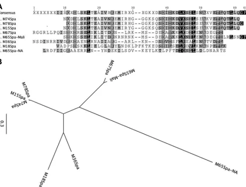
determined previously (7,8). An analysis of the predicted N-ter-minal amino acid sequences of the six newly sequenced Spa pro-teins (GenBank accession no.KJ794096toKJ794101) and the pre-viously sequenced Spa18 and Spa36 (Fig. 3A) indicated that the Spa sequence from the M65 Malian strain was highly homologous to the Spa sequence from serotype M67 (Fig. 3B). Interestingly, the Spa sequence from the M65 North American strain was com-pletely different from the Spa sequence of the M65 Malian strain. In addition, the predicted sequences of the Spa proteins from M15, M74, and M78 were almost identical, with the exception of a single amino acid substitution in M15Spa.
DISCUSSION
It has been known for decades that M antibodies have the capacity to neutralize the antiphagocytic effect of M protein. Virulent GAS multiply in nonimmune human blood because the M protein spe-cifically binds numerous plasma proteins that block or interfere with the activation and deposition of complement (15). Antibod-ies against the N-terminal regions of the M protein are opsonic and are able to bind to the M epitopes that extend beyond the layer of human plasma proteins covering the cell surface (18). In the current study, we have shown that serotype M65 GAS expressed
three distinct cell wall proteins that contained bactericidal epitopes. Highly specific rabbit antisera against synthetic peptides copying the N-terminal regions of M, Mrp, and Spa reacted with the surface of the organism, activated complement, and promoted phagocytic uptake and killing. A combination of all three antisera against M, Mrp, or Spa resulted in greater bactericidal activity than that with any one antiserum alone. Thus, all three proteins functioned in concert as potential protective antigens.
The seminal studies of Lancefield (5) resulted in the classical view of type-specific immunity to GAS infections. It was thought that each serotype of GAS expressed one M protein with unique opsonic epitopes, and once infected, individuals were protected against subsequent infection by that serotype only. We now un-derstand that GAS, depending on the serotype, display an array of surface proteins that have multiple functions related to protective immunity and/or virulence (15). Previous studies showed that when M and Mrp of serotype 4 streptococci are coexpressed, they function in concert as determinants of virulence, and both contain opsonic epitopes (6). Likewise, M and Spa of type 18 and type 36 streptococci were both required for optimal virulence, and both contained protective epitopes (8,19). In the current study, type 65 streptococci expressed all three proteins that contained opsonic
FIG 3Deduced amino acid sequences of the N-terminal 50 amino acids of different Spa proteins (A) and their sequence relatedness depicted by a phylogenetic tree (B).
on August 17, 2020 by guest
http://cvi.asm.org/
epitopes. It is of interest that the highest level of bactericidal activ-ity was observed when all three antisera were present in the reac-tion mixture. This result suggests that because M, Mrp, and Spa were expressed simultaneously in a mosaic pattern on the bacterial surface, antibodies against all three antigens may be required for optimal opsonization and phagocytic killing. In addition, the ap-parent enhanced functional effect of the antisera against the three distinct surface proteins suggests that competition for antibody binding sites on the bacterial surface did not occur, at least with the antiserum concentrations used in the assays.
Although the roles of M and Mrp in virulence have been stud-ied (6,16,21), the precise function of Spa in mediating resistance to phagocytosis is under investigation. Our previous studies showed that the function of Spa in the Mrp-negative GAS sero-types M18 and M36 resembled that of the M protein. Indeed, the 3=sequences of thesespagenes were shown to be highly homolo-gous to those of theseMgene ofStreptococcus equi, a group C streptococcus (8), suggesting that GAS acquired the gene via hor-izontal transfer, and the 5=region subsequently mutated under immunological pressure from the human host. Using antisera against the conserved C-terminal region of the M18Spa protein, we previously reported that 25/70 GAS serotypes expressed Spa-like proteins on their surface (8), and 24/25 are potentially Mrp positive based on the structure of themgaregulon (10). Analysis of theemmgene cluster of GAS indicates that 61% of all serotypes may express Mrp. The coexpression of M, Mrp, and Spa, coupled with our finding that all three contain opsonic epitopes, has po-tential implications for vaccine development and design. The ad-dition of dominant protective epitopes, in particular those that are shared among different serotypes of GAS, may broaden the poten-tial efficacy of vaccines designed to prevent these infections.
ACKNOWLEDGMENTS
This work was supported by U.S. Public Health Service grants AI-010085 and AI-060592 (both to J.B.D.) from the National Institute of Allergy and Infectious Diseases. S.E.N., a UTHSC medical student, was the recipient of a Medical Research Scholar Award from the UTHSC Department of Medicine.
We thank Harry Courtney for helpful suggestions and critical review of the manuscript.
REFERENCES
1.Dale JB, Fischetti VA, Carapetis JR, Steer AC, Sow S, Kumar R, Mayosi BM, Rubin FA, Mulholland K, Hombach JM, Schödel F, Henao-Restrepo AM.2013. Group A streptococcal vaccines: paving a path for accelerated development. Vaccine31(Suppl 2):B216 –B222.http://dx.doi .org/10.1016/j.vaccine.2012.09.045.
2.Carapetis JR, Steer AC, Mulholland EK, Weber M.2005. The global burden of group A streptococcal diseases. Lancet Infect. Dis.5:685– 694.
http://dx.doi.org/10.1016/S1473-3099(05)70267-X.
3.Bisno AL, Rubin FA, Cleary PP, Dale JB, National Institute of Allergy and Infectious Diseases.2005. Prospects for a group A streptococcal vaccine: rationale, feasibility, and obstacles—report of a National Institute of Allergy and Infectious Diseases workshop. Clin. Infect. Dis.41:1150 – 1156.http://dx.doi.org/10.1086/444505.
4.Dale JB.2008. Current status of group A streptococcal vaccine
develop-ment. Adv. Exp. Med. Biol.609:53– 63.http://dx.doi.org/10.1007/978-0 -387-73960-1_5.
5.Lancefield RC.1962. Current knowledge of the type-specific M antigens of group A streptococci. J. Immunol.89:307–313.
6.Courtney HS, Hasty DL, Dale JB.2006. Anti-phagocytic mechanisms of
Streptococcus pyogenes: binding of fibrinogen to M-related protein. Mol. Microbiol.59:936 –947.http://dx.doi.org/10.1111/j.1365-2958.2005 .04977.x.
7.Dale JB, Chiang EY, Liu SY, Courtney HS, Hasty DL. 1999. New protective antigen of group A streptococci. J. Clin. Invest.103:1261–1268.
http://dx.doi.org/10.1172/JCI5118.
8.Ahmed EA, Penfound TA, Brewer SC, Tennant PA, Chiang EY, Dale JB.
2010. Streptococcal protective antigens (Spa): a new family of type-specific proteins of group A streptococci. Eur. J. Clin. Microbiol. Infect. Dis.29:51–57.http://dx.doi.org/10.1007/s10096-009-0819-0.
9.Dale JB, Penfound TA, Tamboura B, Sow SO, Nataro JP, Tapia M, Kotloff KL.2013. Potential coverage of a multivalent M protein-based group A streptococcal vaccine. Vaccine31:1576 –1581.http://dx.doi.org /10.1016/j.vaccine.2013.01.019.
10. Bessen DE, Sotir CM, Readdy TL, Hollingshead SK. 1996. Genetic correlates of throat and skin isolates of group A streptococci. J. Infect. Dis.
173:896 –900.http://dx.doi.org/10.1093/infdis/173.4.896.
11. Dale JB.1999. Multivalent group A streptococcal vaccine designed to optimize the immunogenicity of six tandem M protein fragments. Vaccine
17:193–200.
12. Hu MC, Walls MA, Stroop SD, Reddish MA, Beall B, Dale JB.2002. Immunogenicity of a 26-valent group A streptococcal vaccine. Infect. Im-mun.70:2171–2177.http://dx.doi.org/10.1128/IAI.70.4.2171-2177.2002. 13. Jones KF, Fischetti VA.1988. The importance of the location of antibody binding on the M6 protein for opsonization and phagocytosis of group A M6 streptococci. J. Exp. Med.167:1114 –1123.http://dx.doi.org/10.1084 /jem.167.3.1114.
14. Jacks-Weis J, Kim Y, Cleary PP.1982. Restricted deposition of C3 on M⫹group A streptococci: correlation with resistance to phagocytosis. J. Immunol.128:1897–1902.
15. Walker MJ, Barnett TC, McArthur JD, Cole JN, Gillen CM, Hen-ningham A, Sriprakash KS, Sanderson-Smith ML, Nizet V. 2014. Disease manifestations and pathogenic mechanisms of group A strep-tococcus. Clin. Microbiol. Rev.27:264 –301.http://dx.doi.org/10.1128 /CMR.00101-13.
16. Courtney HS, Li Y.2013. Non-immune binding of human IgG to M-re-lated proteins confers resistance to phagocytosis of group A streptococci in blood. PLoS One 8:e78719. http://dx.doi.org/10.1371/journal.pone .0078719.
17. Li Y, Courtney HS.2011. Promotion of phagocytosis ofStreptococcus pyogenesin human blood by a fibrinogen-binding peptide. Microbes In-fect.13:413– 418.http://dx.doi.org/10.1016/j.micinf.2010.12.008. 18. Whitnack E, Dale JB, Beachey EH.1984. Common protective antigens of
group A streptococcal M proteins masked by fibrinogen. J. Exp. Med.
159:1201–1212.http://dx.doi.org/10.1084/jem.159.4.1201.
19. McLellan DG, Chiang EY, Courtney HS, Hasty DL, Wei SC, Hu MC, Walls MA, Bloom JJ, Dale JB.2001. Spa contributes to the virulence of type 18 group A streptococci. Infect. Immun.69:2943–2949.http://dx.doi .org/10.1128/IAI.69.5.2943-2949.2001.
20. Shulman ST, Tanz RR, Dale JB, Beall B, Kabat W, Kabat K, Cederlund E, Patel D, Rippe J, Li Z, Sakota V, North American Streptococcal Pharyngitis Surveillance Group.2009. Seven-year surveillance of North American pediatric group A streptococcal pharyngitis isolates. Clin. In-fect. Dis.49:78 – 84.http://dx.doi.org/10.1086/599344.
21. Podbielski A, Schnitzler N, Beyhs P, Boyle MDP. 1996. M-related protein (Mrp) contributes to group A streptococcal resistance to phago-cytosis by human granulocytes. Mol. Microbiol.19:429.http://dx.doi.org /10.1046/j.1365-2958.1996.377910.x.