R E S E A R C H A R T I C L E
Open Access
The unresolved problem of beta-2
microglobulin amyloid deposits in the
intervertebral discs of long-term dialysis
patients
Tsung-Ting Tsai
1*†, Arun-Kumar Kaliya-Perumal
1,2†, Chang-Chyi Jenq
3, Chi-Chien Niu
1, Natalie Yi-Ju Ho
1,
Tung-Ying Lee
4and Po-Liang Lai
1Abstract
Background:Dialysis-related destructive spondyloarthropathy caused by beta-2 microglobulin (β2M) amyloid deposits in intervertebral discs is a major burden for patients undergoing long-term dialysis. This study aimed to quantify the presence of β2M amyloid deposits in the intervertebral disc tissue of such patients and analyze whether there was a significant correlation between β2M accumulation and the duration of dialysis. Methods: Two groups of patients who had undergone surgery for degenerative spinal pathologies were selected: the dialysis group (n= 29) with long-term dialysis and the control group (n= 10) with no renal impairment. Tissue sections were prepared from specimens of intervertebral disc tissue obtained during spinal surgery and analyzed via histological staining, including immunohistochemistry (IHC) and Congo red.
Results:There was a statistically significant multifold increase ofβ2M expression in the disc tissue of long-term dialysis patients when compared to non-dialysis patients, as shown by both IHC (0.019 ± 0.023μm2vs. 0.00020 ± 0.00033μm2, respectively;p= 0.012) and Congo red staining (0.027 ± 0.041μm2vs. 9.240 × 10⁻⁵± 5.261 × 10⁻⁵μm2, respectively;
p= 0.047). We also note a moderate strength positive correlation between the duration of dialysis and positive IHC (r= 0.39;p= 0.015) and Congo-red staining (r= 0.42;p= 0.007).
Conclusions:The problem ofβ2M amyloidosis in long-term dialysis patients remains unresolved even with predominant use of high-flux dialysis membranes. This highlights the insufficiency of current dialysis modalities to effectively filterβ2M.
Keywords:Amyloid, Beta-2 microglobulin, Dialysis, Spondyloarthropathy, Congo red, Intervertebral disc
Background
The incidence and prevalence of end-stage kidney dis-ease (ESKD) has incrdis-eased throughout the world [1–3], becoming a major public health problem with high eco-nomic and social costs [4, 5]. The number of patients receiving dialysis therapy has rapidly increased to an all-time high, especially in developing nations [5]. A large proportion of these patients are elderly and predisposed to increased risks for multiple adverse outcomes, such
as hypotension, anemia, myopathy, and atrial fibrillation [6–9]. One of the major problems in such patients undergoing long-term hemodialysis is beta-2 microglob-ulin (β2M) amyloidosis [10]. The accumulation of β2M amyloid fibrils in the musculoskeletal system can lead to various degenerative conditions, depending on the mus-culoskeletal site of involvement [11, 12]. In the spine, such accumulation can damage the intervertebral discs and their adjacent endplates, leading to spinal instability and neurological compromise [13]. This destructive amyloid spondyloarthropathy is a major cause of disabil-ity in long-term dialysis patients.
There are two types of dialysis treatment, hemodialysis (HD) and peritoneal dialysis (PD), which are extensively
* Correspondence:[email protected] †Equal contributors
1Department of Orthopaedic Surgery, Spine Division, Bone and Joint Research Center, Chang Gung Memorial Hospital and Chang Gung University College of Medicine, Taoyuan, Taiwan
Full list of author information is available at the end of the article
practiced to manage ESKD. However, clinical outcomes may vary depending on different factors, such as age, co-morbidities, and dialysis time. Studies demonstrate that PD offers equivalent or better survival than HD in the first year of treatment, especially in patients with ad-vanced age and diabetes [14]. In addition, there are no reported differences in the occurrence of β2M amyloid-osis and musculoskeletal complications between patients treated with HD and PD [11]. However,β2M levels are known to be influenced by the membrane used for HD treatment. For example, the levels ofβ2M are said to be significantly reduced in patients treated with high-flux HD, which uses polysulfone membranes, when com-pared to patients treated with conventional dialysis using cellulose membranes [15].
Recently, high-flux HD is widely performed to improve the quality and efficacy of dialysis treatment. With the advent of this technique, occurrence of destructive amyl-oid spondyloarthropathy should be less likely, though it has to be studied. To address this question, we quanti-fied the presence ofβ2M amyloid deposits in interverte-bral discs among long-term dialysis patients who had undergone HD or PD and analyzed whether there was a significant correlation between β2M accumulation and the duration of dialysis.
Methods
Retrospectively, we reviewed records of patients with ESKD who were on long-term dialysis therapy, but also underwent spinal surgery for single or multilevel degen-eration with or without spinal instability. We shortlisted the patients for whom formalin-fixed paraffin wax em-bedded disc tissue samples were available for further analysis, resulting in a sample size containing a dialysis group of 29 samples. A small control group of 10 sam-ples with similar demographic characteristics (e.g., age, body-mass index, surgical level and location), but with-out any renal disease, were selected for comparison (i.e., the dialysis group). This selection method was non-randomized as only those patients whose disc tissue samples were available for further analysis were chosen.
We tabulated several basic parameters, including age, gender, underlying disease, and the severity of disc de-generation. Chronic kidney disease, mineral and bone disorder-related parameters, such as serum levels of alkaline phosphatase, calcium, and phosphate, were con-sidered. We also included several dialysis-related param-eters, such as type, duration, and adequacy (Kt/V; in which K= the dialyzer clearance, t= time, and V= urea distribution volume) of the dialysis treatment. The severity of disc degeneration at the surgical level among patients of both groups was graded according to Pfirr-mann’s classification using magnetic resonance imaging (MRI) images [16]. We acquired and prepared slides of
the formalin-fixed paraffin wax embedded disc tissue samples from the Tissue Bank. The disc specimens were fixed in neutral buffered formalin, embedded into paraf-fin wax blocks, and stored at room temperature at the Tissue Bank with no storage time limit. These unstained slides were deparaffinized in xylene, rehydrated through a series of graded alcohol washes, and soaked in distilled water before staining. Congo red and immunohisto-chemistry (IHC) staining were then performed on all ac-quired samples.
Congo red staining
After sample preparation, the sections from all 39 sam-ples were stained with Mayer’s Hematoxylin Solution (MHS1-100ML, Sigma-Aldrich, St. Louis, MO) for 10 min and thoroughly washed for 5 min, after which they were immediately soaked with alkaline sodium chloride solution for 20 min, followed by alkaline Congo red solution for 20 min (HT60 kit, Sigma-Aldrich). The sections were dehydrated with a series of graded alcohol washes and then mounted for evaluation under normal and polarized light microscopy.
Immunohistochemistry staining
After sample preparation, the 39 sections were washed twice in PBS (Sigma-Aldrich) for 5 min. The sections were blocked with a solution of 10% normal serum (FBS; Gibco, Grand Island, NY) and 1% BSA (Sigma-Aldrich) in PBS for 3 h and then incubated with anti-β2M anti-body (ab195531, Abcam, Cambridge, MA) at 1:400 dilu-tion for 30 min. We added 3,3′-diaminobenzidine substrate (Abcam) and incubated the sectioned samples for 5 min, followed by thorough washing with tap water for another 5 min, and finally staining with Mayer’s Hematoxylin Solution for 10 min. The sections were dehydrated with a series of graded alcohol washes and then mounted for evaluation under light microscopy.
Protein extraction and western blotting
separated using SDS-PAGE, transferred onto a polyvinyli-dene difluoride membrane, and soaked with blocking re-agent for 3 min. This was followed by overnight incubation in antibody dilution buffer containing the pri-mary anti-β2M antibody (1:2000; Abcam), as well as anti-beta actin monoclonal antibody (1:5000; Proteintech, Rosemont, IL) as an internal control. After washing away the unbound antibodies with TTBS (1× TBS buffer and 0.1% Tween 20; Sigma-Aldrich) three times for 5 min each, rabbit anti-mouse secondary antibody (1:10,000) was incubated for 2 h, followed by washing with TTBS again, and visualized by Pierce Enhanced Chemiluminescence Western Blotting Substrate (Thermo Fisher Scientific).
Statistics
Quantitative analysis of histological staining was per-formed using Image J (National Institute of Health, Be-thesda, MD) to estimate the proportion of positively stained area in the sectioned intervertebral disc tissue samples. Measurements were done for both IHC and Congo red stained sections among both groups, which were tabulated for comparison. A correlation analysis was performed to evaluate the strength of association between the duration of dialysis and the positively stained area by either of the staining methods. The per-centage of the positively stained area over the total area was calculated.
Statistical analyses were performed using IBM SPSS Statistics for Windows, Version 19.0 (IBM Corp., Armonk, NY). For data analysis, the Student’sttest was used for continuous variables and Fisher’s exact test for categorical variables. Apvalue of < 0.05 was considered statistically significant.
Results
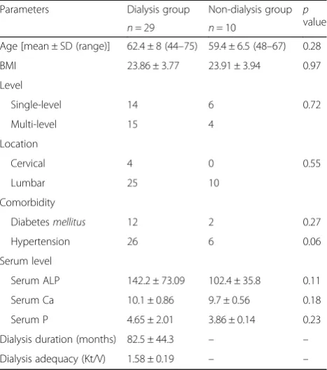
Table 1 summarizes the demographic data among the 39 samples that were selected for the study. The dialysis group had a mean age of 62.4 ± 8 years, and the control non-dialysis group a mean age of 59.4 ± 6.5 years. The numbers of patients with single- or multilevel disc de-generation in both groups were statistically similar. Pre-dominantly, surgeries were done for patients with lumbar degeneration, except for 4 samples among the dialysis group for whom the surgery was done for single-level cervical degeneration. However, this small number did not result in any statistical difference between the groups. The number of patients with associated comor-bid conditions, including diabetesmellitus and hyperten-sion, were similar in both groups. There was no significant difference between the dialysis and non-dialysis patients in terms of their serum-based alkaline phosphatase, calcium, and phosphate levels. For the dia-lysis group, there were 12 grade III, 4 grade IV, and 5 grade V discs. For the non-dialysis group, there were 7
grade III, 2 grade IV, and 1 grade V discs. The severity of disc degeneration also did not show any statistical differ-ence between the groups. Therefore, we considered the groups to be appropriately matched.
For those in the dialysis group, dialysis was done for a mean duration of 82.5 ± 44.3 months, ranging from 17.08 to 194.4 months (Table 1). There were 22 samples on high-flux HD and 7 samples on PD. The mean time inter-val between specimen collection and histological examin-ation was 23.83 ± 19.5 months (range, 0–76 months) in the dialysis group. However, the time interval was only a few months for those in the non-dialysis group.
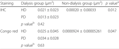
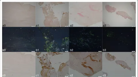
Histology examination of the sections following Congo red staining under polarized light showed areas of apple-green birefringence that indicated amyloid deposits. The mean positively stained area among HD patients of the dia-lysis group was 0.025 ± 0.045μm2, while for PD patients it was 0.034 ± 0.028μm2. The consolidated measurement for the dialysis group (0.027 ± 0.041μm2) proved to be signifi-cantly higher than that of the non-dialysis group (9.240 × 10⁻⁵± 5.261 × 10⁻⁵ μm2; p= 0.047). Similarly, following IHC staining, the HD and PD group were immunopositive, measuring 0.021 ± 0.023 μm2 and 0.013 ± 0.023 μm2, re-spectively. Their consolidated measurement (0.019 ± 0.023 μm2) was also significantly higher than that of the non-dialysis group (0.00020 ± 0.00033 μm2; p= 0.012). However, there was no significant difference in the
Table 1Comparison of demographic data between the dialysis and non-dialysis groups
Parameters Dialysis group Non-dialysis group p value
n= 29 n= 10
Age [mean ± SD (range)] 62.4 ± 8 (44–75) 59.4 ± 6.5 (48–67) 0.28
BMI 23.86 ± 3.77 23.91 ± 3.94 0.97
Level
Single-level 14 6 0.72
Multi-level 15 4
Location
Cervical 4 0 0.55
Lumbar 25 10
Comorbidity
Diabetesmellitus 12 2 0.27
Hypertension 26 6 0.06
Serum level
Serum ALP 142.2 ± 73.09 102.4 ± 35.8 0.11
Serum Ca 10.1 ± 0.86 9.7 ± 0.56 0.18
Serum P 4.65 ± 2.01 3.86 ± 0.14 0.23
Dialysis duration (months) 82.5 ± 44.3 – –
Dialysis adequacy (Kt/V) 1.58 ± 0.19 – –
SDstandard deviation,BMIbody mass index,ALPalkaline phosphatase,Ca
positively stained areas between the HD and PD patients using either IHC or Congo red staining (Table 2).
Assessment of correlation between the duration of dialysis and the positively stained area as measured by either of the staining methods revealed a moderate strength positive correlation between the two (IHC staining, r= 0.39, p= 0.015; Congo red stainingr= 0.42,
p= 0.007). Quantitative results showed the percentage of IHC-stained area over the total tissue area was 2.30% for the dialysis group and 0.01% for the non-dialysis group. The percentage of Congo red stained area over the total area of the sectioned samples was 1.49% for the dialysis group and 0.009% for the non-dialysis group, which sug-gested that the expression ofβ2M amyloid deposits was 230-times and 165-times higher in the disc tissue of pa-tients who had undergone dialysis therapy than in the tissue of non-dialysis patients, based on IHC and Congo red staining, respectively (Fig. 1).
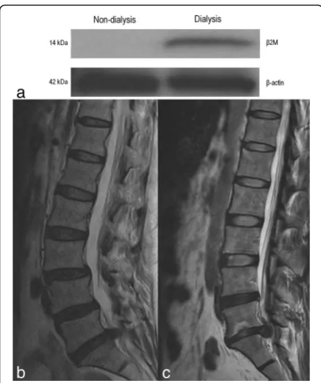
Western blot analysis indicated that β2M protein was present in the intervertebral disc tissue obtained from the patient who had received 13 months of HD therapy, but was absent in the disc tissue obtained from the non-dialysis patient (Fig. 2). Quantifying the band intensity using Image J, the ratio of β2M to β -actin for the dialysis patient was 147.77 times higher than that of the non-dialysis patient, which confirmed a significantly higher expression of β2M protein in the intervertebral disc tissue of the patient who underwent HD treatment when compared to the disc of the patient without dialysis treatment.
Discussion
β2M is constantly produced in the body and eliminated exclusively by the kidneys, mostly by glomerular filtra-tion, after which 99.9% of the excreted proteins are reab-sorbed in the renal tubules, where they are catabolized [17]. When glomerular filtration fails, increased serum levels ofβ2M are observed. This mechanism is the same for the accumulation ofβ2M in dialysis patients, as there is a bio-incompatibility that prevents the dialysis mem-branes from efficiently filteringβ2M. Studies have shown
that β2M levels significantly increase after conventional HD using cellulose acetate membranes [15].
However, the science of dialysis is constantly evolving, and the current standard method is different from the past conventional techniques and membranes. With the advent of high-flux dialysis, β2M levels are significantly decreased after dialysis [13]; hence, destructive amyloid spondyloarthopathy should be less likely to occur. How-ever, even though a type of high-flux dialysis membrane was used in the majority of our patients, we still found
β2M accumulation in their intervertebral discs. This sig-nifies that even with recent advances in dialysis, β2M amyloidosis remains an unresolved issue. Moreover, con-ventional dialysis methods are still used in most develop-ing countries [18], which further contributes to the prevalence ofβ2M amyloidosis.
Literature suggests that dialysis itself is an inflamma-tory stimulus, which can induce cytokine production (e.g., Interlukin-1, Interlukin-6, and tumor necrosis factor-α) and activate complement systems. Subse-quently, the released cytokines stimulate the synthesis of
β2M and its release by macrophages [19]. β2M is often highly expressed in articular cartilage of various joints and the synovial fluid, leading to compromised chondro-cyte function that results in osteoarthritis [20, 21]. Apart from musculoskeletal involvement, β2M amyloid de-posits can also be found in arterioles, venules, and mus-cularis propria of visceral organs, like those of the gastrointestinal tract and heart [22, 23].
Intervertebral discs, facet joints, and ligamentum fla-vum are the most susceptible sites for β2M accumula-tion in the vertebral column [24, 25]. This can lead to destructive spondyloarthropathy (DSA) and consequen-tial disability [26]. DSA is increasingly recognized as a complication of long-term HD [24]. Clinical symptoms depend on the sites affected byβ2M amyloid deposition. Most patients are asymptomatic; however, some might complain about localized back pain, which is often ac-companied by arm pain or numbness [15, 27]. The radiological features include narrowing of disc space and the destruction of adjacent vertebral endplates [15]. This may precede segmental instability or neurological com-promise [28, 29]. Our study included patients pre-dominantly with lumbar disc degeneration and a few patients with cervical disc involvement. This non-random selection may not represent the demographics of the entire population. However, cervical discs are said to be more commonly affected, followed by the lumbar and upper thoracic discs, and then the middle and lower thoracic discs [24].
Disc degeneration can be graded according to Pfirr-mann’s classification system [16]. However, we had 8 samples in the dialysis group who did not fit into any grade. This was due to the difficulty in deciding whether
Table 2Comparison of positively stained areas between the dialysis and non-dialysis groups using IHC and Congo red staining
Staining Dialysis group (μm2) Non-dialysis group ( μm2)
pvaluea IHC HD 0.021 ± 0.023 0.00020 ± 0.00033 0.012
PD 0.013 ± 0.023 pvalueb 0.42
Congo red HD 0.025 ± 0.045 0.0000924 ± 0.00005261 0.047
PD 0.034 ± 0.028 pvalueb 0.63
IHCImmunohistochemistry staining,HDhemodialysis,PDperitoneal dialysis
a
Analyzed between dialysis and non-dialysis groups b
the radiological changes were caused by dialysis-related spondyloarthropathy or spondylodiscitis [27]. Usually, DRS presents as low signal intensity in the MRI of both T1 and T2 weighted images, unlike spondylodiscitis which presents as low signal intensity on T1 and high signal in-tensity on T2 weighted images [15, 27]. However, unusual signal intensity changes as we observed in a few of our cases can also be present; but post-operative culture of the disc material revealed no microorganisms suggestive of in-fection. Therefore, we considered these patients to have dialysis-related spondyloarthropathy.
Histological examination is the gold standard for diag-nosingβ2M amyloidosis. Our histological study included Congo red and IHC staining of disc material sections. Positive Congo red staining, appearing as distinct bi-refringence under polarized light, and immunostaining of amyloid deposits with a labeled anti-β2M antibody are considered pathognomonic for the presence ofβ2M [30]. We also performed western blot analysis, but only for one patient from the dialysis group and one from the non-dialysis group. Our results suggest a multifold in-crease inβ2M expression in the disc tissue of long-term dialysis patients. There was also a moderate strength positive correlation between the duration of dialysis and positive IHC and Congo red staining. These findings
from our pilot analysis provide important insights into our understanding ofβ2M amyloidosis and highlight the need for dialysis membranes that can more effectively filterβ2M molecules to help prevent potential long-term complications [10, 31].
This study was a cross-sectional prevalence measure with certain limitations. Considering the sample size, this study may be underpowered to draw potential con-clusions. Also, not all patients had definite radiological evidence of DRS before surgery. However, β2M depos-ition was noted among all dialysis group patients. Further empirical evidence could be collected by per-forming western blot analysis for all the patients in the study.
Conclusions
We analyzed the presence of β2M amyloid deposits in the intervertebral discs of long-term dialysis patients using Congo red, IHC staining and western blot analysis of surgically harvested tissue. Our results showed a sta-tistically significant multifold increase ofβ2M expression in the disc tissues of long-term dialysis patients when compared to non-dialysis patients. We also inferred a moderate strength positive correlation between the dur-ation of dialysis and positive IHC and Congo red
staining, representing the presence of β2M amyloids. Our findings highlight the issue ofβ2M amyloidosis still prevailing among long-term dialysis patients even with the advent of high-flux dialysis, therefore warranting further study.
Abbreviations
ALP:Alkaline phosphatase; BMI: Body mass index; Ca: Calcium; DSA: Destructive spondyloarthropathy; ESKD: End-stage kidney disease; HD: Hemodialysis; IHC: Immunohistochemistry; MRI: Magnetic resonance imaging; P: Phosphorous; PD: Peritoneal dialysis; SD: Standard deviation;
β2M: Beta-2 microglobulin
Acknowledgements
We thank the Resource Center for Clinical Research, Chang Gung Memorial Hospital, Linkou, for providing statistical assistance. We thank the Tissue Bank, Chang Gung Memorial Hospital, Linkou, for providing the formalin-fixed paraffin wax embedded disc tissue samples.
Funding
The study was supported by the Ministry of Science and Technology, Taiwan, with grant no. MOST 104-2314-B-182-012-MY2.
Availability of data and materials
All data generated or analyzed during this study are included in this published article.
Authors’contributions
TTT initiated the study, collected the data, performed the statistical analysis, interpreted the data, and drafted the manuscript. AKKP participated in statistical analysis, data interpretation, manuscript drafting and revision. CCJ collected the data and helped drafting the manuscript. CCN supervised the study and provided opinions. NYJH prepared the manuscript and helped in the revision. TYL collected, analyzed, and interpreted the data. PLL sanctioned the final manuscript. All authors critically reviewed and approved the final manuscript.
Ethics approval and consent to participate
This study was performed in compliance with the 1964 declaration of Helsinki, its later amendments or comparable ethical standards. Extraction and evaluation of data was approved by the Institutional Review Board (IRB) of Chang Gung Medical Foundation with IRB no. 103-7370B on 2015/02/04.
Consent for publication
Not applicable.
Competing interests
The authors declare that they have no competing interests.
Publisher’s Note
Springer Nature remains neutral with regard to jurisdictional claims in published maps and institutional affiliations.
Author details
1
Department of Orthopaedic Surgery, Spine Division, Bone and Joint Research Center, Chang Gung Memorial Hospital and Chang Gung University College of Medicine, Taoyuan, Taiwan.2Department of Orthopaedic Surgery, Melmaruvathur Adhiparasakthi Institute of Medical Sciences and Research, Affiliated to the Tamil Nadu Dr. MGR Medical University, Tamil Nadu, India. 3Department of Nephrology, Chang Gung Memorial Hospital and Chang Gung University College of Medicine, Taoyuan, Taiwan.4Department of Medical Education, Chang Gung Memorial Hospital, Taoyuan, Taiwan.
Received: 29 September 2017 Accepted: 11 December 2017
References
1. Coresh J, Byrd-Holt D, Astor BC, et al. Chronic kidney disease awareness, prevalence, and trends among U.S. adults, 1999 to 2000. J Am Soc Nephrol. 2005;16(1):180–8.
2. Stengel B, Couchoud C. Chronic kidney disease prevalence and treated end-stage renal disease incidence: a complex relationship. J Am Soc Nephrol. 2006;17(8):2094–6.
3. Varma PP. Prevalence of chronic kidney disease in India—where are we heading? Indian J Nephrol. 2015;25(3):133–5.
4. Joyce AT, Iacoviello JM, Nag S, et al. End-stage renal disease-associated managed care costs among patients with and without diabetes. Diabetes Care. 2004;27(12):2829–35.
5. Khanna U. The economics of dialysis in India. Indian J Nephrol. 2009;19(1):1–4. 6. Stevens LA, Viswanathan G, Weiner DE. Chronic kidney disease and end-stage renal disease in the elderly population: current prevalence, future projections, and clinical significance. Adv Chronic Kidney Dis. 2010;17(4):293–301. 7. Lee YC, Hung SY, Wang HH, et al. Different risk of common gastrointestinal
disease between groups undergoing hemodialysis or peritoneal dialysis or with non-end stage renal disease: a nationwide population-based cohort study. Medicine (Baltimore). 2015;94(36):e1482.
8. Shen CH, Zheng CM, Kiu KT, et al. Increased risk of atrial fibrillation in end-stage renal disease patients on dialysis: a nationwide, population-based study in Taiwan. Medicine (Baltimore). 2016;95(25):e3933.
9. Shu CC, Hsu CL, Wei YF, et al. Risk of tuberculosis among patients on dialysis: the predictive value of serial interferon-gamma release assay. Medicine (Baltimore). 2016;95(22):e3813.
10. Mumtaz A, Anees M, Bilal M, Ibrahim M. Beta-2 microglobulin levels in hemodialysis patients. Saudi J Kidney Dis Transpl. 2010;21(4):701–6. 11. Soyupek F, Demir M, Suslu FE, Baykal B, Sezer MT, Yesildag A. The upper
extremity musculoskeletal complications in dialysis patients: comparison between hemodialysis and peritoneal dialysis. J Back Musculoskelet Rehabil. 2013;26(3):267–371.
12. Wada T, Miyata T, Sakai H, Kurokawa K. Beta2-microglobulin and renal bone disease. Perit Dial Int. 1999;19(Suppl 2):S413–6.
13. Cuffe MJ, Hadley MN, Herrera GA, Morawetz RB. Dialysis-associated spondylarthropathy. Report of 10 cases. J Neurosurg. 1994;80(4):694–700. 14. McDonald SP, Marshall MR, Johnson DW, Polkinghorne KR. Relationship
between dialysis modality and mortality. J Am Soc Nephrol. 2009;20(1):155–63. 15. Acchiardo S, Kraus AP Jr, Jennings BR. Beta 2-microglobulin levels in
patients with renal insufficiency. Am J Kidney Dis. 1989;13(1):70–4. 16. Pfirrmann CW, Metzdorf A, Zanetti M, Hodler J, Boos N. Magnetic
resonance classification of lumbar intervertebral disc degeneration. Spine. 2001;26(17):1873–8.
17. Wibell L. The serum level and urinary excretion of beta2-microglobulin in health and renal disease. Pathol Biol (Paris). 1978;26(6):295–301. 18. Sinha A, Bagga A. Maintenance dialysis in developing countries. Pediatr
Nephrol. 2015;30(2):211–9.
19. Scarpioni R, Ricardi M, Albertazzi V, De Amicis S, Rastelli F, Zerbini L. Dialysis-related amyloidosis: challenges and solutions. Int J Nephrol Renov Dis. 2016;9:319–28.
20. Zhang H, Liew CC, Marshall KW. Microarray analysis reveals the involvement of beta-2 microglobulin (B2M) in human osteoarthritis. Osteoarthr Cartil. 2002;10(12):950–60.
21. Melikoglu MA, Yildirim K, Senel K. Relationship between radiographic grading of osteoarthritis and serum beta-2 microglobulin. Ir J Med Sci. 2009; 178(2):151–4.
22. Ohashi K, Takagawa R, Hara M. Visceral organ involvement and extracellular matrix changes in beta 2-microglobulin amyloidosis–a comparative study with systemic AA and AL amyloidosis. Virchows Archiv. 1997;430(6):479–87. 23. Fuchs A, Jagirdar J, Schwartz IS. Beta 2-microglobulin amyloidosis (AB2M) in
patients undergoing long-term hemodialysis. A new type of amyloid. Am J Clin Pathol. 1987;88(3):302–7.
24. Ohashi K, Hara M, Kawai R, et al. Cervical discs are most susceptible to beta 2-microglobulin amyloid deposition in the vertebral column. Kidney Int. 1992;41(6):1646–52.
25. Ito M, Abumi K, Takeda N, Satoh S, Hasegawa K, Kaneda K. Pathologic features of spinal disorders in patients treated with long-term hemodialysis. Spine. 1998;23(19):2127–33.
26. Inatomi K, Matsumoto T, Tomonaga T, et al. Histological analysis of the ligamentum flavum of patients with dialysis-related spondyloarthropathy. J Orthop Sci. 2004;9(3):285–90.
27. Kiss E, Keusch G, Zanetti M, et al. Dialysis-related amyloidosis revisited. AJR Am J Roentgenol. 2005;185(6):1460–7.
28. Vignes JR, Eimer S, Dupuy R, Donois E, Liguoro D. Beta(2)-microglobulin amyloidosis caused spinal cord compression in a long-term haemodialysis patient. Spinal Cord. 2007;45(4):322–6.
29. Kato Y, Kanaya K, Itoh T. Destructive spondyloarthropathy of the atlantoaxial joint with severe spinal cord compression in long-term hemodialysis patients. J Orthop Sci. 2006;11(6):644–50.
30. Jadoul M, Garbar C, van Ypersele de Strihou C. Pathological aspects of beta(2)-microglobulin amyloidosis. Semin Dial. 2001;14(2):86–9. 31. Traut M, Haufe CC, Eismann U, Deppisch RM, Stein G, Wolf G. Increased
binding of beta-2-microglobulin to blood cells in dialysis patients treated with high-flux dialyzers compared with low-flux membranes contributed to reduced beta-2-microglobulin concentrations. Results of a cross-over study. Blood Purif. 2007;25(5–6):432–40.
• We accept pre-submission inquiries
• Our selector tool helps you to find the most relevant journal
• We provide round the clock customer support
• Convenient online submission
• Thorough peer review
• Inclusion in PubMed and all major indexing services • Maximum visibility for your research
Submit your manuscript at www.biomedcentral.com/submit