BIOCOMPATIBILITY TESTING OF RESORBABLE MATERIALS USING IMPROVED IN -V IT R O TECHNIQUES
by Neelam Gurav
Thesis submitted in fulfilment of the requirements for the degree of Doctor of Philosophy in the Faculty of Science at the University of
ProQuest Number: 10106683
All rights reserved
INFORMATION TO ALL USERS
The quality of this reproduction is dependent upon the quality of the copy submitted.
In the unlikely event that the author did not send a complete manuscript and there are missing pages, these will be noted. Also, if material had to be removed,
a note will indicate the deletion.
uest.
ProQuest 10106683
Published by ProQuest LLC(2016). Copyright of the Dissertation is held by the Author.
All rights reserved.
This work is protected against unauthorized copying under Title 17, United States Code. Microform Edition © ProQuest LLC.
ProQuest LLC
789 East Eisenhower Parkway P.O. Box 1346
Acknowledgement
This thesis is dedicated to my parents
I would like to thank Professor Sandra Downes for all her help and support throughout
this study and being very patient with me. I would also like to thank the Dept of Human Morphology at the University of Nottingham.
Special thanks for help above and beyond the call of duty go to:
Mike Kayser for teaching me everything about microscopy, electron microscopy and
photography and giving me lots of advise and help.
Dr Sanjukta Deb for starting me out on degradable polymers and Dr Mary Anstice for
introducing me to the joys of gel permeation chromatography (GPC) and being a big help
with a subject I knew nothing about A big thankyou also to Paul Clarke at Viscotek for
his help with the GPC equipment
Padmini Sarathachandra my pal, for helping me whenever I was stuck which was quite
often.
Dr Lucy Di Silvio for proof reading the thesis and giving me helpful hints during the writing of the thesis and sharing the office with me without going potty.
Mrs Caroline Clifford for proof reading the thesis without whose input the thesis would
have been in any other language but English.
Dept of Morbid Anatomy and in particular Dr Ann Sandison for putting up with me and
giving me time off to finish writing up. Professor Yousuf Ali, Dr David Lee, Professor George Bently and everyone at the Institute of Orthopaedics who has put up with me during the last few years and let me be part of the team.
Finally I would like to thank my long suffering husband who has helped and supported
Abstract
Interest in degradable polymer systems for use in various biomedical applications has
been increasing since their development in the 1960s. The first polymer to be used was
polyglycolic acid, marketed under the trade name Dexon, as a resorbable suture.
Although the success of resorbable polymers has been mainly in the form of sutures and small pins, their potential use as materials for bone fixation, bone regeneration and drug
delivery vehicles is vast These materials have obvious advantages in that retrieval of the
implant can be avoided causing minimal inconvenience to the patient Extensive in-vivo
biocompatibility testing has been performed on a range of resorbable polymers but there is a lack of information available on events occurring at the cellular level specially during
long term implantation. This can be investigated in depth using in-vitro assays and utilising methods which mimic the long term in-vivo degradation of polymers.
The aim of this study was to determine the short and long term biocompatibility of
degradable polymers using in-vitro cell culture methods. The morphology and proliferation of osteoblast-hke cells and monocyte/macrophages on the polymer surfaces was investigated by light microscopy, electron microscopy and biochemical assays. Polymer degradation by enzymes and other degradation methods was investigated using
gel permeation chromatography and the biocompatibility of polymers at stages of degradation was studied. The effect of the acidic pH caused by the monomers and the
monomers themselves on the viability of osteoblast-like cells and monocyte/macrophages
was studied. Evidence for HOS cells undergoing apoptosis was investigated by transmission electron microscopy and by utilising stains specific for apoptosis when
cultured in the presence of monomers.
This study demonstrated that the accelerated degradation of polymers by heat and gamma
irradiation provides a good method for obtaining polymers for "long-term"
biocompatibility testing. Enzyme solutions also influenced the degradation of the polymers in particular the polymer surfaces. The morphology and proliferation of
osteoblast-like cells varied on the different polymer surfaces depending on surface
structure, crystallinity and the release of degradation products; and in addition the
presence of monomers caused a decrease in the mitochondrial activity of both the cell
types tested. The monocyte/macrophages also had varying morphologies on the different polymers and were stimulated by some of the polymers to a greater extent By using the
C ontents
Title 1
A cknow ledgem ents 2
A bstract 3
C ontents page 4
A bbreviations 5
List of suppliers and m anufacturers 7
List of figures and Tables 8
Chapter 1. General Introduction 13
Chapter 2. Polymer characterisation and degradation 41
Chapter 3. Qualitative evaluation of cells on materials 7 7
Chapter 4. Cell attachment and proliferation on polymer surfaces measured 100
using quantitative assays
Chapter 5. The effect of monomers on cellular activity 123
C hapter 6. The monocyte/macrophage response to degradable polymers 153 and their monomrs
Chapter 7. Future work and general discussion 173
R eferences 180
Abbreviations
APC Antigen Presenting Cells
MHC Major Histocompatibility Complex
MCF Macrophage Chemotactic Factor
U F Lymphocyte Inhibitory Factor
MIF Macrophage Inhibiting Factor
PDMS Polydimethylsiloxane
% -Thymidine Tritiated thymidine
ALP Alkaline Phosphatase
ANOVA Analysis of Variance
CPMs Counts Per Minute
D3-HB D3-hydroxybutyric acid
DAPI 4\6-Diamidino-2-Phenylindole
DMEM Dulbecco's Modified Eagles Medium
DMSO Dimethyl Sulfoxide
DNA Deoxyribonucleic acid
ECACC European Collection of Animal and Cell Cultures
ELISA Enzyme Linked Immunosorbent Assay
PCS Foetal Calf Serum
GA Glycolic Acid
GPC Gel Permeation Chromatography
HMDS Hexamethyldisilazane
HOS Human Osteosarcoma (TE85) cell line
LA Lactic acid
Mn Number average molecular weight
mosm milliosmoles
MTS
3-(4,5-dimethylthiazol-2-yl)-5-(3-carboxylmethoxyphenyl)-2-(4-sulphophenyl)-2H-tetrazolium
MTT 3-(4^-dimethylthiazol-2-yl)-2,5,-diphenyltetrazolium bromide
Mw Weight average molecular weight
NAD Nicotinamide adenine dinucleotide
NADH Nicotinamide adenine dinucleotide reduced form
NGA Neutralised Glycolic acid
NLA Neutralised Lactic acid
NNGA Non-neutralised Glycolic acid
NNLA Non-neutralised Lactic acid
PBS Phosphate Buffered Saline
PDLA Poly D-Lactic acid
PG910 Polyglactin 910 - (90% PLA: 10% PGA copolymer)
PGA Polyglycolic acid
PHB Polyhydioxybutyrate
PHB-PHV Polyhydroxybutyrate-Polyhydroxyvalerate copolymer
PLA Polylactic acid
PLLA Poly L-Lactic acid PMMA Polymethylmethacrylate
PTFE Polytetrafluoroethylene
SEM Scanning Electron Microscopy
SR-PGA Self Reinforced - Poly glycolic acid
TCA Trichloroacetic acid
TCP Tissue Culture Plastic
TEM Transmission Electron Microscopy
THF Tetrahydrofuran
THP-1 Monocyte/Macrophage cell line
TK-HSD Tukey Kramer-Honestly Significantly Different test
TNF Tumour Necrosis Factor
LIST OF SUPPLIERS and MANUFACTURERS
Aldrich, Gillingham, Dorset, UK
Amersham int pic. Little chalfont, Bucks, UK BDH, Poole, UK
Beckton Dickinson UK Ltd, Cowley, Oxon, UK
Biorad, Hemel Hempstead, Herts, UK
Boeringer Mannheim, Lewes, E. Sussex, UK
Calbiochem, Notts, UK
Camlab limited, Cambridge, UK Canberra-Packard, Berks, UK
ELGA, High Wycombe, Bucks, UK
Johnson and Johnson (J & J), (Orthopaedics R&D), Raynham, MA, USA. Life technologies. Paisley, UK
Marlborough Biopolvmers. Stockton-on-Tees, UK. Merck, Poole, UK
Millipore, Watford, Herts, UK Philip Harris Scientific, London, UK
List of Figures and Tables
C hapter 2
Table 2.1 Polymer specifications 48
Table 2.2 Polymer concentrations and formulation 48
Table 2.3 Incubating solutions 49
Figure 2.1 Scaiming electron micrographs of PCL and PHB following incubation in different solutions.
54
Figure 2.2 Scanning electron nticrographs of PHB following incubation in different solutions.
54
Figure 2.3 Scanning electron micrographs of PHB-PHV following incubation in different solutions.
55
Figure 2.4 Calibration curve obtained from polystyrene standards for GPC analysis.
59
Figure 2.5 Molecular weights of untreated PLA granules following incubation in various solutions for 10 and 20 days.
60
Figure 2.6 Molecular weights of gamma irradiated PLA granules following incubation in various solutions for 10 and 20 days.
61
Figure 2.7 Molecular weight of PLA flex bars after incubation in various solutions for 20 months.
62
Figure 2.8 Molecular weight of PCL pellets following incubation in various solutions for 10 days.
63
Figure 2.9 Molecular weight of PCL pellets following incubation in various solutions for 20 days.
64
Figure 2.10 Weight change of PLA expressed as a percent of the initial dry w e i^ t after incubation in various solutions.
66
Figure 2.11 Weight change of PGA expressed as a percent of the initial dry w e i^ t after incubation in various solutions.
66
Figure 2.12 Weight change of PHB expressed as percent initial dry weight after incubation in various solutions.
67
Figure 2.13 pH of solutions incubated with PLA. 68
Figure 2.14 pH of solutions incubated with PGA. 68
Figure 2.15 Lactic acid release from PLA into various solutions. 69
Figure 2.16 D3-Hydroxybutyric acid release from PHB after incubation in various solutions.
70
C hapter 3
Figure 3.1 Scanning electron micrographs of HOS cells on polymer surfaces after 24 hours in culture.
85
Figure 3.2 Scanning electron micrographs of HOS cells on polymer surfaces after 48 hours in culture.
86
Figure 3.3 Scanning electron micrographs of HOS cells on polymer surfaces after 72 hours in culture.
87
Figure 3.4 Scanning electron micrographs of HOS cells on polymer surfaces after 120 hours in culture.
88
Figure 3.5 Scanning electron micrographs of HOS cells on Thermanox, PLA, PGA and PHB surfaces over a 12 day culture period.
90
Figure 3.6 light micrographs of HOS cells on polymer surfaces after 48 hours in culture.
93
Figure 3.7 Transmission Electron Micrographs of HOS cells on polymer surfaces.
95
C hapter 4
Figure 4.1 Total DNA versus cell seeding density. 107
Figure 4.2 Total CPMs versus cell seeding density. 108
Figure 4.3 CPM/pg DNA versus cell seeding density. 108
Figure 4.4 Light micrographs of PCL surfaces. 109
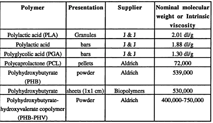
Figure 4.5 Total DNA content of HOS cells cultured on polymers over 0-72 hour culture period.
111
Figure 4.6 Total ^H-thymidine incorporation into cells cultured on polymers over 0-72 hour ciüture period.
111
Figure 4.7 %-thymidine incorporation/pg DNA into HOS cells cultued on polymers over 0-72 hour culture period.
112
Figure 4.8 Total DNA content of HOS cells cultured on polymers over 0-120 hour culture period.
113
Figure 4.9 Total ^H-thymidine incorporation into cells cultured on polymers over 0-120 hour culture period.
113
Figure 4.10 %-thymidine incorporation/pg DNA into HOS cells cultued on polymers over 0-120 hour culture period.
114
Figure 4.11 Response of HOS cells expressed as a percent of cells on TCP when cultured on polymers.
Figure 4.12 Total DNA of HOS cells expressed as a percent of cells on 115 TCP when cultured on PCL, PHB and PHB-PHV following
incubation in various solutions for 5 weeks.
Figure 4.13 Response of HOS cells expressed as a percent of cells on 116 TCP when cultured on PCL following incubation in various
solutions for 5 weeks.
Figure 4.14 Response of HOS cells expressed as a percent of cells on 117 TCP when cultured on PHB following incubation in various
solutions for 5 weeks.
Figure 4.15 Response of HOS cells expressed as a percent of cells on 117 TCP when cultured on Pîffi-PHV following incubation in
various solutions for 5 weeks.
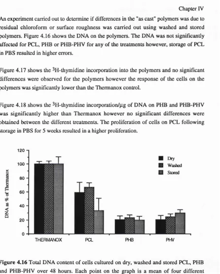
Figure 4.16 Total DNA of HOS cells expressed as a percent of 118 Thermanox when cultured with dry, washed and stored
polymers.
Figure 4.17 Total ^H-thymidine incorporation into HOS cells expressed 119 as a percent of cells on Tkrmanox when cultured with dry,
washed and stored polymers.
Figure 4.18 3jj-thymidine incorporation/|Xg of expressed as a percent of 119 cells on Thermanox when cultured with dry, washed and
stored polymers.
Chapter 5
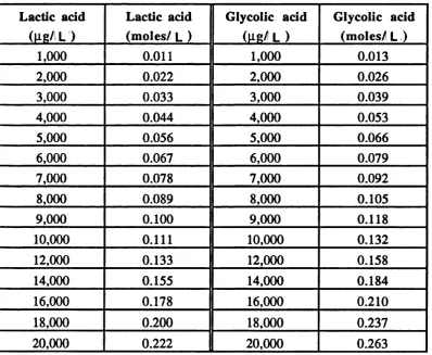
Table 5.1 & LA, GA and D3-HB concentrations in pg/ml and their 127 5.2 equivalent in moles/mL
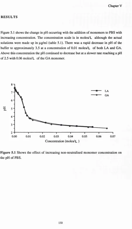
Figure 5.1 Effect of increasing LA and GA monomer concentration on 130 the pH of PBS.
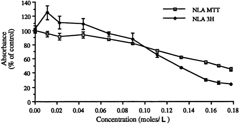
Figure 5.2 MTT conversion and ^H-thymidine incorporation by HOS 132 cells cultured in the presence of NLA.
Figure 5.3 MTT conversion and ^H-thymidine incorporation by HOS 132 cells cultured in the presence of NGA.
Figure 5.4 MTT conversion and ^H-thymidine incorporation by HOS 133 cells cultured in the presence of NNLA.
Figure 5.5 MTT conversion and ^H-thymidine incorporation by HOS 133 cells cultured in the presence of NNGA.
Figure 5.6 MTT conversion and 3H-thymidine incorporation by HOS 133 cells cultured in the presence of D3-HB.
Figure 5.7 Scanning electron micrographs of HOS cells cultured with 136 NLA at various concentrations.
Figure 5.9 Scanning electron micrographs of HOS cells cultured with NNLA at various concentrations.
138
Figure 5.10 Scanning electron micrographs of HOS cells cultured with NNGA at various concentrations.
139
Figure 5.11 Scanning electron micrographs of HOS cells cultured with D3-HB at various concentrations.
140
Figure 5.12 Light micrographs of MTT crystals growing out of HOS ceUs.
142
Figure 5.13 HOS cells stained for apoptosis in the presence of monomers.
143
Figure 5.14 Transmission electron micrographs of HOS cells cultured in the presence of monomers for 24 hours.
144
Figure 5.15 Osmolality changes in growth medium with increasing neutralised monomer content
147
Figure 5.16 Osmolality changes in growth medium with increasing non neutralised monomer content
147
Figure 5.17 MTT by HOS cells cultured in the presence of NLA 149
Figure 5.18 MTT conversion by HOS cells cultured in the presence of NGA.
149
Figure 5.19 MTT conversion by HOS cells cultured in the presence of NNLA and G A
150
Figure 5.19 MTT conversion by HOS cells cultured in the presence of D3-HB.
150
C hapter 6
Figure 6.1 MTS conversion by THP-1 cells cultured on various polymer surfaces.
160
Figure 6.2 SEM micrographs of THP-1 cells cultured on glass after 48 hours.
162
Figure 6.3 SEM micrographs of THP-1 cells cultured on PLA after 48 hours.
162
Figure 6.4 SEM micrographs of THP-1 cells cultured on gamma irradiated PLA gdter 48 hours.
162
Figure 6.5 SEM micrographs of THP-1 cells cultured on PCL after 48 hours.
163
Figure 6.6 SEM micrographs of THP-1 cells cultured on PHB after 48 hours.
Figure 6.7 SEM micrographs of THP-1 cells cultured on PHB-PHV after 48 hours.
163
Figure 6.8 Light micrographs of non-specific esterase stained THP-1 cells after 5 days in culture on Thermanox.
164
Figure 6.9 lig h t micrographs of non-specific esterase stained THP-1 cells after 5 days in culture on PLA.
164
Figure 6.10 Light micrographs of non-specific esterase stained THP-1 ceUs after 5 days in culture on PCL.
164
Figure 6.11 Light micrographs of non-specific esterase stained THP-1 cefis after 5 days in culture on PHB.
165
Figure 6.12 Light micrographs of non-specific esterase stained THP-1 cells after 5 days in culture on PHB-PHV.
165
Figure 6.13 MTS conversion by THP-1 cells cultured with neutralised LA at 0-24 and 24-48 hours.
167
Figure 6.14 MTS conversion by THP-1 cells cultured with non neutralised LA at 0-24 and 24-48 hours.
167
Figure 6.15 MTS conversion by THP-1 cells cultured with neutralised GA at 0-24 and 24-48 hours.
168
Figure 6.16 MTS conversion by THP-1 cells cultured with non neutralised GA at 0-24 and 24-48 hours.
168
Figure 6.17 MTS conversion by THP-1 cells cultured with D3- hydroxybutyric acid at 0-24 and 24-48 hours.
CHAPTER I
Chapter I
Biomaterials have been in use for many centuries but it is only in the last three decades that their use has intensified and their use for novel clinical applications has increased. A
biomaterial is any material of biological or chemical origin which can be used for repairing or reconstructing tissues of the body. Biomaterials generally fall into two main classes,
degradable and non-degradable. Non-degradable materials consist of various metals,
ceramics and cements. They are used for applications that require high mechanical
strength and long term stability. The most common uses of non-degradable materials
include hip replacements and vascular prostheses. Synthetic degradable materials,
however, are relatively new and their clinical use, so far, has been restricted to materials
for drug delivery and as sutures. One of the first degradable materials used in suture form
was catgut, a collagen based material which is still in use today. There were, however, problems encountered with catgut such as non-uniformity in its mechanical properties and
rates of degradation. The material also induced inflammatory responses due to its sheep
origin (Albertsson and Ljungquist, 1981) this led to the development of synthetic degradable polymers which did not have the problems associated with materials of animal
origin. There is now interest in these materials for fracture fixation and tissue regeneration which has been brought about by the development of polymers that can be manufactured
to produce devices with high mechanical strength and "controlled** degradation characteristics.
Polymers are composed of molecules in long sequences which are linked to each other by covalent bonds. The long sequences are also termed macromolecules which can be linear
or branched (Young and Lovell, 1991). The branched polymers are basically linear chains
which have side chains and the degree of branching can be simple or complex depending
on the length of the side chains and the degree of branching. As well as linear and
branched polymers, there are network polymers which are three-dimensional structures in
which chains are connected to other chains. These are also termed crosslinked polymers
and are characterised by the degree of crosslinking. Macromolecules are formed by linking monomer molecules via the process of polymerisation where a monomer is the
basic component of the polymer. Polymers can be homopolymers or copolymers. A
homopolymer is a polymer made up from only one type of monomer whereas a
copolymer is made up of more than one type of monomer. An example of a homopolymer is polylactic acid (PLA), which is made from the polymerisation of lactide and the final
degradation product of this polymer is lactic acid, the monomer component. Several
copolymers of polylactic acid can be formed with polyglycolic acid (PGA) and one of
these is polyglactin which is 90% PLA and 10% PGA; the final degradation products of
this copolymer are lactic acid and glycolic acid. The properties of the various degradable
Chapter I
B iocom patibility testing: in-vitro and in-vivo
The definition of biocompatibility as it currently stands is "the ability of a material to
perform with an appropriate host response in a specific application" (Williams, 1987). This definition came about when materials were developed that were required to have
specific functions as opposed to being totally inert in the body. Biocompatibility
assessment is essential for pre-cUnical testing of materials intended for use as implantable devices. The testing procedures available for the assessment of biocompatibility should
give quantitative data using appropriate cell types. Assessment of biodegradable
polymers, however, is complex due to the degradation of the materials during testing and
the need to artificially age the materials in order to assess longer term effects. Due to the
wide variety of methods for material formulation which have different molecular weights the biocompatibility of many resorbable materials in the long term remains unclear.
Kirkpatrick et al (1997) in a review states that "combinative techniques" involving morphological analysis and molecular biology present a useful set of methods for the biocompatibility testing of materials.
Biocompatible in the in-vitro situation means that the material or any leachable products
from it do not cause cell death or impair cellular functions. Biomaterials used in-vivo should be biocompatible in both the short term and the long term during degradation of
the material and the release of degradation products. These include the bioresorbable materials, and bioactive materials. For example, a material that is intended for use as a
drug delivery vehicle wiU require different properties to one that will be used for tissue replacement A biomaterial designed for hip replacement will require high mechanical
strength, have low corrosion and degradation in addition to having good bone induction, protein adsorption and cell adhesive capabilities. However a material used for use for
vascular reconstruction will need to have good stability, low degradation and low protein
and cell adsorption properties.
In determining the suitability of a biomaterial for a specific application the material and site
of implantation play a major role. Degradable biomaterials are constantly changing with
time as the biomaterial undergoes bulk and surface degradation which leads to changes in the environment around the material Non degradable materials also undergo changes in
their chemical and structural nature but this change is not as dramatic and the time
involved for physical and chemical changes to occur is longer. Appropriate biocompatibility testing is an essential process in the development of implantable devices.
A material must be proved "safe" before it can be implanted into the body.
The Medical Device Directive states that a material to be used for medical devices should
Chapter I
Standardisation (ISO) is a world wide federation of national standards bodies. The ISO
10993 consists of a number of parts under the general title of "Biological evaluation of medical devices". The international standard ISO 10993-5:1992(E), tests for in-vitro cytotoxicity defines a series of guidelines that can help in selection of the most appropriate
test for biocompatibility screening. The three categories are extract tests, direct contact and
indirect contact tests (Wilsnack et al 1973). The other directives of ISO 10993 deal with
other aspects of testing such as in-vivo testing, which include systemic injection tests,
intracutaneous tests and the implantation test in animal models.
The definition of biocompatibility as defined by Williams is widely accepted as the general definition which is applied to all materials used for implantation purposes. However the
designation of "biocompatibiEty" for each current application and newly developing
applications needs to be determined and test methods need to be standardised. This is inevitably a long and difOcult task, but one that is necessary, as currently a whole range
of tests are performed which cannot be compared due to the differences in the
methodologies used. It is first necessary to determine what the requirements of each medical device are and then accordingly choose a material that will fulfil the criteria.
Applications generally fall into the headings; dental, orthopaedic, vascular implants, drug
delivery, transplantation, nerve regeneration, and soft tissue implants. A major requirement for all these applications is that the materials must be non-immunogenic, that
is they should not induce gross inflammatory or immune responses.
There is evidence, to suggest that toxicity at the cellular level is not easy to detect on
experimental animals (Guess et al 1967). They stated that "subtle toxicity" which developed slowly at the cellular level was difficult to demonstrate in animals. It could,
however, be detected by immunological studies using tissue culture models as these allow
the investigation of single effects without interferences from the whole immune system.
Adverse responses in-vivo include decreased cell viability and proliferation of cells around the site of implantation or any reaction not normal for that tissue. An inflammatory
reaction can also occur due to the influx of inflammatory cells to the area. Although some
inflammation is necessary for the wound healing process to proceed normally, if this inflammatory reaction continues it can lead to chronic inflammation and granuloma.
Biocompatibility testing must address all of these parameters using both in-vivo and in-
vitro tests.
In-vivo testing usually involves the implantation of a test device (which has been sterilised by an appropriate method) into an animal model. Such studies yield information on the
long term biocompatibility of materials and the effect of the material on the immune
Chapter I
the difficulty in quantifying speciHc cellular and biological events. However several
methods have been developed which aim to isolate cell-biomaterial interactions in-vivo
one of which is the cage implant system described by Marchant et al (1983). This allowed for the evaluation of the effect of cellular and humoral components of the exudate which
surround polymeric materials following implantation. In this method, the biomaterial was placed inside a stainless steel cylindrical cage which was then implanted subcutaneously
into a rat model. Aliquots of the exudate surrounding the implant were removed using a
syringe and analysed. The types of cells present in the sample can then be identified.
Polymorphonuclear leukocytes (PMNs) and macrophages become phagocytic when
cellular debris or foreign particles are present. When phagocytosis is initiated, the cell
becomes metabolically active and there is a respiratory burst resulting in the production of
hydrogen peroxide and oxygen free radicals and anions. These free radicals are effective
in killing many types of bacteria and have been linked with initiating the degradation of some polymers. The role of the macrophage in the inflammatory process is very important
because it is the longest surviving and most active of the inflammatory cells.
Although in-vivo testing cannot be completely replace in-vitro tests steps are being taken to move away from animal models through the development of better in-vitro tests. In-
vivo testing is also more expensive, lacks control due to animal and species differences and problems are frequently encountered with obtaining ethical permission to carry out
tests.
In-vitro biocompatibility testing involves studying the behaviour of various cell types in response to a particular test agent. The simplest of these can be carried out by monitoring
cell viability and proliferation when in contact with a biomaterial. Cell viability can be
monitored by the incorporation of various dyes such as trypan blue and erythromycin red
which are exclusion or inclusion dyes. Trypan blue stains dying cells blue and
erythromycin red stains non viable cells pink. These are tests which can give rapid
estimations of the number of viable cells present. Coulter counters and cell sorters utilising fluorescent dyes can be used to distinguish dead cells from live ones to give a
more accurate cell count
In-vitro testing using cell culture methods has a number of advantages over in-vivo testing, in that the response of a variety of cells can be compared and evaluated over a
shorter time period in a reproducible and controlled maimer as compared to in-vivo tests.
Chapter I
The "appropriate” cell type is determined according to the application of the material in
question. If the device is designed for orthopaedic fixation osteoblasts will usually be
cultured on the surface and if the material is going to be used for vascular reconstruction
blood compatibility will be investigated. Different material characteristics are needed depending on the final application. For the two applications the material is expected to
behave differently; for example, in orthopaedic fixation, the material will need to have
good protein adsorption characteristics to aid cell adhesion and proliferation, while for the blood contact material it needs to have low protein adsorption and low cell attachment
Although the time points involved for in-vitro testing are short as compared to in-vivo testing, in-vitro testing does yield information on the cellular morphology and retention of
phenotype of the individual cells. In-vivo testing is usually long term and can last several
years. The use of in-vitro and methods which yield polymers that have been artificially degraded the time periods involved for in-vivo testing can be significantly reduced.
Methods such as storage of polymers at high temperature, and in enzyme solutions which
accelerate their degradation compared to in-vivo are termed artificial degradation. This has
been demonstrated by several groups who have artificially degraded polymers before
implantation into animals (Bergsma, 1995). In this study PLLA was predegraded at
elevated temperatures and it was found that the follow-up times could be reduced; the results were comparable with the in-vivo findings which had not utilised pre-degraded particles. Long term biocompatibility testing is particularly important for degradable materials which are constantly changing their chemical and physical structures and
releasing degradation products which may be detrimental to the cells adjacent to the
implant
Before carrying out in-vitro testing it is important to determine the cell type(s) that will be
used for the study. It is important to use an appropriate cell type for that particular
application. For bone implant materials the cells in contact with the device will be
osteoblasts, osteocytes and osteoclasts. For blood contact materials none of these cells
will be in contact with the material in the long term. It is therefore important to use cells
that are most representative of those encountered in the in-vivo situation. Primary cells are
a good choice but obtaining sufficient cells to carry out large numbers of tests can be
problematic as well as obtaining normal human tissue for a particular cell type. Cell lines
are therefore more commonly used, and sometimes more useful as they can replicate
faster and have a well defined phenotype. For other devices fibroblasts, epithelial cells
and hepatocytes can be used while studies of the immune response can utilise
monocyte/macrophages and other cells of the immune system. In-vitro testing allows the
standardisation of test methods and reduces the need for large numbers of experimental
animals. It also allows qualitative determination of signs of cell damage, detected by light
Chapter I
Although quantitative methods are valuable for direct comparisons for biocompatibility
testing, qualitative analysis of cells on materials is crucial for determining the morphology
and behaviour of cells. Differences in the morphology of various cell types can be
observed and cytotoxic effects which lead to dramatic changes in morphology of the cells
can be examined at the light microscope level or in more detail at the electron microscope
level. Microscopy is a vital and often underused tool in determining the biocompatibility
of biomaterials. Light microscopy allows any damage occurring to the nucleus and
cytoplasm to be viewed. The cells can be lysed due to damage caused by high concentrations of degradation products or due to the acidic pH. Scanning electron
microscopy in combination with light microscopy can give valuable information on the morphology of the cells on the polymer surfaces. Most cells have a characteristic shape
but this can be influenced by the substrate to which they are attached. Transmission
electron microscopy is a useful method that can be used to determine changes occurring at
the ultrastructural level such as damage to cellular organelles, for example the Golgi apparatus and the mitochondria.
A major problem with in-vitro cell culture systems is dedifferentiation of cells in culture
which should not be confused with differentiation. A cell differentiates when it stops proliferating and matures to express its phenotype; for example an osteoblast becomes a
mature osteoblast and starts producing alkaline phosphatase, osteopontin and osteonectin. An osteogenic cell line MC3T3-E1, for example was found to be able to differentiate into osteoblasts and osteocytes and formed calcified bone tissue in-vitro (Sudo et al 1983). A
cell may dedifferentiate when it has been passaged in culture a number of times and its
phenotype is lost. Several cells have been found to de-differentiate in-vitro such as chondrocytes from chick embryos which, when cultured in-vitro for long periods, ceased
to synthesize chondroitin sulphate (Holtzer et at 1970). This de-differentitaion of cells is
one of the disadvantages of in-vitro testing and precautions must be taken to monitor cell
dedifferentiation. In our system cells over a passage of 14 were not used for the biochemical or qualitative studies.
The majority of in-vitro biocompatibility tests described in the literature involve the
morphological assessment of cells on materials, usually by light or scanning electron
microscopy. The time courses for cell exposure are usually short term and at the most last
for a few days. The types of cells used for these tests are different in source and species.
This can give conflicting results and makes comparisons between different laboratories
difticulL It has been demonstrated that different cells will behave differently on the same
material surface. Differences were observed between periodontal ligament (PDL)
fibroblasts and L929 cells, (a mouse tibroblast cell line) when measuring toxicity of a
Chapter I
cells were less sensitive than the L929 cells, and ultrastructural differences between the
two cell types were observed. Different cell types, for example fibroblasts and
osteoblasts, were found to respond differently to a range of biomaterials (Hunter et al
1995).
Despite all the problems associated with this type of screening of materials, in-vitro tests
can provide fast, reproducible and useful data on the biocompatibility of a material. This
is possible for in-vivo tests due to the differences in site and species and the size and type
of materials used. In-vitro tests can be used to determine initial attachment of different cell types on various material surfaces and for carrying out "long term" biocompatibility
testing. Cytotoxicity of leachable compounds released from certain materials such as
resorbable polymers can also be measured.
The applications for in-vitro tests are vast, but what is required is a standardised method
of carrying out these tests. A standardised protocol needs to be developed that sets out
tests which provide meaningful results, that can then be used for comparison within groups carrying out similar tests. It is not only the cell types that vary but also the test
materials and cell seeding density. For example polylactic acid may be the material under
investigation but the method of formulation and sterilisation may be different A report by
Engelberg & Kohn (1991) stated that new applications of materials require that they be fully characterised and have defined material properties which is currently not the case.
They also stated that there was a lack of reliable data and tests carried out and materials
could not be compared due to the variability in the samples.
Cells death and apoptosis
Cell death is a major concern with regard to biocompatibility testing. If cells die on a
device or a material under investigation it is obvious that something leachable from the
biomaterial or the material is killing the cells. There are however different types of cell
death, notably necrosis and apoptosis.
Necrotic cell death usually occurs as a result of injury in the presence of excessive toxins.
Apoptosis, also termed cell suicide or programmed cell death, was first described by Kerr
et al (1972) as a mechanism for the natural elimination of cells from the body and is an important mechanism for tissue homeostasis. When a cell is undergoing apoptosis various
morphological and ultrastructural changes occur in the cell that can be detected by light
microscopy or TEM. In apoptosis the cell first detaches from adjacent cells and then
looses surface structures such as microvilli. The organelles within the cytoplasm shrink
and, as a result, the cell also shrinks. Cytoplasmic, plasma and nuclear membrane
Chapter I
organelles, however, remain intact and the mitochondria remain viable late into the
process. The chromatin condenses and the nucleus can break up. The condensed chromatin is one of the major features utilised in TEM to detect apoptotic cells. The
methods for detecting cells undergoing ap o p to ^ are still being developed. There is also
the problem in obtaining cells that are undergoing apoptosis in-vivo as apoptotic cells are
rapidly phagocytosed.
Biodegradable polymers
There has been increasing interest in degradable polymer systems for use in biomedical
applications such as drug delivery (koosha et al 1989; Wagenaar and Miller 1994), fracture repair (Ewers 1990; Eli et al 1992), tissue remodelling (Freed et al 1993; Gilbert et al 1993; Puelacher et al 1994; Chaput et al 1996) and soft tissue implants (Mooney et al 1996). Degradable materials have certain advantages that make them desirable for some orthopaedic applications. Their degradation rates and tensile strength can be controlled by
varying molecular weight and, for copolymers, varying the ratio of the components can
also dramatically affect their degradation rates (Nakamura gr al (1989). The majority of
degradable materials have been poorly characterised using in-vitro methods which have usually involved the assessment of fibroblast, and some osteoblast and hepatocyte, growth on them. There has been considerable work done with degradable polymers in-
vivo but the mechanisms of cell attachment and proliferation on these polymers have not been fully investigated. Moreover the effect of the cellular activity on the degradation of
the polymer and the effect of the degradation products on the cells are not well
understood.
Polymers can be separated into three groups namely thermoplastics, elastomers, and
thermosets. The thermoplastics, which constitute most of the degradable polymers, are
further divided into crystalline and amorphous (non crystalline) polymers. Thermoplastic
polymers are the most commonly used polymers and can be moulded into any shape by extrusion or injection moulding. Thermoplastics do not crystallise easily upon cooling as
the polymer chains have to form highly ordered structures from their entangled state in
order to become crystalline. What usually occurs with most polymers is some crystallisation in certain regions of the polymers resulting in polymers with crystalline
regions and amorphous regions, thus termed semicrystalline polymers. The melting point (Tm) is used to characterise the crystalline regions and the glass transition temperature
(Tg) is used to characterise the amorphous regions. The Tg is the temperature at which the
Chapter I
Biodegradable polymers are categorised under several generic names some of which
include polyesters, polyurethanes, polyamides, polyureas, polyanhydrides,
polyphosphazanes, polyacrylates and polycyanoacrylates. The polymers most studied
studied and those under intensive research currently are the polyesters which include PLA
and PGA. The first synthetic absorbable material was developed in the 1962 by the
Cyanamid Corporation using polyglycolic acid which was marketed under the trade name Dexon in the late 1960s (Gilding and Reed, 1979). This was followed by Medfit, a
homopolymer of glycolic acid and Vicryl and polyglactin 910, copolymers of polyglycolic acid and polylactic acid as sutures.
Aliphatic polyesters:
The biodegradable polymers studied belong to a group of compounds known as aliphatic
polyesters. These include polylactic acid, polyglycolic acid, polyhydroxybutyric acid and poly e caprolactone. These are the most extensively studied of the degradable polymers
which also include polymalic acid and poly dioxanone. They all belong to the sub group poly a hydroxyacids for which the general formula is -(-O-CHR-CO-)n.
The structures of some of the most commonly used degradable polymers are shown below.
H O
I
II
- ( r O - C - C - ^ H
Polv fflvcolide’) (PGA> -[- O - CH2 - CO
” Î
t-0—c —c —>n
CH3
-]-Chapter I
H H H H H O
^ O — Ç — Ç— Ç — 0 — c — C—
H H H H H
Polv fe- caprolactone^ tPCU -[- O - (CHi); - CO -]■
H H O
I
I II
^ o — c — c — c ^
I
I
CH) H
n
Polv (B-hvdroxvbutvratel PHB -[-O - CH2 CH (CH3) - C O -]■
H H O
I
I
II
- e o — C --- C— c —)
I
I
CH2CH3 H
n
Polyhydroxyvalerate (PHV) O - CH2 CH (CH2 CH3) CO -
]-Poly(lactic) and Poly(glycolic) acids
Polylactic acid and Polyglycolic acid aie the most widely studied and clinically used of the
degradable polymers available. They have been used mainly in the form of sutures,
plates, screws and as vehicles for drug delivery. PGA and PLA, made by simple
polycondensation, yield low molecular weight polymers which are not suitable for
devices that are required to have high mechanical strength. The preferred method of
polymer formation is ring opening polymerisation of cyclic diesters using catalysts, which
yields high molecular weight polymers with longer degradation times (Gilding, 1979).
Diagram of Ring opening polymerisation
HRCr I
n
I I --- —[ —O—CO—C —O—CO—c —] —Chapter I
Polyglycolic acid is a highly crystalline linear aliphatic polyester with a melting point of
224-226°C and a glass transition temperature (Tg) of 36°C. PGA is not easily soluble in organic solvents but is easily degraded in-vivo or under physiological conditions in-vitro.
Due to its hydrophilicity it has a rapid degradation rate and complete resorption in-vivo usually occurs between 2-14 weeks depending on the initial molecular weight of the
polymer. The methods of formulation and sterilisation influence its crystallinity and
permeability which, in turn, affect its degradation. PGA can be copolymerised with other
polymers such as PLA to yield copolymers which have a slower degradation rate and greater mechanical strength (Gilding and Reed 1979; Athanasiou etal 1996).
PGA was the first synthetic polymer to be used as a suture, however its low mechanical
strength and rapid degradation rates have not allowed it to be considered for uses which
require greater stability such as fracture fixation. Recently, however, methods which yield
self reinforced PGA rods have been studied for use as osteosynthetic materials due to the
higher mechanical stability achieved for these polymers. (Tormala et al 1991; 1993)
PLA can exist in two stereoisomeric forms, D-PLA and L-PLA as it is formed from the polycondensation of lactide which is a chiral molecule.
D-PLA
Ï Î
- e O — C — C —);
CH3
L-PLA
CH3 O
I
"
f - o —c — c —
y—
I
H
A racemic mixture of D and L PLA yields D, L-PLA which is amorphous although D-
PLA and L-PLA are semicrystalline. L-PLA is the most widely studied of the forms as it
is hydrolysed into L(+) lactic acid which can be metabolised by the body and is eliminated
Chapter I
for applications in drug delivery where it can form a homogeneous mixture with a protein. L-PLA is used more for orthopaedic fixation devices where high mechanical strength is
required (Engelberg, 1991). D-PLA is not as extensively used as the other forms due to
its low mechanical strength; as this form is more amorphous it is used in drug delivery applications.
There has been a large amount of work carried out on the degradation mechanisms of PLA and PGA and it is understood that degradation is faster in-vivo than in-vitro (Claes
1992). Strength retention of self-reinforced poly L-lactide screws and plates was
investigated in-vivo and in-vitro and loss of strength was faster in-vivo than in-vitro (Suuronen et al 1992). Further work demonstrated that degradation was faster in-vivo and
that hydrolytic degradation in semi crystalline poly L-Lactic acid occurs preferentially at
the amorphous regions resulting in increased brittleness both in-vivo and in-vitro (Laitinen et al 1992). The porosity of the material was also found to affect the degradation
rates where Kobayashi etal (1991) investigated the degradation of three types of films
porous, non porous and a combination film in-vitro and in-vivo. The non-porous film degraded faster than the porous film over a 180 day period and the presence of hydrophilic units in the main polymer chain also resulted in increased degradation rates.
The degradation of PGA occurs mainly as a result of hydrolytic scission and to a lesser
extent by enzymatic degradation (Bostman etal 1992). Kulkami etal (1971) demonstrated that poly L(+) lactic acid of high molecular weight could be used in fibre film or coat form and, when implanted into pigs and rats, was completely metabolised without
accumulation in the body organs. It was also found that as D,L-lactic acid is less ordered than L(+) lactic acid, and it degrades faster (Maduit et al 1996; Gerlach and Eitenmuller
1987). Cutright et al (1971) tested sutures for internal fixation of fractures of the mandible
of rhesus monkeys, degradation was slow and complete degradation was not complete
even after 12 weeks. Further work by Cutright et al (1974) on PLA and PGA showed that
degradation was initiated in growth of capillaries and/or phagocytes. The cylindrical
polymer fragments were replaced by a fibrous connection and the resorption times were
dependent on the ratio of PGA to PLA. A mixture of 25% PLA and 75% PGA was
degraded most rapidly followed by 50% PLA and 50% PGA, 75% PLA and 25% PGA
with 100% PLA taking the longest Paivarinta et al (1993) studied degradation in-vivo and found that there was no PGA remaining after 36 weeks, whereas PLA was virtually
intact at 48 weeks; the materials were used as screws to fix femoral osteotomies in
rabbits.
Two types of microbeads of PLA were prepared by solvent evaporation and implanted
Chapter I
(vinyl alcohol) as a residue from the evaporation process at the surface of the beads
(Anselme et al 1993).
Composites of PLLA which are resorbable have good initial mechanical properties but
these are quickly lost with exposure to an aqueous environment (Andriano et al 1992).
PLLA poly (L-lactide) mesh, sheets, microfilaments and mesh cylinders with fresh
autogenic particulate cancellous bone and marrow (PCBM) were implanted
subcutaniously into dogs. The inflammatory response to PLLA was similar to a polypropylene (PP) control. Three months after implantation the number of histiocytes
and mononucleate giant cells increased in number as the monofilaments degraded.
Copolymers with a higher lactic acid content and purified polymers such as poly L-
Lactide have been shown to have a lower rate of degradation (Kinoshita, 1993).
Poly cap rolactone
Poly e caprolactone (PCL) is a semicrystalline aliphatic polyester that has a glass
transition temperature of -60°C and a melting point of 60°C which is low compared to the other polymers. The monomer of PCL, e-caprolactone, is manufactured by oxidation of
cyclohexane with peracetic acid. The polymerisation of e-caprolactone can be by four
different methods: anionic, cationic, co-ordination and radical. Each method has a different degree of control over the molecular weight, molecular weight distribution and
chemical structure which in turn determine the permeability and degradability of the polymer (Pitt et al 1990). The crystallinity of the polymer varies with its molecular weight; usually polymers with molecular weights of over 100,000 have a low crystallinity of 40% which rises to 80%, with a decrease in molecular weight down to 5000. The
crystallinity is important in determining permeability and degradability as the bulk
crystalline regions are inaccessible to water. PCL is readily soluble in organic solvents
such as tetrahydrofuran (THF), chloroform, toluene, benzene, cyclohexane and
dihydropyran at room temperature. It is readily degraded by microorganisms and is
hydrolysed under physiological conditions. Enzymatic surface erosion is also thought to occur although PCL degrades more slowly than PLA and PGA (Pitt et al 1984; Jarret et al
(1984).
Because of its slow degradation rate PCL is widely considered for long term implantable
drug delivery systems and as degradable packaging and bottles. PCL is currently
undergoing clinical trails as a one year contraceptive device by Capronor in Europe and
Asia. PCL readily forms blends with other polymers such as PLA, cellulose propionate
and cellulose acetate butyrate and can be copolymerised with PLA, and PHV which
influence its degradation rate and mechanical properties (Koleske, 1978; Feng et al 1983).
Chapter I
but it can be used in drug delivery applications where biomechanical considerations are
not of such crucial importance. The exact mechanism of degradation of PCL is not
understood, but chemical hydrolysis is thought to play a large part and workers have reported that the molecular weights of polymers determine the rate of degradation
(Albertsson and Ljungquist 1981; Pitt et al 1981; Piskin 1994). Current research in the field of degradation points towards the role of enzymes, peroxides, free radicals,
phagocytic cells and lipids in the degradation of polymers, but there is no standardised method of measuring or preventing this degradation. Heterochain polymers, in particular
those containing oxygen and nitrogen atoms in the main chain, are generally more
susceptible to hydrolysis. Polymers susceptible to hydrolysis are those in the polyamide,
poly amino acid, some polyurethane and polyester groups (Ali et at 1992; All etal 1993).
PCL is slower to degrade than PLA but it has better permeability.
Upon implantation degradation of the polymer begins by random hydrolytic chain
scission of the ester bonds in the polymer backbone. This reduces the viscosity and the
molecular weight of the polymer (Pitt et al 1981). The same rate is observed in water at 40°C and is not altered with a ten-fold change in surface to volume ratio, indicating a bulk
process. There is no weight loss during the first stage suggesting an autocatalytic process
where the liberated carboxylic end groups catalyse the cleavage of additional ester groups. The second stage is a decrease in rate of chain scission and the initiation of weight loss. There is also an increase in crystallinity leading to brittleness and possibly the break-up of
the polymer producing small particles which can be phagocytosed. The process is autocatalytic and, for polymers with molecular weights of over 5000, weight loss is not
significant but once degradation is initiated weight loss rate depends on particle size.
Enzymatic degradation is thought to play a role in the surface degradation of polymers as
rapid surface erosion is observed following implantation. Increasing the crosslinking
reduces the susceptibility of these polymers to enzymatic attack, by restricting the mobility
of the chains; the ester groups cannot assume a configuration able to interact with the
active site of the enzyme (Pitt et al 1981). Feng et al (1983) combined the two polymers to
modify and vary their properties. In-vitro degradation of the polymer films was investigated in deionised water for up to 70 days and degradation of the copolymers was
found to be faster than for PCL and slower than for PLA homopolymers corresponding to
the ratio of PLA to PCL.
Pitt et al (1981) looked at the degradation of PCL in-vivo and concluded that the degradation mechanism involves random chain scission by hydrolytic cleavage of ester
groups. The rate was not dependent on geometry and enzyme degradation was excluded
as the same rate of degradation was observed in water at 40°C. The kinetics of the chain
scission indicated an autocatalytic process i.e. the generation of carboxylic acid ends by
Chapter I
PCL and concluded that uncrosslinked PLA and PCL degraded in-vivo by a random chain
scission process, aqueous hydrolysis of ester links. Also, long induction periods were
required before any bioerosion occurred (Pitt et al 1984). Crosslinked polyesters are
affected by the same hydrolytic process, but also undergo attack by enzymes at the
surface. This is because the mobility of polymer chains for a non crystalline polymer
allows the ester group to change its orientation in order to interact with the enzyme. This enzymatic attack becomes negligible with an increase in cross linking.
Poly(hydroxybutyrate)
Poly(hydroxybutyrate) (PHB) is a linear, thermoplastic polyester of 3-hydroxybutyric
acid produced by the industrial fermentation of gluco se by Alcaligenes eutrophus (Doyle
et al 1991). In bacteria, its purpose is to provide intracellular storage of carbon and energy. In the fermentation process the growth medium has limited phosphorous thus
arresting bacterial growth. Once this has happened, glucose is added to the growth
medium and, as the cells cannot convert glucose to protein, PHB is formed instead and
accumulates in the cells. The level of PHB can be as high as 80% of the dry mass of
cellular material. The polymer is extracted by aqueous or solvent extraction and polymers with molecular weight of 5 x 10^ - 1.5 x 10^ can be produced (Vert et al 1986). The
solvent extraction method yields polymers of higher molecular weight compared to the aqueous method which is cheaper and safer but yields a lower molecular weight polymer
(Luzier, 1992). PHB-PHV is formed if an organic acid such as propionic acid is added with the glucose. This results in the incorporation of HV units within the HB segments to
form the PHB-PHV copolymer.
PHB can be degraded by soil bacteria and in-vivo it degrades into D-3-hydroxybutyric
acid which is found in normal blood. PHB is available both as a homopolymer and copolymer with polyhydroxyvalerate under the trade name Biopol. The homopolymer is
crystalline, but the copolymers are less crystalline and thus readily processed. The
polyester undergoes hydrolytic and enzymatic degradation in a physiological environment
It is currently used in the production of plastic bottles, mouldings, fibres and films. PHB
has also been blended with plasticizers and found to be degraded by PHB depolymerases
and lipases although the enzymes degraded the polymer films to varying degrees suggesting specificity of the enzymes (Abe et al 1994).
A study by Boeree et al (1993) concluded that injection moulding of PHB resulted in a
material with satisfactory mechanical properties for use in orthopaedic use. PHB/HA composite has been found to be bioactive and have good bonding with the surrounding
tissue (Yasin et al 1990, 1992, Knowles and Hastings 1992). Yasin et al (1989) studied
Chapter I
sodium alginate at various pH values with temperatures at 37“C and 70“C. The hydrolytic degradation was found to be affected by the presence of polysaccharides, pH and
temperature. The highest rate of degradation was found to be in pH 10.6 buffer at 70”C.
The degradation of PHB homopolymer is still not fully established. Work by Miller and
Williams (1987) showed that in-vivo degradation only occurred if the polymer was pre degraded by 10.0 Mrad of y irradiation and the rate was faster in-vivo than in-vitro. Copolymer additions of PHV 8% and 17% did not increase degradation and at high
temperatures retarded the rate of degradation. They concluded that biodégradation of PHB
and its copolymers could not be predicted with confidence and that more work was needed to establish the correct polymer properties for degradation to take place. Work by
Knowles and Hastings (1991) concluded that the slower degradation characteristics of
PHB as compared to PLA and PGA, would make this polymer more suitable for use in
applications where longer resorption times were needed; however, more work is needed
to determine the degradability of the polymer. pH has a considerable effect on the
degradation of PHB and its copolymers. Changes in surface gloss were measured as an
indication of polymer degradation, using a technique called goniophotometry which is the measurement of reflected light as a function of viewing angle. The surface roughness or
gloss factor are measured by analysing the reflectance pattern from an incident beam of light. Solutions with pH 3, 6, 7, 9, 10 and 11 were used to degrade the polymers. A
decrease in molecular weight was observed for all polymers irrespective of pH, but the
alkaline solutions produced a more aggressive degradation resulting in areas with deep surface erosions. The degradation of PHB-PHV was investigated by Holland et al (1987;
1990). The polymer was resistant to degradation for a period of one year after which accelerated degradation was observed and an increase in porosity increased the diffusion,
of the degradation products more effectively.
Immune response
One of the first things encountered by a material following implantation in the body is
blood, which contains cells of the immune system. The immune system is the body's
defence system and its primary aim is to defend the body against invasion by foreign
organisms, such as bacteria and viruses. Before dealing with the invasion it has to first
recognise foreign organisms and then eliminate them. This may lead to hypersensitive
reactions characterised by an inflammatory response. This is characterised by increased
blood flow to the affected area, with an increase in capillary permeability allowing
molecules and cells to cross the endothelium and leukocytes to migrate from the blood into the surrounding tissue.
The immune system may be divided into two categories; specific and non-specific (Roitt,
Chapter I
innate mechanisms can be further subdivided into humoral and cellular defence
mechanisms. A number of plasma proteins are involved in the humoral defence mechanism. These include lysozymes, interferons, and the complement system.
Activation of the complement cascade of proteins leads to the generation of a membrane
attack complex, C3bBb which lyses, bacteria. C3bBb attaches to foreign bodies labelling them for phagocytosis and other leukocytes, causing them to move into the area where the
chemoattractant is concentrated. In addition to being chemoattractants, both molecules are
powerful anaphylatoxins, that is, they bind to and cause degranulation of the mast cells
and basophils with release of vasoactive amines, histamine, leukotrienes and other
inflammatory mediators. The net effect of the innate response is contraction of vascular smooth muscle, increased vascular permeability and emigration of neutrophils and
monocytes from the blood vessels. These blood cells engage in phagocytosis of the
complement-bound foreign body or alternatively, degranulate with further production of
inflammatory mediators with the overall result being the production of a local
inflammatory response.
The protection offered by the specific or acquired immune system is due to a group of cells known as lymphocytes: B lymphocytes and T lymphocytes. Each individual cell has
a receptor on its surface which is specific for a particular antigen. In the specific immune response the antigen is processed by antigen presenting cells (APC). These are a
heterogeneous population of leukocytes, which include B cells and macrophages, that can take up antigen and express part of it on their surface. A sub-set of the T cell population,
helper T cells (Th)» recognise the antigen in combination with class n MHC. This causes them to secrete the T cell growth factor Interleukin 2 (IL2), which stimulates T cell
proliferation. Th also "help” the B cells which recognise the same antigen by stimulating
them to differentiate into plasma cells which secrete antibodies. The stimuli for this differentiation are the ILl from the APC and IL4 from the T cells. Antibodies bind to the
foreign body maiking it for killing by phagocytosis or by some other mechanism.
Delayed-type hypersensitivity occurs when antigen becomes trapped in the macrophage
and cannot be cleared, and is mediated by the specific immune system. This causes Th
cell activation and proliferation which produce a number of lymphokines, for example
macrophage activating factor (MAP), which induce inflammatory reactions, attract and
activate macrophages with the further release of inflammatory mediators. T cells are also
activated by class I MHC to become cytotoxic to the cells carrying the antigen.
Continuous stimulation by persisting antigen leads to further influx of macrophages, their differentiation into giant cells and eventually the formation of chronic granuloma.
Biomaterials when implanted into the body, are essentially foreign bodies, which are
Chapter I
depend on the material under investigation. Therefore the initial host response must
encompass a repair process. Neutrophils are the first cells to reach the site of infection»
attracted by chemotactic factors such as complement» with their predominant role being
phagocytosis. The phagocytosed body is fused with a lysosome and destroyed by lytic enzymes. The neutrophil respiratory burst involves a sharp uptake of oxygen and results
in the generation of toxic oxygen products» such as superoxide anions and hydroxyl radicals. If the material cannot be phagocytosed the neutrophil will release destructive
substances into the extracellular environment in a process known as frustrated phagocytosis. Any of these parameters can be used as an indicator of neutrophil activation
by biomaterials. Once at a site of the biomaterial» there are two ways in which neutrophils
could act» either by respiratory burst» or by the release of lysosomal enzymes.
The monocyte/macrophage response to biomaterials has been the most extensively studied
of all the cells of the immune system (Bonfield and Anderson 1993; Benahamed et al
1997) and have been implicated in causing increased degradation of the materials.
Macrophages have been shown to infiltrate the implant site after implantation of a biomaterial and, once activated» macrophages have a much altered metabolism. This
includes altered phagocytic ability and increased lysosomal enzyme release as well as
secretion of cytokines and growth factors. Macrophage activation by materials can be
assessed by measuring any of the above parameters. The effect of metals on macrophage viability has been determined by a number of researchers but the effect of degradation products and the effect of degradable polymers on macrophages both in-vivo and in-vitro
has not been as extensively studied.
Activated macrophages display altered morphology which can be identified
microscopically. Macrophages cultured with a range of biomaterials were analysed by
scanning electron microscopy for alterations in morphology» attachment and cell density
(Miller et al 1989). After 24 hours in culture the cells displayed an activated morphology;
they were elongated or flattened with numerous filopodia for attachment The highest
density of morphologically active cells were seen on Dacron and polyethylene» while the
lowest were on polydimethylsiloxane (PDMS). Supernatants from these cultures were assayed for interleukin-1 (ILl). A good correlation between IL l secretion and
morphological activation was seen.
In-vivo macrophages are not the only cells which come into contact with the implanted biomaterial and it is not just the biomaterial that has an effect on the cells. There can be an indirect effect of biomaterial-stimulated macrophage secretion products on other cell types
such as fibroblasts (Bontield et al 1991» 1992; Greisler et al 1989) and endothelial cells
(Miller and Anderson» 1989). Supernatants from macrophages cultured on Dacron»