Research and Reports in Neonatology
Respiratory management of the preterm
newborn in the delivery room
Hesham Abdel-Hady Nehad Nasef
Neonatal Intensive Care Unit, Mansoura University Children’s Hospital, Mansoura, Egypt
Correspondence: Hesham Abdel-Hady Neonatal Intensive Care Unit, Mansoura University Children’s Hospital, Gomhoria Street, Mansoura 35516, Egypt
Tel +200 0527 8051 Fax +205 0223 4092 Email [email protected]
Abstract: The survival of preterm infants has improved significantly during the past several
decades. However, bronchopulmonary dysplasia remains a major morbidity. Preterm infants have both structural and functional lung immaturity compared with term infants, making them more likely to require resuscitation and more vulnerable to developing bronchopulmonary dysplasia. Interventions in the delivery room may affect short-term and long-term outcomes for preterm infants. The paradigm of resuscitation of preterm infants has been changing over the past decade from being interventional and invasive to be observational and gentle. Recent developments in respiratory management of preterm infants in the delivery room include oxygen supplementation and monitoring, alveolar recruitment techniques, noninvasive ventilation, new surfactant preparations, and new techniques for administration of surfactant. Providing nasal continuous positive airway pressure (CPAP) rather than intubating has been identified as a potentially better practice. Experimental studies have demonstrated that early application of nasal CPAP is protective for the preterm lung and brain compared with mechanical ventilation. Several observational studies have suggested that early nasal CPAP and avoiding intubation leads to reduced oxygen requirements, intubation rates, duration of mechanical ventilation, and may decrease rates of bronchopulmonary dysplasia. Multicenter, randomized controlled trials support the use of nasal CPAP as a primary strategy in preterm babies with respiratory distress syndrome. This approach leads to a reduction in the number of infants who are intubated and given surfactant without an impact on bronchopulmonary dysplasia rates. On the other hand, half of the infants enrolled in these studies failed nasal CPAP treatment. New techniques for surfactant administration include INSURE (“intubate give surfactant and extubate”), administration through a laryngeal mask airway, nebulized surfactant administration, and minimally invasive surfactant therapy.
Keywords: continuous positive airway pressure, lung injury, oxygen, resuscitation, surfactants
Introduction
Preterm infants are at increased risk of morbidity and mortality because of the immaturity of all their body systems, especially the lungs. Survival of preterm infants has improved over the past several decades, mainly due to new interventions, such as use of prenatal corticosteroids, surfactant replacement therapy, and new modalities of mechanical
ventilation.1–4 This improvement in mortality has not been paralleled by a decrease in
morbidity. The prevalence of complications, such as bronchopulmonary dysplasia, retinopathy of prematurity, severe intraventricular hemorrhage, and periventricular
leukomalacia have not changed substantially.4–6 Moreover, these infants continue to be
at risk of adverse neurodevelopmental sequelae.7 Delivery room practices during the
Dove
press
R E v I E w
open access to scientific and medical research
Open Access Full Text Article
Research and Reports in Neonatology downloaded from https://www.dovepress.com/ by 118.70.13.36 on 27-Aug-2020
For personal use only.
Number of times this article has been viewed
This article was published in the following Dove Press journal: Research and Reports in Neonatology
first minutes of life have a great potential to improve these outcomes. The concept of a delivery room intensive care unit has recently been introduced, emphasizing the need to improve the technical equipment at the delivery room bed “as
if it were a neonatal intensive care unit (NICU) bed”.8
Physiology
Respiratory management of preterm infants in the delivery room represents a great challenge for neonatologists because preterm infants are more likely to need significant resuscitation effort and are more likely to develop complications related
to this effort.9 Accordingly, understanding the physiological
differences between the lungs of preterm infants and term infants is important to be able to provide effective and safe resuscitation. Preterm infants have both structural and functional lung immaturity compared with term infants. Intrauterine lung maturation has been divided into five phases, ie, embryonic (26 days to 6 weeks’ gestation), pseudoglandular (6–16 weeks’ gestation), acinar or canalicular (16–28 weeks’ gestation), saccular (28–34 weeks’ gestation), and alveolar (36 weeks’ gestation
until 18 postnatal months).10 Thus, preterm infants delivered
at 23–34 weeks’ gestation during the acinar, canalicular, and saccular stages of development appear to be most susceptible
to lung injury.10,11 At these stages, the premature lung has
poorly developed supporting structures in the airway and a smaller alveolar surface area.
Together with structural lung immaturity, preterm infants have both quantitative and qualitative immaturity of surfactant. Surfactant from immature lungs contains larger amounts of phosphatidylinositol, smaller amounts of phosphatidylglycerol, and has a lower protein to lipid
ratio compared with surfactant from mature lungs.12 Due
to its lower content of phosphatidylglycerol, surfactant from premature lungs has lower surface activity. The phosphatidylglycerol content of surfactant begins to rise after 35 weeks’ gestation. Surfactant proteins are expressed at varying degrees at different stages of gestation, with surfactant protein A after 32 weeks’ gestation, surfactant protein B after 34 weeks’ gestation, surfactant protein C during early lung development, and surfactant protein D at late gestation. Owing to structural and functional immaturity, the lungs of preterm infants are more vulnerable to barotrauma, volutrauma, atelectrauma, and biotrauma.
Immature antioxidant defense systems increase the susceptibility of preterm infants to oxidative stress and concomitant morbidities of prematurity, including bronchopulmonary dysplasia, retinopathy of prematurity,
and necrotizing enterocolitis.13 Thus, the use of excess
supplemental oxygen for resuscitation of preterm infants in the delivery room may be injurious. Hypothermia, perinatal infection, immaturity of respiratory drive, and weak respiratory muscles are cofactors that further add to the unique features of preterm infants and contribute to the need for neonatal resuscitation.
Resuscitation
Unlike for full term infants, there is no clear and approved consensus among neonatologists regarding delivery room resuscitation and respiratory management of preterm infants. The paradigm of resuscitation of preterm infants has been changing over the past decade from being interventional and invasive to be observational and gentle. This change is attributed to the accumulating evidence that early, even if brief, positive pressure ventilation, large tidal volume, and oxygen therapy initiate a process of inflammation,
remodeling, and injury in premature lungs.14 Recent
developments in respiratory management of preterm infants in the delivery room include limitation of excess oxygen use in resuscitation, limitation of inflating pressure, use of a T-piece resuscitator, recognition of the value of a sustained lung inflation technique, early use of noninvasive ventilation, and the INSURE (“intubate give surfactant and extubate”) method for surfactant administration.
Oxygen therapy and target
oxygen saturation
Over the past few years, evidence has been growing to indicate that routine use of 100% oxygen for neonatal resuscitation is not superior to room air and may be hazardous
to newborn infants.15,16 Although preterm infants are more
susceptible to hyperoxia and oxidative stress with use of pure oxygen, the data suggest that room air may be insufficient to
achieve target oxygen levels.17 In an observational study of
resuscitating preterm infants ,30 weeks’ gestation with room
air, 95% of the enrolled infants failed to achieve target oxygen saturation by five minutes of age and required supplemental
oxygen.18 In two trials, preterm infants ,28 weeks’ gestation
were randomly assigned to receive 30% with a step-up increment versus 90% oxygen with a step-down decrement during initial resuscitation, and no differences were found
in mortality or pulse oximetry percent saturation (SpO2)
values from 1–20 minutes of life, but those resuscitated with 30% oxygen needed fewer days of oxygen supplementation and mechanical ventilation, and had a lower incidence of
bronchopulmonary dysplasia.19,20 Although the updated
Dovepress
Abdel-Hady and Nasef
Research and Reports in Neonatology downloaded from https://www.dovepress.com/ by 118.70.13.36 on 27-Aug-2020
American Heart Association guidelines recommended initial resuscitation of preterm infants with blended oxygen without
specification,21 it seems reasonable to start at a concentration
of 30% and step up gradually according to response.
The target SpO2 range that adequately meets metabolic
demands and limits oxygen toxicity for the preterm infants has garnered the interest of investigators over the past decade.
Four major trials have investigated target SpO2 in preterm
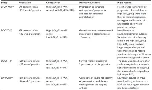
infants (Table 1),22–25 but only one of these studies enrolled
infants in the delivery room. Three levels of saturation were thoroughly evaluated in these trials, including low targeted saturation at 85%–90%, medium targeted saturation at 91%–95%, and high targeted saturation at 95%–99%. Data from the individual trials suggest that high targeted saturation over 95% was associated with bronchopulmonary dysplasia and oxygen dependency. Pooled data from premature
infants ,28 weeks from SUPPORT (the Surfactant, Positive
Pressure, and Pulse Oximetry Randomized Trial) and the BOOST (Benefits of Oxygen Saturation Targeting) II trial showed a higher survival rate in infants assigned to a target oxygen saturation of 91%–95% compared with the low
saturation of 85%–89%.24 Recently, nomograms for oxygen
saturation and heart rate in the first minutes of life for preterm
babies have been developed, showing a slow rise in oxygen
saturation after birth.26,27 SpO
2 values within the 90th and 10th
centile could be considered a valid reference for assessing
improvement and need for oxygen administration.28
Appropriate inflating pressure
of the lungs
There is no consensus about the inflating pressure needed to resuscitate preterm infants. Experimental studies have suggested that lungs are injured if they are inflated with either a high pressure to volumes that exceed total lung capacity, or low pressures to small volumes below a normal
functional residual capacity.29 Overdistension of the lung
(volutrauma) and insufficient lung volumes (atelectrauma) can initiate an inflammatory process, which may progress
to bronchopulmonary dysplasia.30,31 Bjorklund et al showed
in preterm lambs that as few as six large insufflations with tidal volumes of 35–40 mL/kg, probably harmless in normal circumstances, forced into a surfactant-deficient lung immediately after birth can compromise the effect of subsequent surfactant rescue treatment. The immature lambs were more difficult to ventilate and had more widespread lung
injury in histological sections.32 Hoskyns et al reported that
Table 1 Studies addressing target oxygen saturation in preterm infants
Study Population Comparison Primary outcome Main results STOP-ROP22 649 preterm infants
±25.4 weeks’ gestation
High SpO2 (96%–99%)
versus low SpO2 (89%–94%)
Progression to threshold retinopathy of prematurity and need for peripheral retinal ablation
No difference in mortality or progression of retinal disease High SpO2 group were more
likely to remain hospitalized, on oxygen, and have chronic lung disease at 50 weeks postmenstrual age BOOST-I23 358 preterm infants
,30 weeks’ gestation
High SpO2 (95%–98%) versus
low SpO2 (91%–94%)
Growth and neurodevelopmental measures at a corrected age of 12 months
No difference in
neurodevelopmental outcome Six infants died of pulmonary cause in the high SpO2 group
High SpO2 group received longer oxygen therapy, and were more likely to receive supplemental oxygen at 36 weeks postmenstrual age and at home BOOST-II24 1200 preterm infants
,28 weeks’ gestation
High SpO2 (91%–95%)
versus
low SpO2 (85%–89%)
Survival without disability at 2 years corrected for gestation
The study was closed early after a safety analysis demonstrated a higher survival rate in the group that was randomly assigned to a high target SpO2
SUPPORT25 1316 preterm infants
±26 weeks’ gestation
High SpO2 (91%–95%) versus
low SpO2 (85%–89%)
Composite of severe retinopathy of prematurity, death before discharge from the hospital, or both
Low target saturation group were less likely to have severe ROP but had a higher mortality rate before discharge
Abbreviations: STOP-ROP, Supplemental Therapeutic Oxygen for Prethreshold Retinopathy of Prematurity; BOOST, Benefits of Oxygen Saturation Targeting; SUPPORT, Surfactant, positive pressure, and pulse oximetry randomized; SpO2, pulse oximetry percent saturation.
Dovepress Respiratory management of preterm neonates
Research and Reports in Neonatology downloaded from https://www.dovepress.com/ by 118.70.13.36 on 27-Aug-2020
inflation pressures of 25–30 cm H2O in preterm infants rarely led to tidal volumes of more than 4 mL/kg, although higher
pressures were sometimes needed for optimal resuscitation.33
In contrast, Hird et al found that a median inflation pressure
of 22.8 cm H2O was necessary to generate adequate chest
wall expansion in preterm infants, with no infant requiring
a peak inspiratory pressure .30 cm H2O.34 Assessment of
chest rise as a measure of lung expansion is not very accurate
and might lead to delivery of excessive tidal volumes.35
Thus, monitoring of respiratory function has recently been suggested as a useful tool during resuscitation of preterm
infants to minimize lung barotrauma and volutrauma.36
Use of a T-piece resuscitator
The T-piece resuscitator (Neopuff®) is similar to the
flow-inflating bag in providing positive end-expiratory pressure, but with the addition of an adjustable flow control valve. The flow control valve allows for more precise control of the peak inspiratory pressure applied to the infant’s lung, decreases the risk of lung barotrauma, and enables application of prolonged initial inflation breaths and positive end-expiratory pressure to be maintained between inflations, and can give continuous positive airway pressure (CPAP) when it is not being used as a ventilator. The T-piece resuscitator was found more consistently to deliver target pressures and prolonged inflation independent of the operator’s expertise compared
with manual bagging devices.37 In a qualitative review of
30 trials comparing the T-piece resuscitator with self-inflating bags, two on infants and 28 on manikins, no superiority was found for the T-piece resuscitator over the self-inflating bag in the infant trials, but in the manikin studies the T-piece resuscitator was able to deliver inflating pressures closer to predetermined target pressures with minimal variation and to provide prolonged inflation breaths and more consistent tidal
volumes.38 Currently, target ventilation variables with the
T-piece resuscitator include a respiratory rate of 40 breaths
per minute, a peak inspiratory pressure of 20 cm H2O, and a
positive end-expiratory pressure of 4 cm H2O.39
Value of sustained lung inflation
technique
The first breaths in a full term newborn have a prolonged expiratory phase associated with a high positive intrathoracic pressure, and a brief inspiratory component to facilitate the distribution of air within the lungs and to assist in formation
of the functional residual capacity.40 In an experimental study,
it was found that sustained inflation of preterm rabbit lungs for durations of 10 or 20 seconds increased the inspiratory
volume and produced a greater functional residual capacity
compared with durations of one or 5 seconds.41 Sustained
lung inflation for 20 seconds in combination with positive end-expiratory pressure was found to improve the functional
residual capacity of preterm rabbit pups.42 In asphyxiated full
term infants, sustained lung inflation during resuscitation was found to increase their tidal volume and the functional
residual capacity.43 The European Resuscitation Council
has recommended that initial inflation of the lung should be 2–3 seconds in duration to achieve a sustained lung inflation
technique.44,45 In preterm infants, sustained lung inflation
for 5 seconds compared with traditional lung inflation for 2 seconds did not reduce lung injury, as measured by
inflam-matory cytokines.46 Lindner et al compared sustained lung
inflation with nasal intermittent positive pressure ventilation (IPPV) for treatment of preterm infants with respiratory dis-tress syndrome in the delivery room, and found no significant difference in the need for mechanical ventilation or rate of
adverse effects.47 te Pas and Walther found a decrease in need
for intubation and bronchopulmonary dysplasia in preterm infants of more than 28 weeks’ gestation treated by sustained
lung inflation at a pressure of 20 cm H2O for 10 seconds using
a T-piece resuscitator compared with a self-inflating bag and
mask in the delivery room.48 In preterm infants with
respira-tory distress syndrome, sustained lung inflation at a pressure
of 25 cm H2O for 15 seconds using a T-piece resuscitator
was found to decrease the need for mechanical ventilation compared with using a T-piece resuscitator without sustained
lung inflation, without inducing evident adverse effects.49
However, issues like appropriate duration and pressure, hemodynamic changes, and risk of barotrauma, should be further evaluated before recommending routine use of sus-tained lung inflation in the resuscitation of preterm infants.
Continuous positive airway pressure
Mechanism of action
CPAP is a technique of respiratory support that generates a continuous distending pressure to the lung and is delivered to spontaneously breathing infants. Usually, a pressure of
4–8 cm H2O is used to distend the airways and alveoli.
CPAP improves oxygenation without increasing peripheral
arterial carbon dioxide tension (PaCO2) through stabilization
and recruitment of collapsed alveoli. The functional residual capacity is increased, resulting in an increased alveolar surface area for gas exchange and a decrease in intrapulmonary shunting. CPAP enhances the release of surfactant and helps maintain the surfactant present on the alveolar surface. It stabilizes the chest wall and dilates
Dovepress
Abdel-Hady and Nasef
Research and Reports in Neonatology downloaded from https://www.dovepress.com/ by 118.70.13.36 on 27-Aug-2020
the airways, reduces inspiratory resistance, increases lung compliance, counteracts paradoxical movement, and reduces thoracoabdominal asynchrony of the chest wall and reduces
the work of breathing.50,51
Evidence for early use of CPAP
in preterm infants
CPAP was first used as a method of supporting breathing
in preterm infants in 1971.52 Even after the introduction
of surfactant replacement therapy and new mechanical ventilation strategies in the management of respiratory distress syndrome, bronchopulmonary dysplasia remained a frequent sequel of prematurity, and mechanical ventila-tion was described as the important predisposing factor for bronchopulmonary dysplasia; thus, by avoiding intubation and mechanical ventilation, many of these infants will not
develop bronchopulmonary dysplasia.53 Today, there is
increased interest in the use of nasal CPAP in extremely low birth-weight (ELBW) and very low birth-weight (VLBW)
infants,54 and providing nasal CPAP rather than intubation
has been identified as a potentially better practice according
to basic quality improvement criteria.55,56 The evidence for
early application of nasal CPAP in this population comes from experimental, observational/retrospective, and random-ized controlled trials.
Experimental studies
Experimental studies demonstrated that early application of CPAP is protective for the preterm lung and brain compared with mechanical ventilation. The ventilatory strategy of “open up the lung and keep it open” will minimize lung damage, therefore starting nasal CPAP directly after resuscitation is
a gentle, noninvasive attempt to keep the lung open.57 Jobe
et al showed in preterm lambs that nasal CPAP after birth results in lower indicators of acute lung injury than con-ventional mechanical ventilation during the first 2 hours of
life.58 More recently, the same research group randomized
preterm lambs to bubble CPAP (5 or 8 cm H2O) or
conven-tional mechanical ventilation for a period of 2–6 hours. They demonstrated larger lung volumes and better oxygenation with
CPAP 8 cm H2O compared with CPAP 5 cm H2O, and lung
dry/wet ratios were greater for both CPAP groups than for the mechanical ventilation group. Lambs that demonstrated terminal respiratory failure on CPAP within 2 hours had smaller surfactant pools (supporting early administration of surfactant to the most immature infants who are likely to be
surfactant-deficient).59 The value of surfactant replacement
therapy coupled with CPAP has been demonstrated in another
study showing that animals treated with surfactant prior to CPAP had less acute lung injury, suggesting that surfactant administration may modulate pulmonary inflammation and that CPAP alone without surfactant may not provide optimal
pulmonary protection.60 The lungs of premature baboons
maintained on CPAP for 28 days demonstrated thin-walled airspaces with minimal fibroproliferation and scattered secondary crests. Pressure-volume curves were nearly identical to those observed at term gestation, but significantly better than those from preterm baboons ventilated with low
tidal volume (“gentle”) positive pressure ventilation.61 In
contrast with these findings, another study62 demonstrated
comparable acute effects on lung and systemic inflammation
of CPAP (8 cm H2O) and gentle conventional mechanical
ventilation (tidal volume 8 mL/kg), leading to the conclusion that CPAP given shortly after birth fails to attenuate lung injury, supporting the results of randomized controlled trials of early use of CPAP where no reduction of bronchopulmonary
dysplasia in preterm infants was observed.63–66 Early CPAP
was associated with less cerebral injury,67 higher cerebellar
weight,68 and less damage to specific classes of neurons in
the visual cortex expressing gamma aminobutyric acid69
compared with delayed CPAP preceded by positive pressure ventilation in premature baboons.
Observational/retrospective studies
Several observational/retrospective studies have evaluated
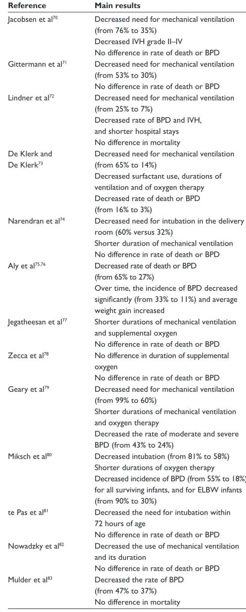
early use of CPAP in preterm infants70–83 (Table 2), with most
of these studies suggesting that early use of nasal CPAP and avoiding intubation leads to reduced oxygen requirements, intubation rates, and duration of mechanical ventilation, and some studies reporting decreased rates of bronchopulmonary
dysplasia.72,73,75,76,79,80,83 Other observational studies compared
the rates of bronchopulmonary dysplasia in centers using early nasal CPAP with centers preferring intubation and
mechanical ventilation. The pioneering work of Avery et al84
found that NICUs using less intubation and mechanical ventilation reported less bronchopulmonary dysplasia
compared with other NICUs. Van Marter et al53 observed
marked differences in the incidence of bronchopulmonary dysplasia (4% at the Babies Hospital, Columbia University, versus 22% at two Boston hospitals) despite similar mortality rates. Infants at the Babies Hospital were less likely to be mechanically ventilated, and those who did were ventilated for significantly shorter time periods. In multivariate logistic regression analyses, initiation of mechanical ventilation was associated with an increased risk
of bronchopulmonary dysplasia.53 Another study showed
Dovepress Respiratory management of preterm neonates
Research and Reports in Neonatology downloaded from https://www.dovepress.com/ by 118.70.13.36 on 27-Aug-2020
Table 2 Studies evaluating respiratory outcomes after application of early nasal CPAP in preterm infants
Reference Main results
Jacobsen et al70 Decreased need for mechanical ventilation
(from 76% to 35%) Decreased IvH grade II–Iv
No difference in rate of death or BPD Gittermann et al71 Decreased need for mechanical ventilation
(from 53% to 30%)
No difference in rate of death or BPD Lindner et al72 Decreased need for mechanical ventilation
(from 25% to 7%)
Decreased rate of BPD and IvH, and shorter hospital stays No difference in mortality De Klerk and
De Klerk73
Decreased need for mechanical ventilation (from 65% to 14%)
Decreased surfactant use, durations of ventilation and of oxygen therapy Decreased rate of death or BPD (from 16% to 3%)
Narendran et al74 Decreased need for intubation in the delivery
room (60% versus 32%)
Shorter duration of mechanical ventilation No difference in rate of death or BPD Aly et al75,76 Decreased rate of death or BPD
(from 65% to 27%)
Over time, the incidence of BPD decreased significantly (from 33% to 11%) and average weight gain increased
Jegatheesan et al77 Shorter durations of mechanical ventilation
and supplemental oxygen
No difference in rate of death or BPD Zecca et al78 No difference in duration of supplemental
oxygen
No difference in rate of death or BPD Geary et al79 Decreased need for mechanical ventilation
(from 99% to 60%)
Shorter durations of mechanical ventilation and oxygen therapy
Decreased the rate of moderate and severe BPD (from 43% to 24%)
Miksch et al80 Decreased intubation (from 81% to 58%)
Shorter durations of oxygen therapy Decreased incidence of BPD (from 55% to 18%) for all surviving infants, and for ELBw infants (from 90% to 30%)
te Pas et al81 Decreased the need for intubation within
72 hours of age
No difference in rate of death or BPD Nowadzky et al82 Decreased the use of mechanical ventilation
and its duration
No difference in rate of death or BPD Mulder et al83 Decreased the rate of BPD
(from 47% to 37%) No difference in mortality
Abbreviations: NCPAP, nasal continuous positive airway pressure; BPD, bronchopulmonary dysplasia; IvH, intraventricular hemorrhage; ELBw, extreme low-birth weight infants.
that rates of bronchopulmonary dysplasia and retinopathy of prematurity were significantly lower at the Neonatal Regional Tertiary Care Center, University of Vienna, compared with the Vermont Oxford Neonatal Network during the period 1994–2002, and this was attributed to more use of nasal CPAP and less use of mechanical ventilation during early
respiratory management of VLBW infants.85
Improved respiratory outcomes for ELBW infants treated with a predominantly nasal CPAP approach were evident at Middlemore Hospital, New Zealand, compared with the Australian and New Zealand Neonatal Network data over a 3-year period (1998–2001). These infants spent a longer time on nasal CPAP and had less time on oxygen. Fewer of the Middlemore cohort were on oxygen at both 28 days, and 36 weeks corrected gestation, and fewer infants were
discharged home on oxygen.86 On comparing the resuscitation
and ventilator techniques between two NICUs in Stockholm, Sweden and Boston, MA, a less aggressive approach with more nasal CPAP administration in the delivery room was used in Stockholm, which did not decrease the risk for bronchopulmonary dysplasia at 36 weeks; however, at 40 weeks, fewer infants were on oxygen support, and a strong association was found between site, mean airway pressure, and mechanical ventilation with bronchopulmonary dysplasia, indicating that CPAP could have a beneficial role
in outcome.87 In contrast with these findings, a recent study
investigated whether the wide variation in frequency of bronchopulmonary dysplasia between Italian hospitals is due to differences in delivery room intubation rates in VLBW infants. Rates of bronchopulmonary dysplasia, delivery room
intubation, and mechanical ventilation for .24 hours differed
significantly between hospitals. It was demonstrated that
delivery room intubation and brief (,24 hours) mechanical
ventilation did not increase the risk of bronchopulmonary
dysplasia.88
Five studies have evaluated neurodevelopmental outcomes in ELBW and VLBW infants managed with early
CPAP.51,89–92 Neurodevelopmental follow-up at 5 years of
age in a cohort of ELBW infants treated mainly with nasal CPAP during the neonatal period in Denmark (the ETFOL study) showed similar proportions of adverse effects on the brain compared with those in published follow-up studies
of preterm infants treated with mechanical ventilation.51
Massaro et al demonstrated that ELBW infants managed with early nasal CPAP have neurodevelopmental morbidity at 3–6 months corrected age comparable with that of the National Institute of Child Health and Human Development
Dovepress
Abdel-Hady and Nasef
Research and Reports in Neonatology downloaded from https://www.dovepress.com/ by 118.70.13.36 on 27-Aug-2020
Neonatal Research Network cohort.89 Dahl and Kamper
demonstrated that VLBW infants treated with a regimen of early nasal CPAP/minimal handling had a relatively low incidence of handicap and impairment, with nearly 90% attending ordinary schools and near-average performance in mathematics and reading/spelling, which was not significantly
different from their siblings.90 Wintermark et al demonstrated
that early application of nasal CPAP and avoidance of mechanical ventilation in VLBW infants showed no adverse effects on neurodevelopment and growth, and a significantly higher developmental quotient was found in the nasal CPAP
group at a corrected age of 18 months.91 Thomas et al
demonstrated that ELBW infants receiving nasal CPAP had better Bayley Score of Infant Development Version II scores at a corrected gestational age of 18–22 months, and lower rates of bronchopulmonary dysplasia and death compared with those receiving mechanical ventilation at 24 hours of age and, after adjusting for acuity differences, the ventilatory strategy at 24 hours of age independently predicts the
long-term neurodevelopmental outcome.92
Randomized controlled trials
Observational studies alone do not provide strong enough evidence to justify and promote new clinical practices. Early randomized controlled trials that evaluated the role of CPAP in preterm infants were done in the presurfactant era and included small numbers of cases. Two Cochrane reviews of these trials showed a reduction in mortality of preterm
infants .1500 g with nasal CPAP, no significant differences
in bronchopulmonary dysplasia, and a greater benefit from early application. They concluded that these trials provided insufficient information to make recommendations for clinical
practice and called for a large randomized controlled trial.93,94
Finer et al investigated the feasibility of randomizing ELBW
infants ,28 weeks’ gestation to nasal CPAP/positive
end-expiratory pressure or no nasal CPAP/positive end-end-expiratory pressure during resuscitation immediately after delivery, avoiding routine delivery room intubation for surfactant administration, and initiating nasal CPAP on admission to the neonatal unit. In the first group, nasal CPAP of 5 cm
H2O or IPPV (if necessary) with peak inspiratory pressure of
15–25 cm H2O was given, and in the second group, no nasal
CPAP and no positive end-expiratory pressure during IPPV was used. Nasal CPAP/positive end-expiratory pressure in the delivery room did not affect the need for intubation at birth or during the subsequent week, although this preliminary study
was underpowered for detecting such a difference.95
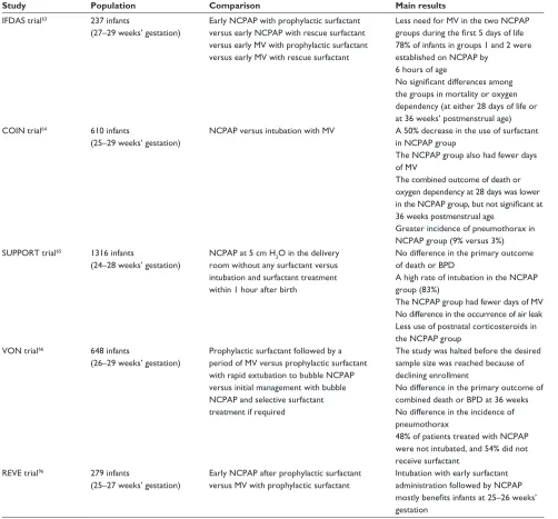
Recently, five large multicenter, randomized controlled trials of appropriate design have tested the hypothesis that not intubating preterm babies with respiratory distress syndrome
soon after birth would have a positive effect on outcome63-66,96
(Table 3). These trials support the use of CPAP as a primary strategy in preterm babies with respiratory distress syndrome who are breathing spontaneously. This approach led to a reduction in the number of infants intubated and given surfactant. On the other hand, half of the infants enrolled in these studies failed CPAP treatment. It can be argued that such babies might have been disadvantaged because of the delay in receiving surfactant therapy. As far as we know, no meta-analysis of the results of these studies has been done until now, and none of these studies evaluated neurodevelopmental outcomes in the infants who participated in this research. Unfortunately, two of these trials were not published in full
and are available only in abstract form.63,96
The strategy of early nasal CPAP in VLBW infants may be justified based on the current clinical evidence, because it has been shown to be safe, and reduces the need for
mechanical ventilation.95 However, to rely on nasal CPAP
and “not to intubate” with the aim of reducing the incidence of bronchopulmonary dysplasia is not justified due to the low quality of evidence from retrospective/observational
studies,72,73,75,76,79,80,83 and the lack of evidence from
random-ized controlled trials that not intubating in the delivery room
reduces bronchopulmonary dysplasia.63–66,96
Early nasal CPAP is not successful in all VLBW infants, and these infants will ultimately be ventilated mechanically. Forty-six percent of infants in the COIN (Continuous Positive Airway Pressure or Intubation at
Birth) trial63 and 83% of infants in SUPPORT65 failed
nasal CPAP. Ammari et al demonstrated that nasal CPAP was successful in 76% of infants with a birth weight of
#1250 g and 50% of infants with a birth weight of #750 g,
and that nasal CPAP failure was associated with a need for positive pressure ventilation at delivery, alveolar-arterial
oxygen tension gradient .180 mmHg on the first arterial
blood gas, and severe respiratory distress syndrome on
the initial chest x-ray.97 Another study reported a delivery
room nasal CPAP failure rate of 51% in preterm infants (26–27 weeks). Medical history and initial blood gas values were poor predictors of subsequent nasal CPAP failure. Moreover, it was found that a threshold fraction
of inspired oxygen (FiO2) of $0.35–0.45 compared with
$0.6 for intubation would shorten the time to surfactant
delivery without a relevant increase in intubation rate.98 A
Dovepress Respiratory management of preterm neonates
Research and Reports in Neonatology downloaded from https://www.dovepress.com/ by 118.70.13.36 on 27-Aug-2020
Table 3 Randomized controlled trials evaluating early NCPAP in preterm infants
Study Population Comparison Main results
IFDAS trial63 237 infants
(27–29 weeks’ gestation)
Early NCPAP with prophylactic surfactant versus early NCPAP with rescue surfactant versus early Mv with prophylactic surfactant versus early Mv with rescue surfactant
Less need for Mv in the two NCPAP groups during the first 5 days of life 78% of infants in groups 1 and 2 were established on NCPAP by
6 hours of age
No significant differences among the groups in mortality or oxygen dependency (at either 28 days of life or at 36 weeks’ postmenstrual age) COIN trial64 610 infants
(25–29 weeks’ gestation)
NCPAP versus intubation with Mv A 50% decrease in the use of surfactant in NCPAP group
The NCPAP group also had fewer days of Mv
The combined outcome of death or oxygen dependency at 28 days was lower in the NCPAP group, but not significant at 36 weeks postmenstrual age
Greater incidence of pneumothorax in NCPAP group (9% versus 3%) SUPPORT trial65 1316 infants
(24–28 weeks’ gestation)
NCPAP at 5 cm H2O in the delivery room without any surfactant versus intubation and surfactant treatment within 1 hour after birth
No difference in the primary outcome of death or BPD
A high rate of intubation in the NCPAP group (83%)
The NCPAP group had fewer days of Mv No difference in the occurrence of air leak Less use of postnatal corticosteroids in the NCPAP group
vON trial66 648 infants
(26–29 weeks’ gestation)
Prophylactic surfactant followed by a period of Mv versus prophylactic surfactant with rapid extubation to bubble NCPAP versus initial management with bubble NCPAP and selective surfactant treatment if required
The study was halted before the desired sample size was reached because of declining enrollment
No difference in the primary outcome of combined death or BPD at 36 weeks No difference in the incidence of pneumothorax
48% of patients treated with NCPAP were not intubated, and 54% did not receive surfactant
REvE trial96 279 infants
(25–27 weeks’ gestation)
Early NCPAP after prophylactic surfactant versus Mv with prophylactic surfactant
Intubation with early surfactant administration followed by NCPAP mostly benefits infants at 25–26 weeks’ gestation
Abbreviations: Mv, mechanical ventilation; NCPAP, nasal continuous positive airway pressure; IFDAS, Infant Flow Driver and Surfactant; COIN, CPAP or Intubation trial; SUPPORT, Surfactant, Positive Pressure, and Pulse Oximetry Randomized Trial; vON, vermont Oxford Network trial; REvE, REduction of vEntilation trial.
clinical prediction score for nasal CPAP failure in VLBW infants was developed recently. On multivariate analysis,
the authors found three variables, ie, gestation ,28 weeks,
preterm premature rupture of membranes, and product of
CPAP pressure and FiO2$1.28 at initiation to maintain
saturation between 88% and 93%, to be independently
predictive of failure.99 In another study, failure of the
INSURE method was reported in 32% of preterm infants (23–28 weeks) receiving nasal CPAP. INSURE failure
was significantly associated with the PaCO2 and mean
arterial-to-alveolar oxygen tension ratio as well as a severe radiological grade in preterm infants with respiratory
distress syndrome.100
Alternative methods of noninvasive respiratory support in VLBW infants include heated, humidified high-flow nasal cannulation, bilevel nasal CPAP, and nasal IPPV. Although the humidified high-flow nasal cannula has been widely
used in many NICUs,101 there are scant data regarding its
efficacy and safety.102 Bilevel nasal CPAP was associated with
better respiratory outcomes versus nasal CPAP, and allowed
Dovepress
Abdel-Hady and Nasef
Research and Reports in Neonatology downloaded from https://www.dovepress.com/ by 118.70.13.36 on 27-Aug-2020
earlier discharge in preterm infants with respiratory distress
syndrome.103 Early use of nasal IPPV in preterm infants
with respiratory distress syndrome reduced the need for
intubation and mechanical ventilation,104 and reduced the
risk of bronchopulmonary dysplasia105,106 compared with
nasal CPAP. Although these findings are promising, further studies are needed to assess the potential benefits of these noninvasive ventilation techniques.
Surfactant
Surfactant therapy has been an excellent evidence-based measure of treatment for preterm respiratory distress
syndrome since its introduction in 1990. In both VLBW107
and larger108–110 preterm infants, it has been shown to lead to
a significant decrease in the risk of neonatal mortality and
morbidity.111 Surfactant replacement therapy has decreased
the incidence of pneumothorax and intraventricular
hemorrhage, and improved survival of preterm infants.112
Recent developments in surfactant therapy for respiratory distress syndrome in preterm infants include comparative trials of natural versus synthetic surfactants, exploration of alternative modes of administration of surfactant, time of surfactant administration, and evaluation of the concomitant use of surfactant with short-term or noninvasive ventilation as adjuvant therapy.
Natural versus synthetic surfactant
Natural and synthetic surfactant preparations commonly used for neonatal respiratory distress syndrome are shown in Table 4. Natural surfactants are generally derived from either lung lavage or minced lung of bovine or porcine origin. Synthetic surfactants are complex combinations of
dipalmitoylphosphatidylcholine and other phospholipids, neutral lipids, lipoprotein, or alcohols.
Natural surfactant
Individual trials have compared the efficacy and safety of various natural surfactants in the treatment of respiratory distress syndrome. Most reported differences in effective-ness between natural surfactant preparations were from short-term studies. Poractant alfa was found to have a more
rapid onset of action than beractant in most studies.113–115
In a meta-analysis of five trials that compared poractant alfa with beractant in the treatment of preterm infants with respiratory distress syndrome, there was no difference in the primary outcome of bronchopulmonary dysplasia between the two different surfactant types (31.5% versus 29.9%, relative risk 0.98, 95% confidence interval 0.75–1.29), but a significant decrease in mortality rate was noted with the use of poractant versus beractant (relative risk 0.51, 95%
confidence interval 0.3–0.89).116 However, this difference in
mortality was attributed to the use of higher doses of porac-tant alpha (200 mg/kg) compared with beracporac-tant (100 mg/kg) in three of the studies included in this meta-analysis. When the results were reproduced after exclusion of these trials,
the difference in mortality was not statistically significant.117
Calfactant was found to have a more rapid effect with fewer doses, without having an effect on overall incidence of bronchopulmonary dysplasia or mortality when compared
with beractant.118,119 Consistent with previous reports, the
use of bovine lipid extract surfactant was associated with a more rapid improvement in oxygenation, but no effect on
short-term outcome when compared with beractant.120 On the
basis of the currently available evidence, no preparation can
Table 4 Surfactant products for neonatal respiratory distress syndrome
Surfactant Type Preparation Origin Composition
Poractant alfa Natural Curosurf® Porcine lung minces, lipid extraction with
purification using liquid-gel chromatography
DPPC + PG + SP-B + SP-C
Calfactant Natural Infasurf® Calf lung lavage with lipid extraction DPPC + PG + SP-B + SP-C
Beractant Natural Survanta® Bovine lung minces, lipid extraction, supplemented
with DPPC, palmitic acid and tripalmitin
DPPC + PG + SP-B + SP-C
Alveofact® Cow lung lavage fluid with lipid extraction DPPC + PG + SP-B + SP-C
BLES Natural BLES® Bovine lipid extract surfactant DPPC + PG + SP-B + SP-C
Colfosceril Synthetic Exosurf® Synthetic DPPC in hexadecanol and tyloxapol
(as spreading agents)
DPPC
Lucinactant Synthetic Surfaxin® Recombinant SP-B analog DPPC + POPG + palmitic acid +
recombinant SP-B analog Lusupultide Synthetic venticute® Recombinant SP-C analog DPPC + PG + palmitic acid +
recombinant SP-C analog
Abbreviations: BLES, bovine lipid extract surfactant; DPPC, dipalmitoylphosphatidylcholine; PG, phosphatidylglycerol; POPG, palmitoyloleylphosphatidylglycerol; SP-B, surfactant protein B; SP-C, surfactant protein C.
Dovepress Respiratory management of preterm neonates
Research and Reports in Neonatology downloaded from https://www.dovepress.com/ by 118.70.13.36 on 27-Aug-2020
clearly be considered superior to another for the treatment of respiratory distress syndrome in preterm infants in terms of morbidity and mortality.
Synthetic surfactant
Although synthetic surfactants have theoretical advantages over natural surfactants, to date natural surfactants have been shown to be superior to synthetic surfactant in clinical
trials due to the presence of protein B and C.124,125 The use
of natural preparations was associated with lower inspired oxygen concentrations, ventilator pressures, mortality, and rates of respiratory distress syndrome complications in preterm infants. When used for treatment of respiratory distress syndrome, colfosceril was associated with slower weaning and more pneumothorax but no difference in major clinical outcomes compared with calfactant. When used for prophylaxis, calfactant was more effective than colfosceril in reducing the severity of respiratory distress syndrome and the number of associated deaths, but had no impact on overall
mortality or bronchopulmonary dysplasia.124,125
New-generation synthetic surfactants containing peptides that mimic surfactant protein B (lucinactant) or surfactant protein C (lusupultide) were developed to improve the efficacy of synthetic surfactants. Two large multicenter, randomized controlled trials compared the safety and efficacy of lucinactant with that of other surfactant preparations. The SELECT (Safety and Effectiveness of Lucinactant versus Exosurf in a Clinical Trial) study showed significant improvement in respiratory distress syndrome, related mortality through 14 days of life, and bronchopulmonary dysplasia when lucinactant was compared with colfosceril, as well as a reduction in total days on ventilation when compared
with beractant.126 Similarly, the STAR (SURFAXIN® Therapy
Against Respiratory distress syndrome) trial found that lucinactant was noninferior to poractant with regard to survival without bronchopulmonary dysplasia. Infants treated with lucinactant required significantly fewer doses
compared with poractant.127 In summary, new-generation
synthetic surfactant seems to have some advantages over previous synthetic surfactants containing only phospholipids, presumably because of the surfactant protein mimic, and is at least as good as currently used animal-derived surfactants.
Mode of administration
Intratracheal administration is presently the only approved means of delivering surfactant, due to its liquid nature. Accordingly, therapeutic use of surfactant is limited to infants
who are intubated and/or undergoing mechanical ventila-tion. Furthermore, the process of surfactant administration requires a skilled attendant for endotracheal intubation. Tracheal intubation has potential risks, such as tracheal tube malpositioning or dislocation, hypoxia, bradycardia, and infection. Further, patients requiring tracheal intubation in an elective situation need to be treated with analgesic and
seda-tive drugs that may have depressive respiratory effects.128
Alternative noninvasive techniques for administration of surfactant without tracheal intubation include administra-tion through a laryngeal mask airway, nebulized surfactant, and minimally invasive surfactant therapy. Evidence for use of a laryngeal mask airway for surfactant administration
comes from case reports and small uncontrolled trials.129,130
A Cochrane review, which included only one small trial, compared late rescue laryngeal mask airway surfactant with
no treatment in preterm infants $1200 g with respiratory
distress syndrome, and administration of surfactant by a laryngeal mask airway was associated with no significant difference in need for subsequent mechanical ventilation and endotracheal surfactant, pneumothorax, days on IPPV, and days on IPPV or oxygen, but may have a short-term effect
in reducing the requirement for oxygen.131
Administration of aerosolized surfactant has gained more interest because it is theoretically more gentle, noninvasive, and homogeneously distributed. Jorch et al demonstrated
that CPAP and nebulized Alveofact® compared with CPAP
alone resulted in an improved alveolar-arterial gradient and
better PaCO2 levels in infants at 28–35 weeks’ gestation.132
A comparison of nebulized Curosurf® and CPAP versus CPAP
alone in infants at 23–36 weeks’ gestation found no difference in clinical outcome, but demonstrated that nebulized
surfactant was safe and well tolerated.133 Similarly, Finer
et al showed the safety of nebulized lucinactant (Aerosurf®)
in 17 preterm infants at 28–32 weeks’ gestation.134 However,
issues regarding development of nebulizer devices capable of administration, effective administration, dosing strategies,
and cost-effectiveness remain unresolved.135
Kribs et al described another technique of administering surfactant, known as the MIST (minimally invasive surfactant therapy) technique, whereby surfactant is administered via a thin endotracheal catheter during spontaneous breathing
together with application of nasal CPAP.136 They reported that
use of this technique increased between 2001 and 2004, and was associated with a significant increase in survival
with-out bronchopulmonary dysplasia.136 A multicenter German
study involving 1541 infants at ,31 weeks’ gestational age
Dovepress
Abdel-Hady and Nasef
Research and Reports in Neonatology downloaded from https://www.dovepress.com/ by 118.70.13.36 on 27-Aug-2020
demonstrated a decreased need for mechanical ventilation during the first 72 hours and a lower rate of bronchopulmonary dysplasia and rate of death or bronchopulmonary dysplasia in infants treated with this new technique compared with
those who received standard care.137 In a recent randomized
controlled trial, preterm infants (26–28 weeks’ gestation) randomized to this new technique required less mechanical ventilation and had fewer median days on mechanical
ventila-tion compared with a standard treatment group.138 In another
report, it was demonstrated that there was no significant differ-ence regarding neurodevelopmental outcome in school-aged children treated after implementing this technique, even though surviving infants were more immature and survival rates had
increased.139 Dargaville et al modified this technique using a
16-gauge vascular catheter inserted through the vocal cords
under direct vision without the need for Magill forceps.140
The early use of noninvasive ventilation alone or in con-junction with surfactant may be as effective as intubation in preventing and treating respiratory distress syndrome in
very premature infants. It has been suggested54 that the high
survival rates and low incidence of bronchopulmonary dys-plasia experienced in some centers in Scandinavia and the US were further improved when nasal CPAP was combined with early surfactant treatment and early extubation to nasal
CPAP.128,141 The INSURE method, developed in Scandinavia,
combines endotracheal intubation and surfactant application
with early extubation to nasal CPAP.142 Infants are usually
anesthetized with morphine, sometimes in combination with thiopental, followed by naloxone application to reverse
respiratory depression.141,143
Time of administration
Surfactant administered within 2 hours of life to
infants #28 weeks’ gestation reduced the incidences of
pulmonary interstitial emphysema, bronchopulmonary
dysplasia, pneumothorax, and mortality.144 Preterm infants
with respiratory distress syndrome who received surfactant within 30 minutes of life showed reductions in pulmonary interstitial emphysema, death, intraventricular hemorrhage grade III–IV, and the combined outcome of bronchopulmonary dysplasia and death in comparison with infants who receive
surfactant later on.145 However, most of the trials which
studied the value of early versus late surfactant therapy were
conducted before the era of routine antenatal steroids.146
Accordingly, the previously observed difference between early and delayed surfactant therapy may be smaller in current practice.
CURPAP, an international randomized controlled trial to evaluate the efficacy of combining prophylactic surfactant and early nasal continuous positive airway pressure in very preterm infants, reported no difference in need for mechanical ventilation, bronchopulmonary dysplasia, or pneumothorax among infants born at 24–28 weeks’ gestation who were randomized to receive prophylactic or early selective surfactant with nasal CPAP, suggesting early rescue surfactant is as
good as prophylactic surfactant.147 These results indicate that
preterm infants (24–28 weeks’ gestation) can be treated with nasal CPAP from birth, and that rescue INSURE should be used if nasal CPAP fails. In contrast, a multicenter study from Colombia randomized 279 preterm infants (27–31 weeks’ gestation) with respiratory distress syndrome in the first hour of life to either an INSURE to CPAP protocol or to CPAP alone. The INSURE to CPAP group had lower mechanical ventilation requirements and less pneumothorax. No differences in rates
of bronchopulmonary dysplasia or mortality were found.148 In
a recent multicenter study from South America, 256 VLBW infants (800–1500 g) were randomized to a CPAP/INSURE group (if respiratory distress syndrome did not occur, CPAP was discontinued after 3–6 hours; if respiratory distress syndrome
developed and the FiO2 was .0.35, the INSURE protocol was
indicated) or to an oxygen/mechanical ventilation group (in the presence of respiratory distress syndrome, supplemental
oxygen without CPAP was given, and if FiO2 was .0.35,
surfactant and mechanical ventilation were provided). They demonstrated a reduced need for mechanical ventilation and less use of surfactant in the CPAP/INSURE group, and no differences in mortality, pneumothorax, bronchopulmonary dysplasia, and other complications of prematurity between
the two groups.149
Conclusion
The lungs of preterm infants are vulnerable to injury, especially immediately after birth. Improvements in respira-tory support for preterm infants in the delivery room have great potential to improve the short-term and long-term out-come. There is a need to avoid routine intubation of ELBW infants in the delivery room. New techniques for monitoring and critical assessment of postnatal adaptation are currently available and may help to improve decision-making during resuscitation. New synthetic preparations and noninvasive techniques for surfactant administration may prove to be better than conventional techniques. Further research is urgently required to assess the potential benefits of these interventions.
Dovepress Respiratory management of preterm neonates
Research and Reports in Neonatology downloaded from https://www.dovepress.com/ by 118.70.13.36 on 27-Aug-2020
Disclosure
The authors report no conflicts of interest in this work.
References
1. Costeloe K, Hennessy E, Gibson AT, Marlow N, Wilkinson AR. The EPICure study: outcomes to discharge from hospital for infants born at the threshold of viability. Pediatrics. 2000;106(4):659–671. 2. Markestad T, Kaaresen PI, Ronnestad A, et al. Early death, morbidity,
and need of treatment among extremely premature infants. Pediatrics. 2005;115(5):1289–1298.
3. Tommiska V, Heinonen K, Lehtonen L, et al. No improvement in outcome of nationwide extremely low birth weight infant populations between 1996–1997 and 1999–2000. Pediatrics. 2007;119(1): 29–36.
4. Stoll BJ, Hansen NI, Bell EF, et al. Neonatal outcomes of extremely preterm infants from the NICHD Neonatal Research Network. Pediatrics. 2010;126(3):443–456.
5. Fanaroff AA, Stoll BJ, Wright LL, et al. Trends in neonatal morbidity and mortality for very low birthweight infants. Am J Obstet Gynecol. 2007;196(2):147. e1–e8.
6. Landmann E, Misselwitz B, Steiss JO, Gortner L. Mortality and morbidity of neonates born at ,26 weeks of gestation (1998–2003). A population-based study. J Perinat Med. 2008;36(2):168–174. 7. Tyson JE, Parikh NA, Langer J, Green C, Higgins RD. Intensive care
for extreme prematurity – moving beyond gestational age. N Engl J Med. 2008;358(16):1672–1681.
8. Vento M, Cheung PY, Aguar M. The first golden minutes of the extremely-low-gestational-age neonate: a gentle approach. Neonatology. 2009;95(4):286–298.
9. Fowlie PW, McGuire W. Immediate care of the preterm infant. BMJ. 2004;329(7470):845–848.
10. Langston C, Kida K, Reed M, Thurlbeck WM. Human lung growth in late gestation and in the neonate. Am Rev Respir Dis. 1984;129(4): 607–613.
11. Baraldi E, Filippone M. Chronic lung disease after premature birth. N Engl J Med. 2007;357(19):1946–1955.
12. Hallman M, Kulovich M, Kirkpatrick E, Sugarman RG, Gluck L. Phosphatidylinositol and phosphatidylglycerol in amniotic fluid: indices of lung maturity. Am J Obstet Gynecol. 1976;125(5):613–617. 13. O’Donovan DJ, Fernandes CJ. Free radicals and diseases in premature
infants. Antioxid Redox Signal. 2004;6(1):169–176.
14. Hillman NH, Moss TJ, Kallapur SG, et al. Brief, large tidal volume ventilation initiates lung injury and a systemic response in fetal sheep. Am J Respir Crit Care Med. 2007;176(6):575–581.
15. Saugstad OD, Aune D. In search of the optimal oxygen saturation for extremely low birth weight infants: a systematic review and meta-analysis. Neonatology. 2011;100(1):1–8.
16. Saugstad OD, Ramji S, Soll RF, Vento M. Resuscitation of newborn infants with 21% or 100% oxygen: an updated systematic review and meta-analysis. Neonatology. 2008;94(3):176–182.
17. Rabi Y, Singhal N, Nettel-Aguirre A. Room-air versus oxygen adminis-tration for resuscitation of preterm infants: the ROAR study. Pediatrics. 2011;128(2):e374–e381.
18. Dawson JA, Kamlin CO, Wong C, et al. Oxygen saturation and heart rate during delivery room resuscitation of infants ,30 weeks’ gestation with air or 100% oxygen. Arch Dis Child Fetal Neonatal Ed. 2009; 94(2):F87–F91.
19. Escrig R, Arruza L, Izquierdo I, et al. Achievement of targeted saturation values in extremely low gestational age neonates resuscitated with low or high oxygen concentrations: a prospective, randomized trial. Pediatrics. 2008;121(5):875–881.
20. Vento M, Moro M, Escrig R, et al. Preterm resuscitation with low oxygen causes less oxidative stress, inflammation, and chronic lung disease. Pediatrics. 2009;124(3):e439–e449.
21. Kattwinkel J, Perlman JM, Aziz K, et al. Neonatal resuscitation: 2010 American Heart Association Guidelines for Cardiopulmonary Resuscitation and Emergency Cardiovascular Care. Pediatrics. 2010;126(5):e1400–e1413.
22. [No authors listed]. Supplemental Therapeutic Oxygen for Prethreshold Retinopathy Of Prematurity (STOP-ROP), a randomized, controlled trial. I: primary outcomes. Pediatrics. 2000;105(2):295–310. 23. Askie LM, Henderson-Smart DJ, Irwig L, Simpson JM.
Oxygen-saturation targets and outcomes in extremely preterm infants. N Engl J Med. 2003;349(10):959–967.
24. Stenson B, Brocklehurst P, Tarnow-Mordi W. Increased 36-week survival with high oxygen saturation target in extremely preterm infants. N Engl J Med. 2011;364(17):1680–1682.
25. Carlo WA, Finer NN, Walsh MC, et al. Target ranges of oxygen saturation in extremely preterm infants. N Engl J Med. 2010;362(21): 1959–1969.
26. Dawson JA, Kamlin CO, Wong C, et al. Changes in heart rate in the first minutes after birth. Arch Dis Child Fetal Neonatal Ed. 2010;95(3): F177–F181.
27. Dawson JA, Kamlin CO, Vento M, et al. Defining the reference range for oxygen saturation for infants after birth. Pediatrics. 2010;125(6): e1340–e1347.
28. Vento M, Aguar M, Brugada M, et al. Oxygen saturation targets for preterm infants in the delivery room. J Matern Fetal Neonatal Med. 2012;25 Suppl 1:45–46.
29. Dreyfuss D, Saumon G. Ventilator-induced lung injury: lessons from experimental studies. Am J Respir Crit Care Med. 1998;157(1): 294–323.
30. Jobe AH, Ikegami M. Mechanisms initiating lung injury in the preterm. Early Hum Dev. 1998;53(1):81–94.
31. Auten RL, Vozzelli M, Clark RH. Volutrauma. What is it, and how do we avoid it? Clin Perinatol. 2001;28(3):505–515.
32. Bjorklund LJ, Ingimarsson J, Curstedt T, et al. Manual ventilation with a few large breaths at birth compromises the therapeutic effect of subsequent surfactant replacement in immature lambs. Pediatr Res. 1997;42(3):348–355.
33. Hoskyns EW, Milner AD, Boon AW, Vyas H, Hopkin IE. Endotracheal resuscitation of preterm infants at birth. Arch Dis Child. 1987;62(7): 663–666.
34. Hird MF, Greenough A, Gamsu HR. Inflating pressures for effective resuscitation of preterm infants. Early Hum Dev. 1991;26(1):69–72. 35. Milner AD. Resuscitation at birth. Eur J Pediatr. 1998;157(7):
524–527.
36. Schmolzer GM, Morley CJ, Wong C, et al. Respiratory function monitor guidance of mask ventilation in the delivery room: a feasibility study. J Pediatr. 2012;160(3):377–381. e2.
37. Finer NN, Rich W, Craft A, Henderson C. Comparison of methods of bag and mask ventilation for neonatal resuscitation. Resuscitation. 2001;49(3):299–305.
38. Hawkes CP, Ryan CA, Dempsey EM. Comparison of the T-piece resuscitator with other neonatal manual ventilation devices: A qualitative review. Resuscitation. January 4, 2012. [Epub ahead of print.] 39. Hussey SG, Ryan CA, Murphy BP. Comparison of three manual
ventilation devices using an intubated mannequin. Arch Dis Child Fetal Neonatal Ed. 2004;89(6):F490–F493.
40. Karlberg P, Cherry RB, Escardo F, Koch G. Respiratory studies in newborn infants. I. Apparatus and methods for studies of pulmonary ventilation and the mechanics of breathing. Principles of analysis in mechanics of breathing. Acta Paediatr. 1960;49:345–357.
41. te Pas AB, Siew M, Wallace MJ, et al. Effect of sustained inflation length on establishing functional residual capacity at birth in ventilated premature rabbits. Pediatr Res. 2009;66(3):295–300.
42. te Pas AB, Siew M, Wallace MJ, et al. Establishing functional residual capacity at birth: the effect of sustained inflation and positive end-expiratory pressure in a preterm rabbit model. Pediatr Res. 2009;65(5): 537–541.
Dovepress
Abdel-Hady and Nasef
Research and Reports in Neonatology downloaded from https://www.dovepress.com/ by 118.70.13.36 on 27-Aug-2020