DEVELOPMENT
Konstadina Kostakopoulou
A thesis submitted for the degree of Ph.D
Department of Anatomy and Developmental Biology
University College London
All rights reserved
INFORMATION TO ALL USERS
The quality of this reproduction is dependent upon the quality of the copy submitted.
In the unlikely event that the author did not send a complete manuscript and there are missing pages, these will be noted. Also, if material had to be removed,
a note will indicate the deletion.
uest.
ProQuest 10042813
Published by ProQuest LLC(2016). Copyright of the Dissertation is held by the Author.
All rights reserved.
This work is protected against unauthorized copying under Title 17, United States Code. Microform Edition © ProQuest LLC.
ProQuest LLC
789 East Eisenhower Parkway P.O. Box 1346
known as progress zone. The progress zone is a region of mesenchymal cells influenced by an interaction with the apical ectodermal ridge that is proposed to maintain cells in a positionally labüe, undifferentiated and prohferative state. The aim o f this research was to investigate the function of the Msx-1 gene, which is expressed at the progress zone and is correlated with outgrowth. Msx-1 expression in the progress zone is maintained by apical ridge signals and as the bud grows out and cells leave the progress zone, they switch off*Msx-1 e?q)ression and differentiate into cartilage and other tissues.
To test the hypothesis that Msx-1 is involved in mamtaining the characteristics of progress zone cells, recombinant retroviruses were prepared encoding Msx-1, When chick limb buds were infected with the viruses in ovo by grafting virus-infected cells to the buds there was no effect on limb development, even though northern hybridisation and immunocytochemistry showed that the viruses directed synthesis of correctly processed chicken Msx-1 mRNA and MSX-1 protein. Similarly, there was no effect on the behaviour o f cultured limb bud mesenchyme cells. These results suggest that Msx-1 act in concert with other factors to maintain progress zone characteristics.
who have aided me or encouraged me during the various stages o f researching and writing the thesis. I would like to thank my supervisors Professor Lewis Wolpert, Professor Cheryll Tickle and Professor Paul Brickell not only for their guidance and
A bstract 2
Acknowledgements 4
Contents 5
List of figures 11
List of Tables 14
A bbreviations 16
Chapter 1- General Introduction to limb development
1.1 Initiation o f limb development 18
1.2 Cell interactions 18
1.2.1 Apical ectodermal ridge signalling and progress zone 19
1.2.2 Ectodermal signals 23
1.2.3 Polarising region signalling 24
1.3 Models for limb development 25
1.3.1 Zone o f Polarising Activity (ZPA) model 25
1.3.2 Polar Co-Ordinate Model 26
1.3.3 A con^arison of principles 26
1.4 Signalling molecules and molecular responses 28 1.4.1 Apical ectodermal ridge signals 28
1.4.2 Ectodermal signals 31
1.4.3 Polarising region signals 31
1.4.4 Progress zone signals 34
1.5.2.2 Expression of Msx-1 during early development 44 1.5.2.3 Other sites of Msx-1 expression 44 1.5.3 Regulation of Msx-1 expression in bmb bud 46 1.5.4 Function o f Msx-1 in the chick limb bud 48
1.6 Aims o f the thesis 51
Chapter 2- Ectopic expression of
cMsx-1
in the developing
chick wing bud
2.1 Introduction 53
2.1.1 Retrovirus life cycle 5 5
2.1.2 Types of retroviral vectors 57
2.1.3 Advantages and limitations 61
2.1.4 Aim o f this chapter 62
2.2 Material and Methods 63
2.2.1 Bacterial strains 63
2.2.2 Oligonucleotides 63
2..2.3 Enzymes 63
2.2.4 Radiochemicals 63
2.2.5 Miscellaneous 63
2.2.6. General Procedures 64
2.2.7 List of Buffers and Culture Media 64
2.2.8 General Techniques 65
2.2.8.8 DNA Sequencing 70
2.2.9 Construction o f Recombinant Plasmids 71 2.2.9.1 Preparation of the Insert and Vector DNA 71
2.2.9.2 Ligation Reactions 72
2.2.9.S Transformation o f E. Coli with Plasmid DNA 72
2.2.9.4 Colony Screening 73
2.2.9.5 Southern Blotting 73
2.2.9.6 Labelling o f DNA Probes 74
2.2.9.7 Hybridisation of Random Primed DNA Probes 75
2.2.9.8 PCR Amplification 76
2.2.10 RNA Techniques 76
2.2.10.1 RNA Probes 76
2.2.10.2 Labelling of RNA Probes 77
2.2.10.3 Isolation o f Total RNA 78
2.2.10.4 Agarose Gel Electrophoresis of RNA 79 2.2.10.5 Northern Blotting and Hybridisation 80 2.2.10.6 In situ //ybridisation o f Whole Embryos 81
2.2.10.6.1 Pretreatment o f Embryos 81 2.2.10.6.2 Hybridisation and Post-Hybridisation Treatments 82
2.2.11 Tissue Culture 82
2.2.11.1 Chick Embryo Fibroblasts (CEF) from Line (O) Chick Embryos 83
2.2.11.2 Passaging o f Cells 84
2.2.11.3 Transfection Protocols 85
2.2.12 Embryology 89 2.2.12.1 Production o f Cell Aggregates 89 2.2.12.2 Grafting o f Cell aggregates into Chicken Embryo Wing Buds 89 2.2.12.3 Alcian Green Staining for Cartilage 90
2.2.12.4 Micromass Cultures 90
2.2.12.5 Alcian Blue Staining o f Micromass Cultures 91
2.3 Results 92
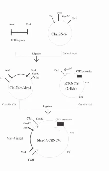
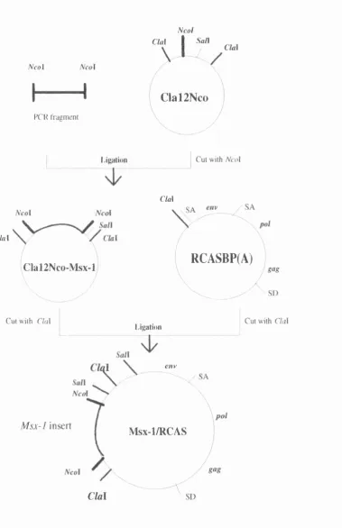
2.3.1 Characterisation o f a chicken Msx-1 cDNA clone 92 2.3.2 Construction o f recombinant retroviral plasmids encoding chicken Msx-1 92 2.3.3 Production o f infectious recombinant retrovirus particles 101 2.3.4 Northern hybridisation and immunocytochemistry 103 2.3.5 Ectopic expression o f recombinant retroviruses in vitro 107 2.3.6 Ectopic expression o f recombinant retroviruses in vivo by grafting transfected
cells into chick limb buds 110
2.4 Discussion 113
2.4.1 Review o f uses o f retroviral vectors in chick embryos 115
Chapter 3-Regeneration of wing bud stumps of chick
embryos in response to FGF-4
3.1 Introduction 118
3.2 Materials and Methods 123
3.2.1 Limb bud amputation and FGF-4 apphcation 123 3.2.2 Whole mount staining for cartilage 123
3.2.3 Muscle Pattern- Histology 124
3.2.4 Embryonic Neurofilament Staining 124
3.3 Results 125
3.3.1 FGF-4 apphcation can promote development of an^utated early wing buds 125 3.3.2 FGF-4 promotes development o f distal skeletal elements after excision o f tips o f
3.4 Discussion 136 3.4.1 Annulations can be rescued by FGF-4 136 3.4.2 FGF-4 restores development o f muscle pattern after annulation o f
wing buds 137
3.4.3 Relationship of chick wing bud regeneration to the nerve supply 139
Chapter 4- Reactivation
oi Msx-1, Shh
and
Hoxd-13
in
response to FGF-4 signals in regenerating chick wing bnds
4.1 Introduction 143
4.2 Material and Methods 146
4.2.1 Limb bud Annulation and FGF-4 or SHH Application 146
4.2.2 Dil Injection 146
4.2.3 Transplantation of Mouse Limb Proximal Cells to Distal Chick Wing Bud 147
4.2.4 Preparation of Digoxigenin-Labelled Riboprobes and Whole mount in situ
Hybridisation 147
4.3. Results 148
4.3.1 Identification o f the origin of cells which contribute to the
regenerating limb 148
4.3.2 Msx-1 expression in regenerating chick limb buds 151
4.3.3 Shh expression in regenerating chick limb buds 158 4.3.4 Shh bead implantation and Msx-1 expression in the regenerating chick
limb buds 158
4.3.5 Hoxd-13 expression in regenerating chick limb buds 158 4.3.6 Reactivation of Msx-1 expression in mouse limb tissue 161
4.4 Discussion 164
4.4.3 Rescue may require several signals 167
Chapter 5- General discussion
5.1 Msx-1 and limb development 171
5.2 The role o îMsx-1 in limb regeneration 173
5.3 Prospects for stimulating regeneration 178
List of Figures
Figure 1.1 Experiments demonstrating reciprocal inductions between the apical ectodermal ridge (AER), the underlying limb bud mesenchyme and the zone o f
polarising activity (ZPA) :20
Figure 1.2 Molecules expressed at the tip o f the limb and in ventral and dorsal
ectoderm :29
Figure 1.3 In situ hybridisation to Shh, Fgf-4 and Hoxd-13 transcripts in whole mounts o f chick wing buds at different developmental stages :30
Figure 1.4 Proposed model o f molecular interactions in the polarising region signalling pathway :38
Figure 1.5 Distribution o ï Msx-1 transcripts in whole mount preparations o f wing buds at stages 20-21 and 24 :43
Figure 2.1 Life cycle o f Rous Sarcoma Virus :56 Figure 2.2 Adaptor plasmids :59
Figure 2.3 Map of the RCAS series of vectors :60
Figure 2.4 Map of the rephcation defective retroviral vector pCRNCM:60
Figure 2.5 (A) Diagrammatic representation o f the chicken M sx-1 cDNA clone {qMsx
-1) showing the positions of the primers used for sequencing. (B) Complete nucleotide sequence o f qMsx-1 :93
Figure 2.6 Conq)arison o f the predicted nucleotide sequence o f cMsx-1 with the pubHshed cMsx-1 sequence :94
Figure 2.7 PCR primers :96
Figure 2.8 Procedures for cloning cMsx-1 into pCRNCM retroviral vector :97 Figure 2.9 Diagnostic cuts and sequence analysis of Msx- 1/pCRNCM :98
Figure 2.10 Procedures for cloning cMsx-1 into RCASBP(A) retroviral vector ;99 Figure 2.11 Diagnostic cuts of Msx-l/RCAS :100
Figure 2.13 Map of the genome of replication defective recombinant retrovirus Msx- 1/pCRNCM :104
Figure 2.14 Map o f the genome of rephcation con^etent retrovirus Msx-l/RCAS :105 Figure 2.15 Total RNA and Northern blots o f all different constructs :106
Figure 2.16 Immunocytochemical staining for chicken MSX-1 in CEF(O) cells transfected with Msx-l/RCAS :108
Figure 2.17 Micromass cultures of stage 20 chick wing bud mesenchyme : 109 Figure IAS In situ hybridisation to transcripts of rephcation defective Msx-
1/pCRNCM virus in vivo fohowing grafting of Q2bn transfected cehs into chick limb buds:111
Figure 2.19 In situ hybridisation to transcripts o f rephcation competent Msx-l/RCAS virus in vivo fohowing grafting of CEF(O) transfected cehs into chick limb buds :112 Figure 3.1 Successive stages in the regeneration o f opposite limbs in a newt fohowing amputation through lower and upper arms :119
Figure 3.2 Effect o f FGF-4 apphcation on development o f skeletal structures fohowing amputation o f chick wing buds at different stages :127
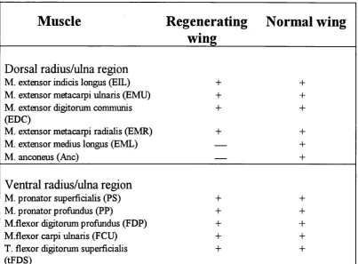
Figure 3.3-Muscle pattern of regenerating wing buds :132
Figure 3.4 Innervation pattern o f regenerated limbs 24-96 hours after amputation fiom the tip o f stage 24 wing bud by whole mount staining with a monclonal antibody against neurofilaments :135
Figure 4.1 Locations o f Dil injections in limb buds after amputation and posterior FGF- 4 apphcation ;149
Figure 4.2 Contribution o f cehs to the regenerating limb :150
Figure 4.3 Msx-1 and Shh expression after wing bud amputations and FGF-4 apphcation :153
Figure 4.4 Competence o f mesenchymal cehs to express Msx-1 and Shh in response to FGF-4 :157
Figure 4.6 Diagram showing location of pieces o f mouse mesenchyme cut from 10- to 11.5- day mouse limb buds and their behaviour when grafted beneath the apical ridge o f stage 18-22 chick wing buds :162
Figure 4.7 Expression oiMsx-1 in mouse forelimb bud tissue grafted distally into chick
List of tables
Table 2.1 Recent applications o f retroviral vectors ;54 Table 3.1 Antibody reactivity in the anq)hibian limb :122
Table 3.2 Skeletal pattern of early stage wings following removal o f the hmb bud and FGF-4 application :126
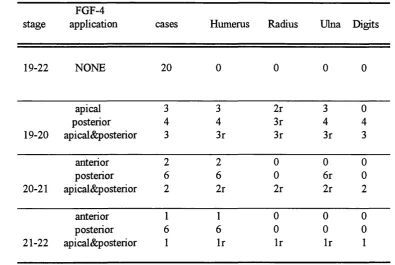
Table 3.3 Skeletal pattern of late stage wings following amputation and FGF-4 application :129
Table 3.4 Muscles in the regenerating wings :131
Table 4.1 Induction o f Msx-1 expression following removal o f whole limb buds at early stages and FGF-4 application :152
Table 4.2 M sxl and Shh expression following amputation and application of FGF-4 of late stage wing buds 155
Abbreviations
AEMF AEC AER AP Antp BMP bp CEF CMV cMsx-1 ControFpCRNCM Control/RCAS dix dpp ds D IT En-1 evx-l FGF hh LTR kb: msx Msx-l/pCRNCM Msx-l/RCAS M yoDapical ectoderm maintenance factor
apical ectodeiTnal cap
apical ectodermal ridge
alkaline phosphatase
antennapaedia
bone moiphogenetic proteins
base pairs
chick embryonic fibroblast
cytomegalovirus immediate early gene promoter chicken Msx-1 antisense Msx-l/pCRNCM antisense Msx-l/RCAS distaless decanpentaplegic double stranded dithiothretol engrailed 1 evenskipped 1
fibroblast growth factor hedgehog
long terminal repeat
kilobase
muscle segmentation homeobox-class homeobox gene sense Msx-l/pCRNCM
sense Msx-l/RCAS
PBS phosphate buffered saline
PET phosphte buffered saline with 0.1 % Tween
PCR polymerase chain reaction PFA p araformaldehyde
ptc patched
RA retinoic acid
RSV Rous Sarcoma Virus shh sonic hedgehog
t2? talpid
TCA trichloroacetic acid
TPA tumour promoting phorhol ester T/V trypsin/versin
ZPA zone of polarising activity
Chapter 1
1.1 Initiation of limb development
Embryonic chick limbs arise as thickenings in the mesoderm o f the body wall at approximately stage 16 (Hamburger and Hamilton, 1951). These thickenings occur in precise locations (wing buds develop opposite somites 15 to 20 and leg buds develop opposite somites 26 to 32), and their appearance is correlated with a decrease in the mitotic rate in the intervening flank mesoderm (Searls and fanners, 1971). Little is known about what controls the first stages o f limb
development, but it is likely that the mesoderm carries the information required for defining the position and the type of the limb. Transplantation experiments show that cells are determined to form a limb before a limb bud is visible. As early as stage 12, pre-limb mesoderm transplanted mto the flank region gives rise to an additional limb, whereas transplantation of ectoderm does not lead to ectopic limb formation (Saunders and Reuss, 1974).
Members of the Fibroblast Growth Factor (FGF) family have been implicated in limb initiation. The early observations that grafts of tissues such as nasal placode or otic vesicle could induce additional limbs in amphibians (Balinsky 1952, 1957, in Cohn et al., 1995) can now be attributed to the expression o f FGFs in these tissues (see Cohn et al., 1995). Recently it has been shown, that members of the
fibroblast growth factor family can act as a signal to initiate chick limb development (Cohn et al., 1995; Ohuchi et al., 1995; Mima et al., 1995; Crossley et al., 1996; Vogel et a l, 1996). The position in which limbs develop is probably
determined as the main body is being laid down and may involve expression of Hox genes (see later). Consistent with this idea is the shift in limb position when Hoxb5 is functionally inactivated (Rancourt et a l, 1995).
1.2- Cell interactions
limb axes appear to be set up at different times and by different mechanisms. The mesenchymal limb bud cells have two origins: lateral plate mesenchyme, which gives rise to the connective tissue including cartilage, and cells from the somites which migrate into the early limb-forming region and give rise to myogenic cells (Chevallier et al., 1977; Christ et al., 1977). Cellular condensation precedes overt differentiation o f both cartilage and muscle and begins in the proximal-most region o f the bud. Differentiated tissues are thus laid down in a proximo-distal direction. The competed appendicular chick skeleton is divided into three zones called the stylopod, zeugopod, and autopod. In the wing bud, the stylopod is made up o f the humerus, the zeugopod contains both the radius and ulna, and the autopod conq)rises of three digits, arranged in the pattern 2 3 4, running anterior to posterior (Figure 1. lA).
Estabhshment of limb pattern requires three major sets of cell interactions: a) an epithelial-mesenchymal interaction at the tip o f the limb between the apical ectodermal ridge and the underlying mesenchyme (progress zone) b) a second interaction between the covering ectoderm of the limb bud and the underlying mesenchyme and c) a mesenchymal-mesenchymal interaction between the polarising region and the nearby cells at the tip of the limb bud.
1.2.1 Apical ectodermal ridge signalling and progress
zone
Polarising Activity (ZPA). (A) Normal limb development. The location o f the AER and the ZPA are shown schematically in a stage 20 chicken wing bud. By day 10, the limb
bud resembles the adult wing in that it has a well defined humerus, radius, ulna, and digits 2, 3 and 4. (B) Removal o f the AER early in limb development results only in the formation o f humerus with more distal elements lost. (C). Effect of transplanting an ectopic ZPA to anterior margin o f a host limb bud. The presence of two ZPAs leads to the formation o f complete supernumerary mirror-symmetric structures. A, anterior; P,
AER
ZPA
A
-A E R
\
I
hours thereafter (Saunders and Reuss, 1974). O f additional interest is the fact that ectoderm from other parts of the body, hke neural tube, is able to form an apical ridge (Kieny and Brugal, 1970; in Saunders, 1977).
A major fimction o f the apical ridge is to mediate bud outgrowth, and it maintains a zone o f undifferentiated cells, the so-called progress zone, at the tip o f the limb bud. Removal of the ridge from a limb bud, results in cessation o f the outgrowth and results in truncations; the degree of truncation being dependent on the developmental stage of the embryo at the time o f operation. When the removal is carried out on the wing bud at stage 18, only the most proximal part o f the humerus is formed (Figure I.IB ; Saunders, 1948; Summerbell, 1974). When the operation is performed at stage 28, only the terminal phalanx o f digit 3 is absent (Summerbell et al, 1973). Thus the presumptive wing parts acquire the power of differentiation in proximo-distal sequence under the influence of the apical ridge. In addition, grafting an additional apical ridge to the dorsal surface of a limb bud brings about formation of a supernumerary limb (Saunders et al, 1976). In the chicken mutant eudiplopodia, a second ridge arises in the dorsal limb bud ectoderm proximal to the normally placed ridge and a supernumerary limb tip grows out at that site (Goetinck, 1964).
The inductive signal coming from the apical ridge is non-specific with respect to developmental stage and the type of structures the mesenchyme cells will form.
Irrespective of the age o f the ridge, the limb pattern is dictated by the mesenchyme (Rubm and Saunders, 1972). Tissue recombination experiments in which the apical ridges are interchanged between leg and wing mesoderms (Zwilling, 1955) or inversion o f the antero-posterior axis of the apical ridge with
AU these findings are in agreement with the thesis, first proposed by ZwiUing and Hansborough (1956), that mesodermal factors (Apical Ectoderm Maintenance
Factor -AEMF) are responsible for maintaining the apical ridge in its elevated configuration which then induces, in turn, mesodermal outgrowth and patterning. In addition to evidence aheady cited in support of an inductive role o f the apical ridge, it has also been found that when mica sheets were inserted into chick wing mesoderm subjacent to the apical ridge, the latter flattened and the resulting limbs were terminaUy deficient (Saunders, 1949). Recombination experiments carried
out on polydactylous mutants, where autopodal parts are dupHcated to varying degrees, resulted in autopodal duphcations only when normal limb ectoderm was recombined with polydactylous mesoderm (Zwilling and Hansborough, 1956). FinaUy, when the apex of a wing bud is amputated and replaced on its own stump after being rotated 180®C about the proximo-distal axis, the preaxial ectoderm of the grafted tip thickened, foUowed by the outgrowth o f a secondary set of posterior digits. The same result was obtained when a porous filter was interposed between the stump and graft, but not when non porous material was used (Saunders and Gasseling, 1963).
1977). However, Saunders found that the necrosis that foUows apical ridge removal does not affect the autopod mesoderm but rather, a band o f mesoderm several layers below the surface (Saunders, 1977).
Mesenchyme cells in the progress zone remain in a labile state with respect to positional identity by the influence of the overlying apical ridge: this being the
only region o f the limb where positional identity is labile (Summerbell et al., 1973). It has been postulated that information along the proximo-distal axis o f the limb is designated by the amount o f time a cell has spent in the progress zone. Therefore with increasing time, which is likely to be measured in terms o f the number of cell divisions, the positional identity specified within the progress zone
becomes more distal (Summerbell et al, 1973). When distal tips from older wings are grafted on to proximal stumps from younger wings, limbs consisting o f a humerus attached to digits developed, with the normally intervening radius and ulna absent. Similarly, young tips grafted on to old stumps resulted in the formation o f wings containing two humerus elements followed by a radius and ulna and digits. Thus Httle, if any, regulation occurred at the junction o f the grafts, instead each part developed autonomously (Summerbell and Lewis, 1975).
1.2.2- Ectodermal signalling
Ectodermal signals appear to be important in controlling the pattern across the
wings. Furthermore, when an additional apical ridge is grafted either to the dorsal or ventral surface of the limb bud, the resulting supernumerary wing tip is of bidorsal or biventral polarity respectively (Saunders et al., 1976). These
observations suggest that both dorsal and ventral ectoderm have some morphogenetic significance and could act as a signal source for the dorsoventral patterning.
1.2.3- Polarising Region signalling
The third set of interactions in the developing limb is between the zone of polarising region (ZPA) and the cells in the progress zone. The signal firom the polarising region controls the pattern of differentiation across the antero-posterior axis o f the limb bud. The signalling o f the polarising region was first discovered when this region was grafted to the anterior margin o f a second bud (Figure 1.1C; Saunders and Gasseling, 1968). This operation evokes the production o f a mirror- image symmetrical wing patterns from the host tissue in response to the graft. The results o f a series of grafting experiments show that the identity o f a digit that develops depends on the distance from the polarising region. The digit next to the polarising region is always more posterior in character than the digit further away (Tickle et al., 1975). In addition, the type o f additional digits that develop is dependent on the number of polarising region cells grafted. When a small number o f cells were grafted just an additional digit 2 develops (Tickle, 1981).
The spatial and temporal distribution of the cells with polarising activity in the limb and flank o f the chick embryo has been systematically mapped. Polarising
Honig and Summerbell, 1985). Maintenance of the polarising region occurs in the presence o f the apical ridge. In the absence of the ridge, polarising activity is reduced (Vogel and Tickle, 1993). At late stages, where the apical ridge losses its abihty to induce outgrowth, and polarising activity is no longer detectable, polarising region cells either contribute to tissues that make up the limb (Bowen
et al., 1989) or die.
Finally, the signalling activity of the polarising region is not species specific: posterior tissue firom the limb buds of such diverse amniotes as turtles, mice and humans are all capable of inducing duplicated wing patterns (MacCabe and Parker
1976; Tickle et al., 1976; Fallon and Crosby, 1977).
1.3- Models for limb development
During the last twenty years or so, the interpretations o f morphogenetic phenomena in limb development (and regeneration) have been based upon the concept o f positional information (Wolpert 1969). In developing limbs, cells first acquire positional values with respect to certain reference points within the
system and then interpret these values in terms o f the appropriate cytodifferentiation. Built on this idea, two rather divergent models, for explaining how patterning occurs have emerged. The zone o f polarising activity (ZPA) model based largely on studies of developing chick limbs and the polar co ordinate model based primarily on studies on regenerating amphibian limbs.
1.3.1 Zone of Polarising Activity (ZPA) model
respect to the antero-posterior axis, results in the formation o f posterior structures; the lower concentration of morphogen on the preaxial side specifies positional values characteristic of anterior elements.
1.3.2- Polar Co-Ordinate Model
This model differs firom the polarising zone model in that cellular interactions, eventually resulting in supernumerary formation, occur at a local level and are not related to long-range signalling mechanisms.
According to this model, the limb is represented by a series of concentric circles.
Cells have two positional values, one with respect to the circle which specifies position on the limb circumference, and the other on a particular circle, which specifies position along the proximal-distal axis. The model postulates that confirontation of ceUs possessing non-adjacent positional values (through grafting or wound healing), triggers their division and the intercalary regeneration of missing circumferential values. Two rules govern the restoration o f pattern. The "shortest intercalation" rule states that intercalation of circumferential values takes place via the shortest route on the circle between non neighbouring cells. The "distahsation" rule states that if the circumferential values of cells in the new circle are the same as those o f adjacent cells in the old circle, the new cells must adopt a more distal value (Bryant and Muneoka, 1986). These simple principles o f the polar co-ordinate model account for many respects o f limb outgrowth in the development and regeneration, for regulation o f the pattern foUowiug the deletion of parts, for the production of supernumerary limbs, and for the regulation of growth and final size.
1.3.3- A comparison of principles
change their fate, by long range signalling. The most direct evidence for a long-
range signal comes from grafting experiments where leg anterior mesenchyme is interposed between a polarising region graft and wing anterior tissue. Leg tissue maintains its character in wings and when placed at wing buds tips develops into toes. In addition, an anterior chick wing digit, digit 2, develops from the wing tissue which is separated from the polarising graft by leg mesenchyme (Honig,
1981).
In contrast, the polar co-ordinate model presupposes the existence o f certain initial key positional values, and cells respond to discontinuity by dividing and acquiring new positional values by intercalation. This is consistent with the finding that cell proliferation in the anterior cells after a polarising region graft is increased (Cooke and Summerbell, 1980). However, it is difficult to explain the loss of digit 2 when two polarising regions are grafted at the anterior margin of the limb (Wolpert and Hombruch, 1981).
Iten and Murphy (1980) have challenged the notion o f a special role for posterior tissue and therefore the polarising region zone model, by demonstrating that when anterior tissue is grafted posteriorly extra digits also arise. However, the effects of irradiation o f grafts o f anterior and o f posterior (polarising region) tissue differ
enormously. Smith et al (1978, 1979) showed that an irradiated polarising region is still capable of signalling positional information and specifying additional digits
1983). Therefore, the simplest explanation is that the cells in anterior grafts placed posteriorly give rise to the additional digits in response to signals from the nearby polarising region.
1.4- Signalling molecules and molecular responses
1.4.1- Apical ectodermal ridge signals
Members o f the FGF family have recently been identified as the molecular signals most likely to account for apical ridge function. The FGF family currently comprises nine members, Fgf-1-9, but not all o f them are expressed in developing limbs. Transcripts o f two members of the FGF farmly, Fgf-4 and Fgf-8, are expressed in the apical ridge of chick limb buds, while FGF-2 protein appears to be more widespread (Niswander and Martin, 1992; Crossley and Martin, 1995; Crossley et al., 1996; Vogel et al, 1996; Savage et a l, 1993). Fgf-8 transcripts are found throughout the ridge whereas Fgf-4 transcripts are more abundant in the posterior ridge (Figure 1.2A; Figure 1.3).
G O
P r
Fgf-2, Fgf-8
(A)
Fgf-4, Fgf-2, Fgf-8
Progress zone molecules
Msx-1, Msx-2, Wnt-Sa, Evx-1, Cek-8
Wnt-7a
Lmx-1
Shh
V
Wnt-5a
Bmp-4
Shh
Fgf-4
only reconstructed when mesenchymal cells are exposed to both the ridge signal (FGF) and a polarising signal (Niswander et al., 1993b).
Many other genes are known to be expressed by apical ridge cells including other signalling molecules such as Bone Morphogenetic Proteins-2 and -4 (BMP-2, -4)
and the product o f the Wnt-5a gene, and a number o f transcription factors including distaless, engrailed (and earher in ventral limb ectoderm), Msx-2, and Msx-1 (reviewed by Tickle and Eichele, 1994). Although their roles have not been fuUy investigated, the products o f these genes might be involved in controlling the expression of FGFs or controlhng ridge morphology (Tickle and Eichele, 1994).
1.4.2- Ectodermal signals
Signalling from the limb bud ectoderm appears to control dorso-ventral patterning and is mediated by Wnt-7a gene (Yang and Niswander, 1995). Wnt-7a is expressed in the dorsal ectoderm (Parr et al., 1993; Yang and Niswander, 1995; Figure 1.2B). Functional inactivation of Wnt-7a in mice o f a transgenic strain resulted in paws with a double ventral pattern (Parr and McMahon, 1995). In addition, Engrailed-1 {En-1\ a homeodomarn-containing transcription factor, is expressed in ventral limb ectoderm and is essential for ventral patterning (Joyner
and Martin, 1987). Loss oïEn-1 function in mice results in dorsal transformations of ventral paw structures, and in subtle alterations along proximo-distal limb axis (Loomis et al., 1996). In contrast to the dorsaUy restricted expression of Wnt-7a in the wild type limb buds, in En-1 mutant limbs, VJnt7a is expressed in ventral as well as dorsal limb ectoderm (Loomis et al., 1996)
1.4.3- Polarising region signals
protein (Lopez-Maitinez et al., 1995) at the anterior margin o f chick wing buds leads to the induction o f dupHcated wing patterns virtually indistinguishable from those obtained by polarising region grafts.
Limb buds can synthesise RA from its precursor (retinol) in vivo (Thaller and Eichele, 1988). Retinol has a uniform distribution in the hmb bud, whereas there is a differential distribution of RA, with high levels distributed posteriorly, in the zone o f polarising activity (Thaller and Eichele, 1987). For many years, retinoids
have been regarded as major candidates for the polarising region morphogen. However, recent research has shown that RA is part o f a signalling cascade and anterior cells are induced to become polarising cells. Transplantation experiments in which pieces of mesenchyme located distally to the RA bead at the anterior margin were grafted to the anterior margin of a host wing bud produced additional digits (Summerbell and Harvey, 1983; Wanek et al., 1991). In addition, Noji et al., (1991) showed that when a RA bead was placed anteriorly in a wing bud, a higher level of retinoic acid receptor-B (RAR-B) expression was induced around the bead. This was not observed around a grafted ZPA, and this suggests that it is unlikely that exogenous RA is identical to the signal from the ZPA.
Apphcation of RA to the anterior margin induces expression o f Shh. Shh induction always requires a ridge signal (FGFs) (Riddle et al., 1993; Niwsander et a l, 1994). The product of Shh is the more recent contender for the role of
(reviewed by Johnson and Tabin, 1995). Studies have shown that cleavage is necessary for hedgehog protein to function in Drosophila. Deletion of the carboxy-temhnal portion of the protein has little effect on activity of the protein,
whereas abohshing the secretion of the amino-tenninal half leads to a complete loss o f signalling (reviewed by Fietz et al, 1995). Extending these studies to spinal cord of vertebrate embryos. Fan et al. and Roelink et al. (1995) likewise demonstrated that the amino-terminal processed form o f SHH is sufficient to direct long-range effects (sclerotome differentiation and proliferation and motor neuron induction) as well as short-range effects (floor plate induction within the neural tube).
In chick limb buds, transcripts of Shh co-localize with the polarising region (Riddle et a l, 1993; Figure 1.3) but it is not clear whether all the cells in this region express Shh. In mouse and chick limb buds, reduced levels of Shh expression are correlated with loss of posterior digits (Yang and Niswander,
1995; Parr and McMahon, 1995).
It is still unclear whether Shh acts as a long-range graded signal or activates a cascade o f local interactions. \iShh acts as a long-range signal, it is expected to diffiise in a dose-dependent manner and cells exposed to high concentrations of SHH should produce posterior identity digits whereas cells exposed to low SHH concentration should produce anterior digits. It would also be interesting to find out the range over which Shh signals in the limb bud. However, Shh is known to be able to induce the production o f secondary signals.
cells lead to an ectopic domain of Bmp-2 in the anterior mesenchyme (Francis et al., 1994; Laufer et al., 1994). Activation of Bmp-2 by Shh in the chick limb bud is consistent with the finding that in Drosophila, dpp is secreted in response to hh in imaginai discs and is responsible for the effects of hh on disc patterning (Basler and Struhl, 1994). However, it is particularly intriguing that apphcation of Bmp-2 alone (Francis et al., 1994) or grafl;ing cells expressing BMPs (Duprez et al., 1996a) to developing limb buds do not produce ah the effects o i Shh signalling on
digit pattern perhaps indicating that multiple BMPs may required as heterodimers or multiple secondary factors are required in concert to transduce the initial SHH signal. However, it is likely that BMPs have other roles in hmb development. For example, another member o f the BMP family, BMP-7, is also expressed in developing limbs and functional inactivation of this gene results in polydactyly (Luo et al., 1995). Furthermore, inactivation o f a BMP receptor in chick hmb buds leads to interdigital webbing (Zou and Niswander, 1996).
An immediate downstream gene whose expression induced by Shh is the transmembrane protein patched {PTC). In Drosophila, hh regulates gene expression by antagonising the action of the patched gene product; patched also being itself a transcriptional target o f hh signahing (Ingham et al., 1991). PTC is expressed at high levels m the posterior mesenchyme of vertebrate hmb bud and it is likely that high levels of PTC expression mark cehs that are actively responding to Shh signalling (Marigo et al, 1996; Goodrich et a l, 1996). Induction o f PTC by Shh seems likely to be more direct than Bnq)-2 induction since it does not require signals fi-omthe apical ridge (Marigo et al, 1996).
1.4.4 Progress zone signals
the two related homeobox containing genes, Msx-1 and Msx-2. Their expression patterns and their role in hmb development are discussed in section 1.5.
Mesenchymal cehs in the progress zone also express the transcription factors Evx- 1 (Niswander and Martin, 1993a) m d AP-2 (Mitcheh et al., 1991), the growth factor Wnt-5a (Gavin et al., 1990) as weh as the ^/z-related receptor tyrosine kmase Cek-8 (Patel et al., 1996).
1.4.5 Response
of Hox
genes to polarising zone signals
Vertebrate homeobox genes were initiaUy isolated on the basis that they encoded
DNA sequences which were identical to the homeobox o f the Drosophila Antennapaedia gene. Four homologous clusters o f Antp-VîkQ homeogenes have been identified in chick, Hoxa, Hoxb, Hoxc and Hoxd. These clusters are thought to have arisen by duphcation during the course o f evolution and exhibit a high degree o f homology both with each other and with the Drosophila homeogenes. In both vertebrates and Drosophila, the anterior boundary of expression of each gene corresponds to the relative order o f genes in each chromosome, with (3') genes exhibiting the most anterior expression and the (5') genes sequentially exhibiting more posterior expression.
Expression domains of homeobox genes in 5' regions o f the Hoxa and Hoxd clusters appear to correlate with regions that give rise to distract skeletal elements along the proximo-distal and antero-posterior axis o f developing limb buds. Specifically, the expression domain of 5* Hoxa genes appear to identify different
the anterior base o f the bud express Hoxd-9 and Hoxa-9 only. As the limb grows, expression expands anteriorly so that the final boundaries o f Hox expression domains are perpendicular to the proximo-distal axis (Yokouchi et al., 1991).
Hox genes are activated in response to signals that specify limb pattern. Hoxd genes are induced in anterior mesenchyme by apphcation o f RA, polarising region grafts or SHH expressing cehs (Izpisua-Belmonte et al., 1991; Francis et al., 1994; Laufer et al., 1994). Importantly, induction oîH oxd gene expression takes place in the same 3' to 5' sequence as in normal development with Hoxd-11 being turned on before Hoxd-13. In addition, FGF-4 apphcation mamtains Hoxd-13 expression in the distal posterior mesenchyme after apical ridge removal. In contrast, expression of Hoxd-11 is relatively stable after ridge removal (Vogel et al., 1995a). This is consistent with the observed expression pattern o f H oxdl3 and Hoxd-11 in the developing hmb. Expression of Hoxd-13 remains at the tip o f the bud near the ridge (Figure 1.3), whereas expression o f Hoxd-11 is locahsed to regions o f the mesenchyme that do not appear to be influenced by the ridge. The requirement of apical ridge signals for proper Hoxd expression is also shown by the fact that Hoxd genes can be activated in anterior cehs in the absence o f the ridge only by applying two beads, one soaked in RA and one soaked in FGF-4 (Niswander et al, 1994).
Additional evidence supporting the crucial role of Hox genes in patterning and growth o f the hmb comes fi*om experiments in which a particular gene is overexpressed or fimctionahy inactivated. When Hoxd-11 is expressed throughout
colinearity of the Hox genes, such that inactivation of the 3' genes has a more proximal phenotypic boundary (afiFecting both the zeugopod and autopod of the limb) than that of the more 5' genes (affecting only the autopod). For example, Hoxd-11 targeted disruption results in limb defects including malformations o f the radius and ulna, inappropriate fusions between wrist bones, and regional malformations at the distal end of the forelimb (Davis and Capecchi, 1994). Mice with mutation in the Hoxd-13 gene exhibit more distal abnormahties such as growth retardation of several metacarpals and phalanges and the presence o f an extra posterior digit (DoUe et al., 1993). More recently, double "knock-outs" have been produced in which two paralogous genes in two different clusters have been inactivated in mice and more severe defects locahsed to predicted regions o f the limb have resulted. Thus, inactivation of Hoxd- 11 and Hoxa-11 leads to almost complete absence of zeugopod while proximal and distal structures are unaffected (Davis et al., 1995). However, functional redundancy can also occur even between non-paralogous Hox genes (for example between Hoxd-11 and Hoxa-10\
see Favier et al., 1996).
Recently, a transcription factor Lmx-1, has been shown to be activated in dorsal limb ectoderm by Wnt-7a signalling (Riddle et al., 1995; Vogel et al., 1995b; Figure 1.2B). When Lmx-1 is ventraUy expressed in chick limb buds this leads to
ectopic dorsal structures. It was suggested that the limb pattern could be specified by the combination of expression of Hox genes and Lmx-1.
1.4.6 Molecular model of limb development
anterior
proximal
distal
G F -4 (AER)
WNT-7a (dorsal ectoderm)
posterior
Shh (ZPA)
1.4). Recently, it has been suggested that Bmp-2 may mediate the induction of Fgf-4 expression by SHH (Duprez et al., 1996a). This is consistent with the fact that the endogenous expression domain o f Fgf-4 is more closely related spatially to the mesenchymal Bmp-2 domain, than to that of Shh.
One approach towards understanding early patterning mechanisms in the limb is to study chick and mouse limb mutants (reviewed by Cohn and Tickle, 1996). Recent analysis o f the molecular mechanisms by which the preaxial abnormalities
in polydactylous mutants revealed alterations in gene expression of signalling molecules based on the polarising region. In talpid^ (/a-^) chick mutants, where many and sometimes fused morphologically similar digits are formed but not in a mirror -image pattern, Shh is still restricted posteriorly. However, BMP and Hoxd genes are expressed uniformly throughout distal hmb limb bud cell mesenchyme and Fgf-4 is expressed throughout the apical ridge, instead o f being posteriorly restricted as in normal limb buds (Francis-West et al., 1995). It has been suggested that BMP-2 in talpid mesenchyme might induce FGF-4 expression throughout the ridge, and that BMP-2 and FGF-4 would then induce Hoxd-13 expression. AH these genes are downstream of Shh. Some similar features are also found in limb buds that develop from reaggregated anterior leg bud mesenchyme (Hardy et al., 1995), where Bmp-2 and Hoxd-13 expression are found together, and the apical ridge is maintained in the absence of Shh expression.
another mouse mutant, limb deformity {Id), in which digits are reduced and fused.
These mutant limb buds show a severe decrease in the expression o f Shh, Fgf-4 is not expressed in the posterior ridge, and Hoxd-12 expression is delayed (Chan et al., 1995b; Haramis et al., 1995). The Id gene is expressed in the apical ridge of normal limb buds (Jackson-Grusby et al., 1992) and one possibility is that It maintains Fgf-4 expression in the ridge.
1.5 Muscle segment homeobox-containing genes
{Msx)
1.5.1 Isolation and characterisation
of Msx
genes
In general, Hox genes are organised in clusters and individual genes in each cluster have distinct expression domains along the antero-posterior axis (McGinnis and Krumlau^ 1992). However, there are classes o f homeobox genes which are scattered within the genome and appear to play more diverse roles in growth and development. One such class is the Msx gene family, identified by a distinct and highly conserved homeodomain related to the single msh {muscle segment homeobox) gene m Drosophila. The striking sequence conservation of these genes in and around the homeobox, and their expression early m the difierentiation of various organs suggested that they might have a fundamental developmental role, which has been conserved during the evolution o f the vertebrates.
otherwise highly conserved homeodomain, members o f vertebrate Msx genes are divided into Msx-l-Vks (including the Msx-3 gene, which is partially characterised and has a homeobox sequence that places it in the Msx-1 subclass) and Ms'x-2-lîke subclasses (Bell et al., 1993).
The first characterised vertebrate wj/z-like gene was the mouse Msx-1 gene (formerly known as Hox7) which was identified independently by PCR anq)lification o f genomic DNA (HtU et al., 1989) and by hybridisation to the Drosophila msh gene (Robert et al, 1989). Mouse Msx-1 maps to the proximal region o f chromosome 5, IcM distal to the mouse homologue of D4S43, a human probe closely linked to Huntington's disease, and 2-3cM distal to the mouse En-2 gene (Hill et al., 1989; Robert et al., 1989).
The chicken Msx-1 homolog was originally isolated by screening a chick embryonic cDNA Hbrary with the mouse Msx-1 cDNA probe (Coelho et al, 1992a; Nohno et a l, 1992) or with a DNA fragment containing the homeobox of the Drosophila msh gene (Suzuki et a l, 1991). Comparison o f the deduced amino acid sequences o f chicken Msx-1 with those o f the mouse Msx-1 and chicken Msx-2 genes revealed that the chicken Msx-1 homeodomain is identical to the homeodomain o f mouse Msx-1 and diflfers only by two amimo acids from the homeodomain o f by chicken Msx-2. Moreover, there are several specific regions outside the homeodomain which are highly conserved among the Msx-1 cognates but are not conserved in the corresponding region encoded by the Msx-2 genes (Coelho et a l, 1992a; Suzuki et a l, 1991; Nohno et al, 1992). Therefore, since Msx-1 and Msx-2 homeodomains are highly similar, the conserved non- homeodomain sequences specific for either Msx-1 and Msx-2 proteins might be those regions involved in determining the fimctional specificities o f the two
1.5.2 Expression of
Msx-1
during chick embryonic
development
1.5.2.1 Expression
of Msx-1
in chick limb bud
When the limb bud starts to develop (stages 16-18), Msx-1 is expressed throughout the whole mesoderm (Robert et al., 1991). As development proceeds, (stages 20-21), the domain of Msx-1 expression becomes restricted, and transcripts o f Msx-1 are detected in the mesoderm beneath the apical ridge extending furthest proximally at the anterior of the bud (Robert et al, 1991; Coehlo et al., 1992a, Suzuki et al., 1991; Figure 1.5A). There is a clear gradation in Msx-1 transcripts abundance with the highest levels o f transcripts in mesenchyme immediately adjacent to the apical ridge, but the ridge itself expresses A/5X-7 at low levels (Davidson et al., 1991; Suzuki et a l, 1991; Nohno et al., 1992; Brown et al, 1993). At later stages of development (stages 24-25) Msx-1 transcripts are present throughout the proximal anterior periphery of the limb bud. In contrast, in the posterior periphery, expression o î Msx-1 is limited to a small group of mesodermal cells at the edge of the wing bud in the mid- proximal region (Robert et al., 1991; Suzuki et al., 1991; Coelho et a l, 1992a; Figure 1.5B). This region corresponds to the posterior necrotic zone in which programmed cell death occurs. Finally, in older limb buds (stages 31-32), Msx-1 transcripts are located throughout the necrotic mesenchyme between the developing digits of the limb where programmed cell death occurs (Coelho et al.,
1992a; Nohno et al., 1992; Robert et a l, 1991; Suzuki et a l, 1991).
wing bud. (B) As the limb grows out Msx-1 is expressed in the posterior and anterior
1.5.2.2 Expression
of Msx-1
during early development
The early stages o f development are marked by the widespread expression of Msx-1. Msx-1 is first detected during gastrulation, where the estabhshment o f the basic structure o f the fixture organism is laid down. In particular, Msx-1 transcripts are first detected in the primitive streak region as well as in the extraembryonic tissues (Suzuki et al., 1991). As neurulation starts, Msx-1 expression is detected in all along the neural axis but the signal is restricted to a narrow dorsal region. In this respect, Msx-1 is unique among the vertebrate homeobox containing genes, in that its anterior boundary is at the anterior end of the neural tube. Later in development, transcripts of Msx-1 are found in neural crest cells and their derivatives (Suzuki et al., 1991).
1.5.2.3 Other sites
of Msx-1
expression
The early expression patterns of Msx-1 undergo dramatic changes during organogenesis so that Msx-1 becomes expressed in characteristic, spatially restricted patterns in a wide range o f organs (Davidson, 1995). Some o f these sites are briefly outlined below:
Facial processes and sense organs
development proceeds, transcript levels in lateral nasal process and frontonasal
mass increase but mandibular primordia continue to express most strongly (Suzuki et al, 1991; Brown et al, 1993; Nishikawa et a l, 1994; Mina et al,
1995).
In the developing ear, Msx-1 is expressed in the otic vesicle at the stage when the invaginating placode is sthl open to the outside. Transcripts o î Msx-1 are locahsed
in the dorsal one-third of the placode near the neural tube where it is also expressed (Suzuki et al, 1991).
H eart
Msx-1 expression is detected in the heart in individual cells delaminating from the antrioventricular junction (AV) and forming the mesenchymal population o f the endocardial cushions. Endocardial cushion tissue of the outflow tract (OFT) is not expressing Msx-1 but strong Msx-1 expression is found in the distal regions which probably correspond to the developing OFT valve leaflets (Suzuki et a l, 1991;
Chan-Thomas et a l, 1993).
Feather Buds
Msx-1 is an early marker of epithehal placodes for skin appendages. In the feather follicle, Msx-1 is expressed in the collar and barb ridge epitheha which are regions of continuous ceU proliferation (Noveen et al, 1995).
These temporal and spatial patterns o f expression are highly conserved in mammals (Davidson, 1995). In addition to the above sites, Msx-I expression has also been described in the tooth germs, eye and mammary glands of mouse
embryos.
cup that gives rise to the cihary body in the adult retina (Monaghan et al, 1991). In mammary glands, Msx-1 transcripts are locahsed to the mammary epithehum as weU as the developing alveoh (Friedmann et a l, 1996).
1.5.3 Regulation
of Msx-1
expression in limb buds
Several lines of evidence suggest that Msx-1 expression is under the control o f the apical ridge. Transplantation of non-expressing Msx-1 proximal limb cells underneath the apical ridge o f a host limb, resulted in the re-expression of Msx-1 in the transplanted mesenchyme within 5 hours (Davidson et a l, 1991). In large
grafts, the part directly under the apical ridge expressed a high level of transcripts, whilst the most proximal part often exhibited no transcripts, thus showing that there is a gradient of expression consistent with the apical ridge as a source o f the inducing factor. Quail ridge cells grafted on to non-expressing proximal chick limb bud cells can induce Msx-1 expression in the underlying mesenchyme (Robert et al, 1991). Normally expressing distal tissue when grafted proximally, loses expression completely after 5 hours, thus further showing that Msx-1 expression is position dependent (Davidson et a l, 1991). Removal of the ridge is rapidly followed by cessation of Msx-1 expression in the posterior subridge mesenchyme. It is not clear whether expression of Msx-1 in distal anterior mesenchyme requires a continuous ridge signal (Ros et a l, 1992; Vogel et al, 1995a). One report found that anterior mesenchyme continues to express Msx-1 (Ros et a l, 1992), whereas another group of workers did not detect any Msx-1 expression in the anterior mesenchyme after ridge removal (Vogel et a l, 1995a).
addition to apical ridge-containing ectoderm, ectoderm from the anterior but not proximal posterior border can induce strong Msx-1 e?q)ression in cultured limb bud cells (Coelho et a l, 1993a). Thus it appears that, in addition to the apical
ridge, ectoderm from different chick limb bud regions has the abihty to regulate Msx-1 expression. However, similar experiments with mouse cells gave different results. When the apical ridge is excised from mouse limb explants, Msx-1 expression levels remained high in limb mesenchyme, whereas removal o f the entire limb ectodermal jacket caused a complete loss of Msx-1 expression. Thus there are some differences between the role of apical ridge in terms o f regulation
0Ï Msx-1 expression in the chick and mouse (Wang and Sassoon, 1995).
In contrast to the above, at later stages of development (stage 29), in chick embryos Msx-1 expression is maintamed in the subridge mesoderm after removal o f the interdigital apical ridge (Ros et a l, 1994a). This observation suggests that expression of Msx-1 in the interdigital mesoderm is stabUised and is no longer dependent on the influence of the apical ridge.
Further evidence to support the hypothesis that Msx-1 expression is involved in the outgrowth-promoting effect of the apical ridge and may required for limb outgrowth comes from the analysis o f its expression pattern in chick limb
grafting an apical ridge from a normal embryo onto limbless mesenchyme (Robert
et al., 1991; Coelho et al, 1991). In a second chick limb mutant, eudiplopodia, which generates an ectopic apical ridge on the dorsal surface o f the limb bud, Msx-1 expression is found underneath both ridges (Robert et al., 1991).
Recently, members of the FGFs, which have been demonstrated to be expressed by the apical ridge, have been in^hcated in the molecular pathway in which Msx genes function. Apphcation of FGF-4 beads to posterior and apical mesenchyme after ridge removal can restore an almost normal pattern o f Msx-1 expression (Vogel et al., 1995a) and Msx-1 expression appears to be also maintained in cultured limb mesenchyme cells by both FGF-2 and FGF-4 (Watanabe and Ide, 1993; Wang and Sassoon, 1995). Other growth factors have also been imphcated in the control of Msx-1 expression. Bmp-2 and Bmp-4 are co-expressed with the Msx genes at numerous sites, including the limb bud, tooth germ, primitive streak and hindbrain. BMP-4 protein has been shown to induce Msx-1 expression in proximal limb tissue (Wang and Sassoon, 1995). In addition, recombinant BMP-2 or BMP-4 protein can induce Msx-1 expression in cultures o f tooth mesenchyme, thereby mimicking the action of dental epithehum on the mesenchyme (Vainio et al., 1993). The functional signifrcance of this induction is supported by the finding that Msx-7-deficient mouse embryos show a failure in tooth development (Satokata and Maas, 1994). These observations suggest possibhities for complex regulation o f the Msx-1 gene by the independent, or co-operative action of members o f the FGF family and BMPs (Davidson, 1995).
1.5.4 Function
o i Msx-1
in the chick limb bud
influence o f the ridge as a result of limb outgrowth. There is some evidence that Msx-1 expression does indeed inhibit cell differentiation. f,imb mesenchymal cells treated with the tumour promoting phorbol ester, TPA exhibit deregulated expression of Msx-1 and also fail to exhibit the extensive gap junctional intercellular communication that normally occurs at the onset o f limb cartilage differentiation (Ferrari et al., 1994). Furthermore, when cells o f a myogenic cell
line are transfected with Msx-1 ^ they do not differentiate into muscle and expression o f muscle specific genes is inhibited (Song et al., 1992). This is in agreement with the finding that dissociation o f limb bud mesenchyme leads to an acceleration of myogenic factor accumulation and a concomitant decrease in Msx- 1 expression (Wang and Sassoon, 1995). In contrast to the above, transfection o f Msx-1 into amphibian limb blastema had no detectable effect on the differentiation
o f myogenic blastema cells (Crews et al., 1995). In similar experiments, expression of the related Msx-2 gene had no effect on growth or differentiation of the myoblast cell line (Song et al., 1991).
limb primary culture. In order to explain the above results, the authors suggested that RA-mediated repression of Msx-1 and Msx-2 requires cellular interactions not present in proximal mesenchyme and/or is mediated by factors that are lost after disruption of cell-cell contacts. Examination o f the expression pattern of Msx-1 and Msx-2 in the limb buds o f two mutant chick embryos, àdplopodia-5
and talpidP, which develop supernumerary digits fi"om anterior limb mesoderm, revealed that the formation of supernumerary digits fi'om anterior limb mesoderm
is preceded by the concomitant suppression of Msx-1 and Msx-2 expression in the anterior mesenchyme (Krabbenhoft and Fallon, 1992; Coelho et al., 1992b, 1993b). This observation is consistent with the finding that expression of Msx-1 and Msx-2 is diminished in the anterior mesoderm o f non-polarised recombinant limbs (Ros et al, 1994b).
Finally, Msx-1 and Msx-2 are thought to be involved in programmed cell death. Both genes are expressed in the posterior necrotic zone and the interdigital mesenchyme suggesting possible involvement of these genes in defining regions of cell death shape the contours of the developing limb bud. However, experimental changes in the fate o f the interdigital tissue firom death programme to chondrogenesis resulted in no modification of the Msx-1 expression pattern suggesting a role for Msx-1 in maintaining interdigital tissue in an undiJfferentiated
state (Ros et al, 1994a). This observation complements recent studies showing that inhibition of interdigital cell death by administration o f FGFs takes place in
the absence of significant modifications in the normal pattern o î Msx-1 expression (Macias et a l, 1996). In contrast, implantation of BMP-4 soaked bead at the tip of the growing digits, which induces an ectopic domain of cell death by ap opto sis in the undifferentiated limb mesoderm, is accompanied by a locahsed extension of
1.6 Aims of the thesis
The aim o f the experimental work described in this thesis was to understand the role 0Î Msx-1 gene in the development of the chick limb. In particular, we wished
Chapter 2
2.1 Introduction
Analysis o f the function of gene expression in animal cells is one of the central themes o f current molecular biology. Procedures for introducing manipulated genes into animal cells, where their fimction can be assayed, have therefore been
the subject o f intensive research. Currently, the methods for achieving this vary with dififerent experimental organisms. For example, in the mouse, there are well-
dominant-negative BMP-2/BMP-4/BMP-7 Type I receptors have been expressed
in the developing chick hindlimb with retroviral vectors, support a role for BMPs in regulating the apoptosis that normally occurs between the digits (Zou and Niswander, 1996).
GENE
Virus Type
Cell/Tissue
References
Alkaline Phosphatase RCASBP Nervous system
Fekete and Cepko, 1993
Bcl-2 RCASBP CEF Givol etal., 1994 Thyroid hormone
rec^tor-a RCASBP CEF, QT6 Hillgartner etal., 1992
Transferrin realto r RCASBP CEF Odorizzi et al., 1994
Hoxa-13
Replication-Competent wiugbud Yokouchi et al., 1995
Hoxd-11 RCAS wing bud Morgan et al., 1992
...
Fsf-2
...
Replication-Defective wing bud Riley et al., 1993
Sonic hed^eho^ RCASBP wing bud Riddle et al., 1993
Sonic hed^eho^ RCASBP somites Johnson et al., 1994
Bmp-2
RCASBP
pCRNCM wing bud Duprez et al., 1996a
Bmp-4 RCASBP
wing bud
lateral plate mesoderm
Duprez et al., 1996b Pourquie et al., 1996 Mutant
BMP receptor RCASBP wing bud
Zou and Niswander, 1996
^nt-7a RCASBP wing bud
Yang and Niswander, 1995
2.1.1- Retrovirus life cycle
Retroviruses are single-stranded RNA viruses that replicate via DNA intermediates. Once the virus particle has bound to the target cell surface, the capsid is released into the cytoplasm and the single stranded RNA is copied by reverse transcriptase into double-stranded linear and then circular DNA, and integrated into the cellular genome at random sites. The provirus, which is the integrated form o f the viral DNA, serves as a template for the synthesis o f the viral RNA (reviewed by Whitcomb and Hughes, 1992). The life cycle is shown in Figure 2.1.
The molecular organisation o f a retrow al RNA strand is (5')R-U5-gag,pol,env-
U3-R(3'). The R region at both ends of the RNA represents a short repeated sequence. This region is required for dimerization o f the two RNA strands. U5 and U3 represent unique sequences, respectively, to the 5' and 3' ends o f the RNA genome. These three sets of end sequences form the long terminal repeats (LTR) in the viral genome and they contain transcriptional promoter sequences (a sequence that directs the RNA polymerase to a specific initiation site), and enhancer sequences (a sequence that facihtates transcription although it need not be located near the initiation site) (reviewed by Whitcomb and Hughes, 1992).
The internal regions of the viral RNA code for the structural proteins of fiiUy infectious retroviruses: gag encodes a polyprotein that is cleaved to form internal virus structural proteins; pol, encodes the reverse transcriptase and the integrase;
and env encodes a membrane glycoprotein that is located in the hpid envelope o f the virus particle. Virus entry into the cell requires the binding o f env protein to receptors present on the cell surface. There are five major envelope glycoprotein
classes, designated A to E, according to their recognition of cellular receptors (reviewed by Weiss et al., 1985). Subgroup A viruses bind to a cell surface