Page 361 www.ijiras.com | Email: [email protected]
The Feasibility Of Hydrogen Bonding In Guanine-Adenine (AG)
On Subsequent Tautomerization Relevant To Available AG
Mispairs: DFT And MP2 Level Of Studies
J. Basumatary
Department of Applied Sciences, Gauhati University, Guwahati
R. Kalita C. Medhi
Department of Chemistry, Gauhati University Guwahati
I. INTRODUCTION
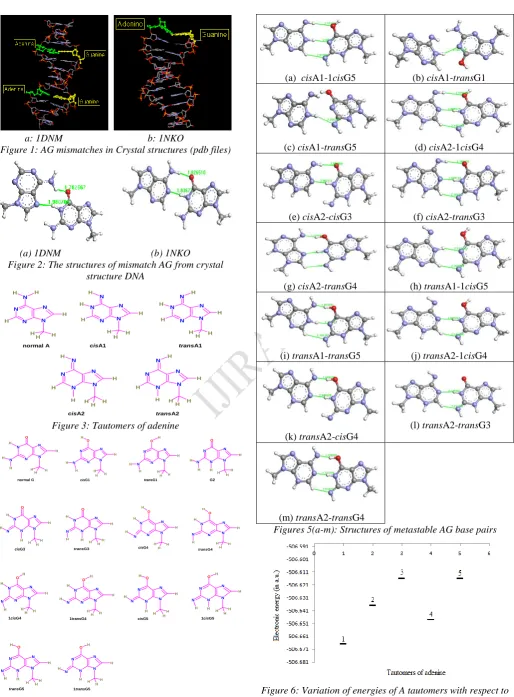
The various modes of hydrogen bonding among purine nucleobases can be described in many ways. Several sites of purine nucleobases, A and G can be involved in bonding at different orientations and also interact with surrounding ions and molecules (1-5). Tautomerization pathway on subsequent destabilization of complementary base pairs AT and GC to generate many tautomers of A and G have been shown in several studies, which could be the reason to form metastable AG pairs due to instantaneous interaction between A and G tautomers(5-8). This pathway has been considered as one of the mechanisms of AG pairing and also the driving factors for generating available AG mismatches (Figures 1(a-b). The hydrogen bonding in these AG mismatches are clearly indicated in Figures 2 (a-b) and both structures obtained from crystallographic database are similar. Some of the dominant tautomers of A and G are shown in Figures 3 and 4. The energetic of these tautomeric forms, and their orientations to form hydrogen bonds together might contribute to generate AG pairs in DNA (Figure 5)(5-14). In such situation pairing of A and G tautomers may take place at any region in the two
strands of DNA or with another strand. Moreover, understanding of the mostly found mismatches could rely on the acid-base characteristics between hydrogen bonding sites and the type of hydrogen bonds between non-complementary base pairs (5-7,15-18). It is also important to calculate the interaction capacity of these mismatches based on the acid-base properties of the interaction sites as well as on the type of hydrogen bonds. The information may be useful for exploring how the AG mismatches occur in DNA sequences.
Understanding the basis of forming mismatches of nucleobases is rather vast, but certain aspects such as detailed mechanisms of forming H-bonds between unstable tautomers species can be looked depending on the basicity of the donor sites of A and G tautomers. It is also known that the exocyclic amino group of G can efficiently form different H-bonds if normal GC base pair is destabilized. Similarly, A tautomers have several sites for bonding with G tautomers (Figures 3 and 4). Moreover energetic of these tautomers can distinctively identify the favorable combinations of A and G. So, the electron donating ability as well as the efficiency of hydrogen bond formation between A and G tautomers are considered to be the model study for understanding the existence of AG
Abstract: The structures of stable tautomers of Adenine and Guanine are chosen for analyzing the formation of metastable AG pairing. The metastable structures are optimized using density functional theory (B3LYP) with basis sets 6-31+G(d,p) and 6-31++G(d,p). The structures and energetic of these metastable AG pairs are further analysed to extract information on the available AG mismatches. Moreover the thermodynamics parameters of these AG pairs are used to explain the sensitivity and stability of stable geometries. The geometry of cisA2-cisG3 combination is found energetically favourable and also the structure is very similar to available crystal structure.
Page 362 www.ijiras.com | Email: [email protected] mismatches. Some of the tautomers of A and G may be minor
or major, whereas several metastable AG pairs may be formed from any of these tautomers, which is the interest of theoretical chemists.
In many studies on DNA nucleobases and hydrogen bonding DFT (B3LYP) method with medium size basis set have been successfully used (1-9). Moreover, DFT method has been applied in the study of several types of molecules. Hence, this method can be used to study metastable AG mismatches. The method is based on the Kohn-Same (KS) formulation where comparatively large molecules with cubic scaling can be performed in DFT method (18-20). Hence, in the present study we propose to study the formation of several metastable AG pairs with this method and further comparison of the results will be performed with MP2 level of theories.
II. COMPUTATIONAL DETAILS
B3LYP and DFT methods were used for complete geometry optimization of tautomers and metastable AG pairs(21). Initially the geometries of A and G tautomers are collected from literature, and drawn with Gaussview (22). Complete geometry optimization were performed by using B3LYP with 6-31+G(d,p) and 6-31++G(d,p) basis sets. We performed Moller Plesset second order perturbation theory MP2 calculations utilizing a 6-31G+(d,p) basis set on the optimized geometries. The structures were fully optimized on the respective level with10-8 hartree as SCF convergence in energy. The zero point energy (ZPE) were also calculated using the harmonic frequencies calculated at the B3LYP/6-31+G(d,p) level of theory. The vibrational frequencies were calculated by performing a normal mode analysis on the optimized geometries using analytical gradients of the energy. To calculate the geometries and energies of the various metastable AG pairs, we used B3LYP type of density functional theory (DFT)(18-19). It is based on the generalized gradient approximation and a component of the exact Hartree-Fock (HF) exchange. The ground state gas-phase calculations for AG pairs were chosen after considering 6-31+G(d,p) and 6-31G++G(d,p) basis sets, where p-type polarization functions on H atoms is essential in dealing with hydrogen bonding systems.
III. RESULTS AND DISCUSSIONS
The possible impairing of A and G tautomers to form metastable AG pairs after tautomerization is considered as important pathway for explaining the occurrence of certain AG pairs in DNA(Figures 1(a-b)). As we can see in Figures 5, the hydrogen bonding patterns between A and G tautomers may take place entirely in different ways depending on the conformation of nucleotides involved at the time of pairing, since there are several sites of forming hydrogen bonds. Some of them may produce highly stable pairs due to the presence of strong hydrogen bonds dominating for generation stable AG pair. Moreover, the existence of other types of AG pairs may be due to the pre-reactive hydrogen bonding occurred at a particular orientation. However, it is believed that hydrogen
bonding capacity of various sites are not so different for nucleobases and the chances of forming several hydrogen bonds of various types are possible resulting metastable AG pairs(5-9). In most cases the formation of hydrogen bonds involving terminal oxygen and –NH2 groups of A and G
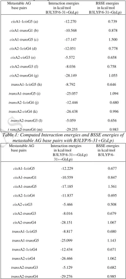
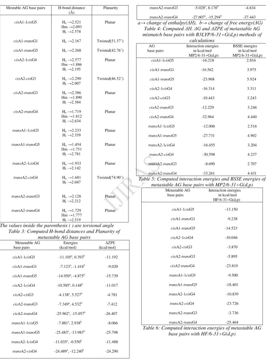
tautomers are found common (Figures 5). The interaction between –CH with terminal oxygen of purine ring is also observed. Hence, the importance of –C-H……O- type of H-bond is also highlighted in these AG pairs (Figure). Beyond the understanding of the energetically controlled mechanism of hydrogen bonding by specific site, it would be important to analyse the mode of associations of A and G tautomers at a particular conformation, and further validate the contribution of H-bonds to the stability of metastable AG pairs. As we have seen in Figure 5, the interaction of A and G tautomers can take place in various degrees of orientations, so the theoretical techniques capable of estimating such weak interactions should be properly chosen. Here, DFT (B3LYP) method using 6-31+G(d,p) and 6-31++G(d,p) basis sets has been used to optimize the structures, and and the interaction energies of all the tautomer combinations are computed(Tables 1-2). The characteristic of hydrogen bonds are shown in Table 3. Here the harmonic vibrational frequency has been analysed, and ZPE and other thermodynamic contributions for the optimized structures are examined (Table 4 ). The BSSE corrections are approximately 1 kcal/mole for all the structures for both 6-31+G(d,p) and 6-31++G(d,p) basis sets. To compare the variations of the results of B3LYP, we have performed MP2 calculations with 6-31++G(d,p). The values of B3LYP are quite different from those of MP2 calculations and the variation of energy trend is similar, the method overestimates BSSE (Table 5).
–C-Page 363 www.ijiras.com | Email: [email protected] H….N=C, in cis or trans orientations (iv) –N-H…N=C- type
is found in cisA1-transG5(c)(Figure 5). So according to these structures the existence of several bonding patterns might be due to the incorporation of A and G in DNA sequences in various possible conformations. Naturally, the type of bonding responsible for AG pairing at that particular orientation is very important. The existence of rare tautomers of Adenine has been discussed in the context of prototropic tautomerization, which on the other hand depends on the acidity/basicity of several sites(5-10). The feasibility of H-atom migration among these respective structures can be understood from these structures and energy level diagrams (Figure 8). It has been indicated that tautomerization of adenine is less feasible compared to guanine. Guanine is the most basic nucleobase and tautomerization processes can occur easily in this nucleobase. In that case guanine tautomers might be more responsible than adenine tautomers in forming metastable AG pairs. The various H-bonds formed between AG are analyzed and the stability of these AG pairs can be understood with respect to these bonds (Table 3). If we consider the structural alignment and conformation of approaching nucleotides of strands of DNA that can bring with A and G closer during pairing may in particular determines what type of metastable pairs can be formed. As we can see that the interaction energies of these AG pairs are not largely different, and formation of any type of bonding are equally possible at a particular conformation. The relative energies of these metastable AG pairs, other thermodynamic properties i.e changes of enthalpy, Gibb’s free energy and zero point energies are shown in Table 4. However, all AG pairs are found feasible for pairing forming a H-bonds during association of A and G, since the interaction energies are negative (Table 3 and Tables 1-2). Even the HF/6-31+G(d,p) energies are quite reasonable for predicting metastable AG pairs(Table 6). The existence of –NH-CH-(iv) bonds might be due to structural alignment of these sites during intermolecular interaction of A and G to form other energetically favorable H-bonds ((i), (ii) and (iii) types.
The optimized geometries of AG pairs were found mostly stable with approximately 10-20 kcals differences in the interaction energies. In some cases proton shifting has been observed, it might depend on the sensitivity of proton at the H bonding sites (Figures). The relative energies of these AG pairs are shown in Figure 8, where some are very close energy values, transA2-transG4 is the most stable pair (Figure 5). From Table 2, we note that AG pairs do not form equivalent H bonds, some has shorter H-bonds, whereas much longer H bonding are also observed (Table 3). It may be one of the reasons for the variation of structures and stability of these AG pairs.
The process of proton transfer once these metastable species are formed might also operate which subsequently transform to available crystal structure AG mismatches. Theoretically it is possible to predict the intermolecular interaction between these unstable species for understanding mispairing pathway through tautomers. Also tautomer preference in A and G association is clearly shown. However the geometries obtained from crystal structures are not the exact form and at the same time what type of tautomer combination is involved cannot be known. The hypothesis of
H-transfer between bases, and bases and surrounding ions and molecules might be relevant in this context. So as far as the stability of these metastable AG pairs computed in the gas phase shows the possibility of AG association through tautomerization pathways.
Metastable AG Interaction energies BSSE energies base pairs in kcal/mol in kcal/mol B3LYP/6-31+G(d,p) B3LYP/6-31+G(d,p)
cisA1-1cisG5 (a) -12.270 0.739
cisA1-transG1 (b) -10.568 0.878
cisA1-transG5 (c) -17.147 1.500
cisA2-1cisG4 (d) -12.051 0.778
cisA2-cisG3 (e) -5.572 0.658
cisA2-transG3 (f) -8.036 0.758
cisA2-transG4 (g) -28.149 1.055
transA1-1cisG5 (h) -8.792 0.646
transA1-transG5 (i) -25.057 1.094
transA2-1cisG4 (j) -12.446 0.680
transA2-cisG4 (k) -26.438 0.996
transA2-transG3 (l) -5.059 0.656
t ransA2-transG4 (m) -29.255 0.983 Table 1: Computed Interaction energies and BSSE energies of
metastable AG base pairs with B3LYP/6-31+G(d,p) Mestastable AG Interaction energies BSSE energies
base pairs in kcal/mol in kcal/mol 31++G(d,p)
B3LYP/6-31++G(d,p)
cisA1-1cisG5 -12.229 0.677
cisA1-transG1 -10.559 0.847
cisA1-transG5 -17.185 1.561
cisA2-1cisG4 -11.837 0.695
cisA2-cisG3 -5.466 0.508
cisA2-transG3 -8.016 0.679
cisA2-transG4 -28.151 1.067
transA1-1cisG5 -8.817 0.680
transA1-transG5 -25.099 1.143
transA2-1cisG4 -12.434 0.671
transA2-cisG4 -26.466 1.062
transA2-transG3 -5.129 0.682
Page 364 www.ijiras.com | Email: [email protected]
Mestable AG base pairs H-bond distance (Å)
Planarity
cisA1-1cisG5
cisA1-transG1
cisA1-transG5
cisA2-1cisG4
cisA2-cisG3
cisA2-transG3
cisA2-transG4
transA1-1cisG5
transA1-transG5
transA2-1cisG4
transA2-cisG4
transA2-transG3
transA2-transG4
Hu →2.521 Hm →2.093
Hl →2.574
Hu →2.167
Hu →2.260
Hu →2.577 Hm →1.886
Hl →2.195
Hu →2.290 Hl →2.007
Hu →2.386 Hm →1.890
Hl →2.384
Hu →1.719 Hm →1.812
Hl →2.634
Hu →2.233 Hl →2.359
Hu →1.454 Hm →1.751
Hl →2.781
Hu →1.933 Hl →2.142
Hu →1.681 Hl →2.047
Hu →2.128 Hl →2.212
Hu →1.729 Hm →1.777
Hl →2.519
Planar
Twisted(51.57˚)
Twisted(42.76˚)
Planar
Twisted(46.52˚)
Planar
Planar
Planar
Planar
Planar
Twisted(74.90˚)
Planar
Planar
The values inside the parenthesis ( ) are torsional angle Table 3: Computed H-bond distances and Planarity of
metastable AG base pairs
Metastable AG Energies ΔZPE base pairs (kcal/mol) (kcal/mol)
cisA1-1cisG5 -11.105a, 0.393b -11.192
cisA1-transG1 -7.123a, -1.416b -9.020
cisA1-transG5 -14.950a, -4.875b -15.739
cisA2-1cisG4 -10.585a, 0.148b -11.017
cisA2-cisG3 -4.138a, 5.527b -4.781
cisA2-transG3 -7.349a, 4.532b -7.412
cisA2-transG4 -25.962a, -15.057b -26.407
transA1-1cisG5 -7.881a, 2.938b -8.066
transA1-transG5 -25.483a, -13.983b -25.798
transA2-1cisG4 -11.035a, -0.550b -11.488
transA2-cisG4 -24.489a, -12.240b -24.290
transA2-transG3 -5.028a, 8.176b -4.634
transA2-transG4 -27.607a, -15.294b -27.443 a→ change of enthalpy(ΔH), b→ change of free energy(ΔG)
Table 4: Computed ΔH, ΔG and ΔZPE of metastable AG mismatch base pairs with B3LYP/6-31+G(d,p) methods of
calculations
AG Interaction energies BSSE energies base pairs in kcal/mol in kcal/mol MP2/6-31+G(d,p) MP2/6-31+G(d,p)
cisA1-1cisG5 -16.218 2.816
cisA1-transG1 -16.562 3.975
cisA1-transG5 -23.968 5.924
cisA2-1cisG4 -16.314 3.311
cisA2-cisG3 -10.443 3.243
cisA2-transG3 -12.229 3.246
cisA2-transG4 -32.964 4.440
transA1-1cisG5 -12.006 2.516
transA1-transG5 -27.731 4.902
transA2-1cisG4 -16.455 3.204
transA2-cisG4 -30.598 4.237
transA2-transG3 -8.690 2.707
transA2-transG4 -33.261 4.431 Table 5: Computed interaction energies and BSSE energies of
metastable AG base pairs with MP2/6-31+G(d,p) Metastable AG Interaction energies
base pairs in kcal/mol HF/6-31+G(d,p)
cisA1-1cisG5 -13.150
cisA1-transG1 -9.238
cisA1-transG5 -14.523
cisA2-1cisG4 -10.046
cisA2-cisG3 -3.870
cisA2-transG3 -5.895
cisA2-transG4 -23.819
transA1-1cisG5 -9.500
transA1-transG5 -18.401
transA2-1cisG4 -10.839
transA2-cisG4 -23.726
transA2-transG3 -3.736
transA2-transG4 -25.464
Page 365 www.ijiras.com | Email: [email protected]
a: 1DNM b: 1NKO
Figure 1: AG mismatches in Crystal structures (pdb files)
(a) 1DNM (b) 1NKO Figure 2: The structures of mismatch AG from crystal
structure DNA
Figure 3: Tautomers of adenine
Figure 4: Tautomers of guanine
(a) cisA1-1cisG5 (b) cisA1-transG1
(c) cisA1-transG5 (d) cisA2-1cisG4
(e) cisA2-cisG3 (f) cisA2-transG3
(g) cisA2-transG4 (h) transA1-1cisG5
(i) transA1-transG5 (j) transA2-1cisG4
(k) transA2-cisG4
(l) transA2-transG3
(m) transA2-transG4
Figures 5(a-m): Structures of metastable AG base pairs
Figure 6: Variation of energies of A tautomers with respect to A. H N N N H H N N H H H H normal A H H H N N N N N H H H H cisA1 H H H N N N N N H H H H transA1 H H N N N N N H H H H H cisA2 H H N N N N N H H H H H transA2 H N N O N N H N H H H H H G2 H N N N O N N H H H H H H cisG1 H O N N N H H N N H H H H
1cisG4
H H N N N O H N
H HH
N H cisG5 H N N N O H N N H H H H H
1transG5
N N N N N H H H O H H H H cisG4 O H N N N H H N N H H H H transG3 O H N N N H H N N H H H H cisG3 H N N N O N N H H H H H H transG1 N N N N N H H H O H H H H transG4 H O N N N H H N N H H H H
1transG4
H H N N N O H N
H HH
N H transG5 H N N N O H N N H H H H H
Page 366 www.ijiras.com | Email: [email protected]
1. G 2. cisG1 3. transG1 4. G2 5. cisG3 6. transG3 7. cisG4 8. transG4 9. 1cisG4 10. 1transG4 11. cisG5 12. 1cisG5 13. transG5 14. 1transG5
Figure 7: Variation of energies of G tautomers with respect to G.
1. cisA1-1cisG5 2. cisA1-transG1 3. cisA1-transG5 4. cisA2-1cisG4 5. cisA2-cisG3 6. cisA2-transG3 7. cisA2-transG4 8. transA1-1cisG5 9. transA1-transG5 10. transA2-1cisG4 11. transA2-cisG4 12. transA2-transG3 13. transA2-transG4
Figure 8: Comparison of interaction energies of metastable AG base pairs
IV. CONCLUSION
The geometries and energies computed with B3LYP and MP2 calculations can reasonably predict the formation of several metastable AG mismatches. The results of two basis sets 6-31+G(d,p) and 6-31++G(d,p) used in B3LYP can predict the favorable association of metastable AG mismatches and most are found at close energy levels. The
cisA2-cisG3 combination is one of the energetically favorable combination and structurally similar to crystal structure. The most favorable combination is transA2-transG4.
REFERENCES
[1] Bernhard Lippert, Deepali Gupta,(2009) Promotion of rare nucleobase tautomers by metal binding, Dalton
Trans., 4619-4634.
[2] Ignacio Faustino, Anna Avino, Ivan Marchan, F. Javier Luque, Ramon Eritja, Modesto Orozco,(2009),Unique Tautomeric and Recognition Properties of Thioketothymines? J. Am. Chem. Soc., 131, 12845-12853. [3] Jiande Gu, Anmin Tian, Wai-Kee Li, Ning-Bew Wong, (2000) Intramolecular Proton Transfer in the Tautomers of C8 Oxidative Adenine: A DFT Study, J. Phys. Chem.
B, 104, 10692-10698.
[4] Victor I. Danilov, Victor M. Anisimov, Noriyuki Kurita, Dmytro Hovorun, (2005),MP2 and DFT studies of the DNA rare base pairs: The molecular mechanism of the spontaneous substitution mutations conditioned by
tautomerism of bases, Chemical Physics Letters, 412, 285-293. AG) 1DNM (crystal structure).
[5] Webster, G.D., Sanderson, M.R., Skelly, J.V., Neidle, S., Swann, P.F., Li, B.F., Tickle, I.J.(1990), Crystal structure and sequence-dependent conformation of the A.G mispaired oligonucleotide d(CGCAAGCTGGCG). Proc.Natl.Acad.Sci.USA
87:6693-6697DOI: 10.2210/pdb1dnm/pdb NDB: BDL022.
[6] Johnson, S.J., Beese, L.S. (2004), Structures of mismatch replication errors observed in a DNA polymerase. Cell (Cambridge, Mass.) 116: 803-816.
DOI: 10.2210/pdb1nk0/pdb. NDB: PD0376
[7] Zhibo Yang, M. T. Rodgers, (2004),Theoretical studies of the unimolecular and bimolecular tautomerization of cytosine, Phys. Chem. Chem. Phys., 6, 2749-2757. [8] Alejandro Perez, Mark E. Tuckerman, Harold P.
Hjalmarson, O. Anatole von Lilienfeld, (2010), Enol Tautomers of Watson-Crick Base Pair Models Are Metastable Because of Nuclear Quantum Effects, J. Am.
Chem. Soc. 132, 11510-11515.
[9] C. Fonseca Guerra, F. M. Bickelhaupt, S. Saha, F. Wang, Adenine Tautomers: (2006), Relative Stabilities, Ionization Energies, and Mismatch with Cytosine, J.
Phys. Chem. A, 110, 4012-4020.
[10] Adchara Padermshoke, Yukiteru Katsumoto, Misako Aida, (2006), Dimerization and Double Proton Transfer-Induced Tautomerism of 4(3H)-Pyrimidinone in Solution Studied by IR Spectroscopy and Quantum Chemical Calculations, J. Phys. Chem. B, 110, 26388-26395. [11] Sodio C. N. Hsu, Tzu-Pin Wang, Chai-Lin Kao, Hui-Fen
Chen, Po-Yu Yang, Hsing-Yin Chen, (2013), Theoretical Study of the Protonation of the One-Electron-Reduced Guanine-Cytosine Base Pair by Water, J. Phys. Chem. B,
117, 2096-2105.
[12] Asit K. Chandra, Danuta Michalska, Rafat Wysokinsky, Therese Zeegers-Huyskens, (2004), Theoretical Study of the Acidity and Basicity of the Cytosine Tautomers and Their 1:1 Complexes with Water, J. Phys. Chem. A, 108,
9593-9600.
[13] Yiqun Huang, Hilkka Kenttamaa,( 2003), Theoretical Estimations of the 298 K Gas-Phase Acidities of the Pyrimidine-Based Nucleobases Uracil, Thymine, and Cytosine, J. Phys. Chem. A107, 4893-4897.
[14] Jiri Sponer, Michal Sabat, Leonid Gorb, Jerzy Leszczynski, Bernhard Lippert, Pavel Hobza, (2000), The Effect of Metal Binding to the N7 Site of Purine Nucleotides on Their Structure, Energy, and Involvement in Base Pairing, J. Phys. Chem. B, 104, 7535-7544. [15] Nino Russo, Marirosa Toscano, Andre Grand, Franck
Jolibois, (1998), Protonation of Thymine, Cytosine, Adenine, and Guanine DNA Nucleic Acid Bases: Theoretical Investigation into the Framework of Density Functional Theory, J. Comput. Chem., 19, 989-1000. [16] Jose P. Ceron-Carrasco, Jose Zuniga, Alberto Requena,
Eric A. Perpete, Catherine Michaux, Denis Jacquemin, (2011), Combined effect of stacking and solvation on the spontaneous mutation in DNA, Phys. Chem. Chem. Phys., 13, 14584-14589.
Page 367 www.ijiras.com | Email: [email protected] Barrier Heights for a Diverse Set of Water-Catalyzed
Proton-Transfer Reactions, J. Phys. Chem. A,116, 4211-4221.
[18] Manuel Rueda, F. Javier Luque, Josep Maria Lopez, Modesto Orozco, (2001), Amino-Imino Tautomerism in Derivatives of Cytosine: Effect on Hydrogen-Bonding and Stacking Properties, J. Phys. Chem. A, 105, 6575-6580.
[19] Jose Ramon Blas, F. Javier Luque, Modesto Orozco, Unique (2004), Tautomeric Properties of Isoguanine, J.
Am. Chem. Soc. 126, 154-164.
[20] Ivonne Otero-Navas, Jorge M. Seminario, Molecular electrostatic potentials of DNA base-base pairing and mispairing, J Mol Model, DOI 10.1007/s00894-011-1028-1.
[21] Frisch MJ, Trucks GW, Schlegel HB, Scuseria GE, Robb MA, Cheeseman JR, Montgomery JA Jr, Vreven T, Kudin KN, Burant JC, Millam JM, Iyengar SS, Tomasi J, Barone
V, Mennucci B, Cossi M, Scalmani G, Rega N, Petersson GA, Nakatsuji H, Hada M, Ehara M, Toyota K, Fukuda R, Hasegawa J, Ishida M, Nakajima T, Honda Y, Kitao O, Nakai H, Klene M, Li X, Knox JE, Hratchian HP, Cross JB, Bakken V, Adamo C, Jaramillo J, Gomperts R, Stratmann RE, Yazyev O, Austin AJ, Cammi R, Pomelli C, Ochterski JW, Ayala PY, Morokuma K, Voth GA, Salvador P, Dannenberg JJ, Zakrzewski VG, Dapprich S, Daniels AD, Strain MC, Farkas O, Malick DK, Rabuck AD, Raghavachari K, Foresman JB, Ortiz JV, Cui Q, Baboul AG, Clifford S, Cioslowski J, Stefanov BB, Liu G, Liashenko A, Piskorz P, Komaromi I, Martin RL, Fox DJ, Keith T, Al-Laham MA, Peng CY, Nanayakkara A, Challacombe M, Gill PMW, Johnson B, Chen W, Wong MW, Gonzalez C, Pople JA (2004) Gaussian 03, Revision C.02. Gaussian Inc, Wallingford CT.