Bosn J Basic Med Sci 2013; 13 (3): 146-152Abstract
Lesatropane, a synthesized chiral tropane (S, S-isomer of satropane), is a novel muscarinic agonist, and is being under preclinical develop-ment in China for the treatdevelop-ment of primary glaucoma. Th e reports concerning that activation of muscarinic acetylcholine receptors (mA-ChRs) could protect cells against apoptosis prompted us to study the neuroprotective eff ects of lesatropane and the mechanism. We found that lesatropane could protect PC cells from glutamate-induced neurotoxicity and reverse the decreased ERK/ activation caused by glu-tamate. Atropine or pirenzepine, antagonist of mAChR or M mAChR, antagonized the protective eff ects of lesatropane respectively and sup-pressed the lesatropane’s eff ects on ERK/. Furthermore, chelerythrine, a PKC inhibitor, partially supsup-pressed ERK/ activation induced by lesatropane. Th e results indicated that the specifi c M mAChR via PKC-ERK/ pathway might be involved in the neuroprotective eff ects of lesatropane. While M mAChR is a therapeutic target of Alzheimer’s disease (AD), the results of this paper contribute to further information concerning the activation of M mAChR as a therapeutic target in AD.
© Association of Basic Medical Sciences of FB&H. All rights reserved
KEY WORDS: muscarinic agonist, neuroprotective eff ects, glutamate, PC cells
glutamate neurotoxicity in PC12 cells
via
PKC-mediated phosphorylation of ERK1/2.
Ke Ma, Zhi-Hui Yang, Li-Min Yang, Hong-Zhuan Chen, Yang Lu*
Department of Pharmacy, Shanghai Jiaotong University School of Medicine, 280 South Chongqing Road, Shanghai 200025, China.
INTRODUCTION
Tropane derivatives (atropine, scopolamine, etc.) usually serve as muscarinic antagonists. Interestingly, lesatropane (Figure .), a chiral tropane derivative (S, S-isomer of satropane) synthesized in our laboratory, is a novel mus-carinic agonist [,], and is being under preclinical develop-ment in China for the treatdevelop-ment of primary glaucoma []. Muscarinic acetylcholine receptors (mAChRs) have five subtypes (M-M). It has been reported that acti-vation of the Gq/-coupled mAChRs (M, M, M) could protect cells against apoptosis induced by DNA damage, oxidative stress, impaired mitochondrial function, serum deprivation or UV irradiation [-]. Glutamate is a major excitatory neurotransmitter in the brain and also could lead to neuronal injury or cell death when it released in excess or overstimulated its membrane receptors []. Previously we reported that activation of the M mAChR
could protect rat retinal ganglion cells against glutamate-in-duced neuronal apoptosis []. It is interesting to confi rm the neuroprotective eff ects mediated through M mAChR using PC cells, since PC cells as a common model to evaluate the neuroprotective effects of certain compounds, endog-enously express mAChRs, predominantly the M mAChR. In this paper, we show that lesatropane protects PC cells from glutamate – induced neurotoxicity and the M mA-ChR is mainly responsible for the neuroprotective effects partially through the PKC-ERK/ pathway. Because of the important role of M mAChR in cognitive processes, it has long been aimed at a high-profi le target for the treat-ment of neurodegenerative disorders like Alzheimer’s
* Corresponding author: Yang Lu
Department of Pharmacy, Shanghai Jiaotong University School of Medicine, 280 South Chongqing Road, Shanghai 200025, China Phone: +86 21 63846590 / 776466; Fax: +86 21 54650067. e-mail: [email protected]; [email protected] (Y. Lu)
Submitted: 8 September 2012 / Accepted: 29 April 2013
Bosn J Basic Med Sci 2013; 13 (3): 147-152
chloride, N,N-dimethyl sulfoxide (DMSO), ,,-triphenyl-tetrazolium chloride solution (TTC), -(,-dimethylthi-azol--yl)-,-diphenyltetrazolium bromide (MTT), and L-glutamic acid monosodium salt were purchased from Sigma (St. Louis, U.S.). Hoechst was purchased from Beyotime (Zhejiang, China). Chelerythrine chloride was from Alexis (Pennsylvania, U.S.) and U was from Up-state (Massachusetts, U.S.). Non-Radioactive Protein Kinase Assay Systems was purchased from Promega (Madison, U.S).
Cell culture and drug treatment
PC cells obtained from the Shanghai Cell Culture Cen-ter (Shanghai, China) were cultured in RPMI me-dium supplemented with fetal bovine serum in a hu-midifi ed atmosphere with CO at C. Th e cells in the medium without serum were pre-incubated with lesat-ropane for h before being exposed to glutamate. Atro-pine or pirenzeAtro-pine was used at μM. U was used at μM and chelerythrine was at μg/ml. They were used respectively min before addition of lesatropane.
Measurement of cell viability
PC cells were plated in -well culture plates at .×
-.× cells/well, and pre-incubated with lesatropane for
h before being exposed to glutamate for h. Cell vi-ability assays were performed using MTT method. MTT in PBS was added to the cultures at a fi nal concentration of . mg/ml. After further incubation at C for h, the me-dium was carefully removed and formazan crystals were dissolved in μl DMSO per well. Th e absorbance at nm was measured on a plate reader (Bio-tek, Vermont, U.S.). Preparation of brain slices and drug treatment Th e brain slices were made as described before []. SD rats with a mean weight g were decapitated. Whole brains were quickly removed and immersed in iced ACSF, which had the following composition (in mM): NaCl , KCl ., CaCl , MgSO , NaHPO ., NaHCO ., glucose (fi nal pH .). Brains were cut coronally into μm thick sections with a vibrating tissue slicer (ZQP-, Xiangshan, China). Cortical slices were quickly isolated from the
sec-containing mM glutamate for min; (III) glutamate in-jury + drug group, in which slices were incubated with μM lesatropane in oxygenated MgfACSF min prior to and during glutamate application. All slices were trans-ferred to oxygenated ACSF for h recovery, and the activ-ity of the slices was evaluated by using TTC staining method.
LDH measurement
The plasma membrane damage of the PC cells was assessed by the release of LDH into the culture me-dium. LDH leakage was calculated of the percentage of LDH in the medium versus total LDH activity in the cells. After the treatment, the LDH released into the culture medium was examined by using an assay kit (Ji-ancheng, Nanjing, China) according to the manufactur-er’s protocol on a plate reader (Bio-tek, Vermont, U.S.).
Hoechst staining
PC cells were plated on coverslips in culture plates and treated as before. After the treatment, the cells were fixated with cold paraformaldehyde for min, and stained with Hoechst ( μg/mL) for min under dim light. For each coverslip, cells were observed under a fluorescence microscope (Zeiss, Oberkochen, Germany) with a UV-A fi lter, and the apoptotic cells were identifi ed.
Immunoblots
Bosn J Basic Med Sci 2013; 13 (3): 148-152 and ERK/ primary antibodies (:, Cell signaling,Mary-land, U.S.). After washing with TBST, the membranes were incubated with horseradish peroxidase-conjugated goat anti-rabbit secondary antibody (:, KPL, Maryland, U.S.) and visualized with the ECL detection kit (Pierce Biotechnology, Illinois, U.S.) according to the manufacturer’s instructions.
PKC activity measurement
Th e non-radioactive protein kinase assay was used to detect the Protein kinase C (PKC). After the treatment, cells were suspended in cold PKC extraction buff er and homogenized using a cold homogenizer. Each sample was combined with the peptag, PKC reaction buffer, peptag C peptide, the sonicated PKC activator buff er, and was incubated at C for min. After the separation of phosphorylated and nonphosphorylated peptag peptides by electrophoresis, the kinase activity was quantitated by densitometric methods.
Statistical analysis
Th e data are presented as the mean ± standard error of the mean. Th e diff erence between the groups was assessed by using Student-Newman-Keuls test, where p<. indicated a signifi cant diff erence.
RESULTS
Lesatropane protects PC cells and cortical slices against glu-tamate neurotoxicity.
Treatment of PC cells with mM glutamate for h re-sulted in a decrease of survival rate by around relative
to the untreated group, which was consistent with the pre-vious report []. Pre-treatment with .-. μM lesat-ropane for h significantly prevented PC cell death caused by over-mentioned glutamate neurotoxicity, and the maximal effect was observed at . μM lesatropane (Figure A). Th e neuroprotective eff ects of lesatropane on cortical slices were also observed. Cortical slices were sub-jected to mM glutamate for min and following h re-covery. Th e survival activity of the brain slices was reduced to .±. and the pre-treatment with . μM lesat-ropane increased the activity to .±. (Figure B).
Neuroprotective eff ects of lesatropane are mediated through M mAChR in PC cells
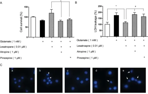
In order to fi nd out if the activation of M mAChR was re-sponsible for the neuroprotection observed, PC cells were pre-treated with atropine (a nonselective mAChR antago-nist) or pirenzepine (a selective M mAChR antagoantago-nist) for min respectively before . μM lesatropane treatment. Th en, the cell viability, LDH release, and the apoptotic status
Bosn J Basic Med Sci 2013; 13 (3): 149-152
apoptotic cells was signifi cantly decreased in the lesatropane group. But, when the PC cells were pre-treated with μM atropine, or μM pirenzepine for min before using lesatro-pane, the protective eff ects of lesatropane on glutamate neu-rotoxicity were suppressed (Figs. A-C), suggesting that M mAChR was involved in lesatropane’s neuroprotective eff ects.
Activation of ERK/ pathway is involved in the neuroprotec-tive eff ects of lesatropane
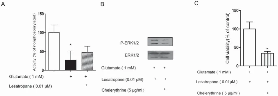
We interested in the intracellular MAPK/ERK/ signal-ing pathway for the neuroprotective eff ects of lesatropane, as ERK/ is an eff ecter of mAChRs in PC cells [,] and the importance of ERK/ in AD has been recognized [,]. Since the activity of ERK/ is dependent on its phosphorylation [], the phosphorylation of ERK/ in PC cells was detected by immunoblot. Treatment of cells with mM glutamate led to a signifi cant decrease of the ERK/ activity. Lesatropane (. μM ) could acti-vate ERK/ and prevent the decrease of ERK/ activity caused by glutamate (Figure A). Th e lesatropane-induced ERK/ activation can be inhibited by atropine or pirenz-epine (Figure B). In the presence of MEK inhibitor U
( μM), the eff ect of lesatropane against glutamate-induced cell death was reduced by . (Figure C). In all, ERK/ signaling plays a beneficial protective role in PC cells against glutamate neurotoxicity and lesatropane exerts its neuroprotective effects via M mAChR-ERK/ pathway.
Lesatropane-induced ERK/ activity is partially dependent on PKC
Bosn J Basic Med Sci 2013; 13 (3): 150-152DISCUSSION
Base on the couple of diff erent G proteins, the mAChRs are generally considered to divide into two distinct classes and the Gq/-coupled M and M mAChRs are mainly report-ed for the pro-survival response in a variety of stimuli [-]. Here, we found that lesatropane signifi cantly reversed the decreased PC cell viability caused by glutamate. Th e protective eff ects of lesatropane against glutamate-induced PC cell death should be principally mediated by the M mAChR, since M mAChR antagonist pirenzepine can in-hibit lesatropane's eff ects and PC cells principally express M mAChR at the mRNA level (data not shown). Although the activation of ERK/ known as an eff ecter of mAChRs,
is usually considered to promote neuronal cell survival or memory preservation [, -], there are still some evi-dences implicating the opposite eff ects [-]. In order to clarify the mechanism of neuroprotective eff ects of lesatro-pane, it is critical to identify the role of ERK/ pathway in activation of M mAChR in glutamate-induced PC cell death. ERK/ activation was markedly reduced after treat-ment with glutamate and the pre-treattreat-ment with lesatropane could reverse the decrease of ERK/ activation. Atropine or pirenzepine inhibited the rescued activation of ERK/ by lesatropane. Meanwhile, MEK inhibitor U abolished lesatropane’s eff ects. So here we found that ERK/ played a pro-survival role in PC cells during glutamate neurotox-icity and the neuroprotective eff ects of lesatropane should FIGURE 4. Activation of ERK1/2 pathway was involved in the neuroprotective eff ects of lesatropane. (A) lesatropane stimulated activa-tion of ERK1/2 in PC12 cells. Cells were treated with 0.01 μM lesatropane for 5 min, 15 min, 30 min, 12 h, 24 h, then harvested and lysed. The activated ERK1/2 (pERK1/2) and total ERK were determined (Western blotting). (B) The activation of ERK1/2 was measured in PC12 cells which were treated with or without 1 μM atropine or pirenzepine 30 min before addition 0.01 μM lesatropane, then 1 mM gluta-mate. (C) PC12 cells were pre-treated with U0126 (20 μM) 30 min before addition of 0.01 μM lesatropane, then 1 mM glutagluta-mate. Cell vi-ability was quantifi ed using the MTT assay after 48 h. * p<0.01 vs lesatropane group. Data represent the mean ± S.E. of n=5 independent observations.
Bosn J Basic Med Sci 2013; 13 (3): 151-152
the view of action of cholinergic activity could promote neuron survival during glutamate-induced cell death. More importantly, the neuroprotective eff ects of lesatropane are mainly mediated by M mAChR which is well accepted target in AD. In fact, a number of M mAChR agonists were reported to relieve the symptoms and intervene in the pathological process [-], our studies may help validate truly selective M mAChR agonists for the treatment of AD.
CONCLUSION
This paper indicated lesatropane could protect gluta-mate induced neurotoxicity through the specific M mAChR via PKC-ERK/ pathway. While M mA-ChR is a therapeutic target of AD, the results of this paper may provide further information of the acti-vation of M mAChR as a therapeutic target in AD.
ACKNOWLEDGMENTS
This study was supported by National Natural Science Founding of China (No. , , ), Key Basic Project of Shanghai Municipal Science and Technol-ogy Commission (No. JC, JC), Scientifi c and Technological Support Projects of Shanghai Municipal Science and Technology Commission (No. ).
DECLARATION OF INTEREST
Th e authors declare no confl ict of interest.
REFERENCES
[] Niu YY, Yang LM, Liu HZ, Cui YY, Zhu L, Feng JM, et al. Activity and QSAR study of baogongteng A and its derivatives as musca-rinic agonists. Bioorg Med Chem Lett. ;():-. [] Zhu L, Yang LM, Cui YY, Zheng PL, Niu YY, Wang H, et al.
Stere-oselectivity of satropane, a novel tropane analog, on iris muscarinic receptor activation and intraocular hypotension. Acta Pharmacol Sin. ;():-.
[] Fu J, Feng X, Yuan H, Yan L, Kuang X, Xia Z, et al. Study of ocular pharmacokinetics of in situ gel system for S(-)-satropane evaluated
tion of muscarinic receptor agonist pilocarpine against glutamate-induced apoptosis in retinal neurons. Cell Mol Neurobiol. ; (): -.
[] Langmead CJ, Watson J, Reavill C. Muscarinic acetylcholine recep-tors as CNS drug targets. Pharmacol Th er. ;():-. [] Yang L, Wang H. [Th e preparation and bioactivities of chiral
ana-logs of baogongteng A]. Yao Xue Xue Bao. ;():-. [] Wang ZJ, Liang CL, Li GM, Yu CY, Yin M. Neuroprotective eff ects
of arachidonic acid against oxidative stress on rat hippocampal slices. Chem Biol Interact. ;():-.
[] Kogo J, Takeba Y, Kumai T, Kitaoka Y, Matsumoto N, Ueno S, et al. Involvement of TNF-alpha in glutamate-induced apoptosis in a diff erentiated neuronal cell line. Brain Res. ;():-. [] Berkeley JL, Levey AI. Muscarinic activation of mitogen-activated
protein kinase in PC cells. J Neurochem. ;():-. [] Wotta DR, Wattenberg EV, Langason RB, el-Fakahany EE. M, M
and M muscarinic receptors stimulate mitogen-activated protein kinase. Pharmacology. ; ():-.
[] Grewal SS, York RD, Stork PJ. Extracellular-signal-regulated kinase signalling in neurons. Curr Opin Neurobiol. ;():-. [] Zhu X, Lee HG, Raina AK, Perry G, Smith MA. Th e role of
mito-gen-activated protein kinase pathways in Alzheimer's disease. Neu-rosignals. ;():-.
[] Seger R, Krebs EG. The MAPK signaling cascade. FASEB J. ;():-.
[] Haring R, Fisher A, Marciano D, Pittel Z, Kloog Y, Zuckerman A, et al. Mitogen-activated protein kinase-dependent and protein kinase C-dependent pathways link the m muscarinic recep-tor to beta-amyloid precursor protein secretion. J Neurochem. ;():-.
[] Budd DC, McDonald J, Emsley N, Cain K, Tobin AB. Th e C-ter-minal tail of the M-muscarinic receptor possesses anti-apoptotic properties. J Biol Chem. ; () : -.
[] Budd DC, Spragg EJ, Ridd K, Tobin AB. Signalling of the M-muscarinic receptor to the anti-apoptotic pathway. Biochem J. ;(Pt ):-.
[] Tobin AB, Budd DC. Th e anti-apoptotic response of the Gq/-coupled muscarinic receptor family. Biochem Soc Trans. ;(Pt ):-.
[] De Sarno P, Shestopal SA, King TD, Zmijewska A, Song L, Jope RS. Muscarinic receptor activation protects cells from apoptotic eff ects of DNA damage, oxidative stress, and mitochondrial inhibi-tion. J Biol Chem. ;():-.
[] Fukunaga K, Miyamoto E. Role of MAP kinase in neurons. Mol Neurobiol. ;():-.
[] Xia Z, Dickens M, Raingeaud J, Davis RJ, Greenberg ME. Oppos-ing eff ects of ERK and JNK-p MAP kinases on apoptosis. Science. ;():-.
[] Sweatt JD. Mitogen-activated protein kinases in synaptic plasticity and memory. Curr Opin Neurobiol. ;():-.
Bosn J Basic Med Sci 2013; 13 (3): 152-152[] Satoh T, Nakatsuka D, Watanabe Y, Nagata I, Kikuchi H, Namura S. Neuroprotection by MAPK/ERK kinase inhibition with U against oxidative stress in a mouse neuronal cell line and rat pri-mary cultured cortical neurons. Neurosci Lett. ;():-. [] Stanciu M, DeFranco DB. Prolonged nuclear retention of activated extracellular signal-regulated protein kinase promotes cell death generated by oxidative toxicity or proteasome inhibition in a neu-ronal cell line. J Biol Chem. ;():-.
[] Almeida RD, Manadas BJ, Melo CV, Gomes JR, Mendes CS, Graos MM, et al. Neuroprotection by BDNF against glutamate-induced apoptotic cell death is mediated by ERK and PI-kinase pathways. Cell Death Diff er. ;():-.
[] Zhu D, Wu X, Strauss KI, Lipsky RH, Qureshi Z, Terhakopian A, et al. N-methyl-D-aspartate and TrkB receptors protect neurons against glutamate excitotoxicity through an extracellular signal-regulated kinase pathway. J Neurosci Res. ;():-. [] Stanciu M, Wang Y, Kentor R, Burke N, Watkins S, Kress G, et al.
Persistent activation of ERK contributes to glutamate-induced oxi-dative toxicity in a neuronal cell line and primary cortical neuron cultures. J Biol Chem. ;():-.
[] Messing RO, Stevens AM, Kiyasu E, Sneade AB. Nicotinic and muscarinic agonists stimulate rapid protein kinase C translocation in PC cells. J Neurosci. ;():-.
[] Takada Y, Yonezawa A, Kume T, Katsuki H, Kaneko S, Sugimoto H, et al. Nicotinic acetylcholine receptor-mediated neuroprotec-tion by donepezil against glutamate neurotoxicity in rat cortical neurons. J Pharmacol Exp Th er. ;():-.
[] Hock C, Maddalena A, Heuser I, Naber D, Oertel W, von der Kammer H, et al. Treatment with the selective muscarinic agonist talsaclidine decreases cerebrospinal fl uid levels of total amyloid beta-peptide in patients with Alzheimer's disease. Ann N Y Acad Sci. ; : -.
[] Nitsch RM, Deng M, Tennis M, Schoenfeld D, Growdon JH. Th e selective muscarinic M agonist AFB decreases levels of total Abeta in cerebrospinal fl uid of patients with Alzheimer's disease. Ann Neurol. ;(): -.
![FIGURE 1. The chemical structure of lesatropane [2].](https://thumb-us.123doks.com/thumbv2/123dok_us/8672635.1731214/1.595.316.525.447.566/figure-chemical-structure-lesatropane.webp)