How Does It Kill?: Understanding the Candidacidal Mechanism of
Salivary Histatin 5
Sumant Puri, Mira Edgerton
Department of Oral Biology, School of Dental Medicine, University at Buffalo, Buffalo, New York, USA
Histatins are salivary cationic peptides that provide the first line of defense against oral candidiasis caused byCandida albicans. This minireview presents a critical evaluation of our knowledge of the candidacidal mechanism of histatin 5 (Hst 5). Hst 5 is the most potent among all histatin family members with regard to its antifungal activity. The mode of action of Hst 5 has been a sub-ject of intense debate. Unlike other classical host innate immune proteins, pore formation or membrane lysis by Hst 5 has largely been disproven, and it is now known that all targets of Hst 5 are intracellular. Hst 5 bindsC. albicanscell wall proteins (Ssa1/2) and glycans and is taken up by the cells through fungal polyamine transporters in an energy-dependent manner. Once inside the fungal cells, Hst 5 may affect mitochondrial functions and cause oxidative stress; however, the ultimate cause of cell death is by volume dysregulation and ion imbalance triggered by osmotic stress. Besides these diverse targets, a novel mechanism based on the metal binding abilities of Hst 5 is discussed. Finally, translational approaches for Hst 5, based on peptide design and synergy with other known drugs, are considered a step forward for bench-to-bed application of Hst 5.
H
istatins (Hsts) were first described as basic components of the secretions of human salivary glands based on their electro-phoretic mobility in cationic gel systems (1). The mean concen-tration of total Hsts secreted from unstimulated parotid glands was shown to be approximately 53g/ml, with Hst 5 being a major component (2) at an estimated range of 15 to 30M in whole saliva (3). The family of salivary Hsts comprises a group of 12 structurally related members of which histatin 1 (Hst 1) and Hst 3 are full-length proteins encoded by closely related loci of two distinct genes,HIS1(encoding Hst 1) andHIS2(encoding Hst 3) (4). The smaller proteins, Hst 2 (derived from Hst 1) and Hsts 4 to 12 (derived from Hst 3), are generated by proteolytic cleavage of parent Hsts by salivary proteases during secretion (5; reviewed in reference6).Early attempts to purify these Hsts from whole saliva, using ion-exchange chromatography or gel filtration, yielded groups of proteins rich in the amino acid histidine; therefore, they were initially referred to as histidine-rich peptides (HRPs) (reviewed in reference1). Further purification efforts using gel filtration fol-lowed by reverse-phase high-performance liquid chromatography finally isolated three major distinct purified components consist-ing of Hsts 1, 3, and 5 (1). Subsequently, a novel and less labor-intensive purification method that exploited the metal binding ability of Hsts, using zinc precipitation under alkaline conditions, was developed (7).
Hsts emerged as important components of the human innate immune system with the discovery of their candidacidal and to a lesser extent bactericidal effects. This coincided with the AIDS epidemic of the 1980s that was accompanied by emergence of oral thrush caused byCandidaspecies in HIV-infected immunocom-promised individuals, leading to a great interest in Hsts as poten-tial therapeutics. Their role in preventing oral candidiasis in this subpopulation was further bolstered in a recent study by Khan et al. (8), showing that reduction in Hst 5 levels in HIV-positive individuals contributes to enhanced prevalence ofCandida albi-cansamong these patients. Besides their microbicidal properties, Hsts 1, 3, and 5 participate in the formation of a protective layer (pellicle) on smooth tooth surfaces, where they prevent microbial
colonization and stabilize mineral-solute interactions (9,10). Re-cently, it has also been suggested that Hsts 1 and 2 may be the primary reason for efficient wound healing in the oral cavity, based on their ability to promote reepithelialization by stimulat-ing epithelial cell migration in a human full-skin wound model (11,12).
Of the many proposed functions of Hsts, the most significant and well researched one is their strong antifungal activity. Among all Hsts, Hst 5 has the most potent fungicidal activity against pathogenic fungi, includingCandida albicans(1,13) and other medically importantCandidaspecies, such asCandida kefyr, Can-dida krusei, andCandida parapsilosis(MIC50, 10 to 20g/ml), as
well asCryptococcus neoformansandAspergillus fumigatus(MIC50,
5 to 6g/ml) (14). Hst 5 potentially inhibits almost 100% of theC. albicanscells from germinating into more-virulent hyphae (13,15).
Although over a decade of studies have been devoted toward understanding the mechanism of fungicidal activity of Hsts, the crucial toxic event is still incompletely defined. Earlier studies pointed toward membranolytic mechanisms for Hst 5 in causing fungal cell death; however, more-recent studies have shown that multiple intracellular targets are the more likely sites of action. This minireview will primarily focus on the evolution of our un-derstanding of the candidacidal mechanism of Hst 5.
STRUCTURE-BASED CLUES TO THE MEMBRANE LYSIS HYPOTHESIS
Domain-based analyses of the complete Hst 5 protein (Asp-Ser- His-Ala-Lys-Arg-His-His-Gly-Tyr-Lys-Arg-Lys-Phe-His-Glu-Lys-His-His-Ser-His-Arg-Gly-Tyr) found that a minimum of 12 residues at the C terminus (“functional domain”) were required
Published ahead of print20 June 2014
Address correspondence to Mira Edgerton, [email protected]. Copyright © 2014, American Society for Microbiology. All Rights Reserved.
doi:10.1128/EC.00095-14
on September 8, 2020 by guest
http://ec.asm.org/
for killing of fungal cells (16). Fungicidal activity was shown to increase proportionally with an increase in length from 12 to 16 residues from the C terminus (16). Specifically, Lys13, Arg12, and Glu16 were identified as important residues by mutational analy-ses (17,18). Extensive structural studies of Hst 5 revealed that these peptides lack any secondary structure and are unordered in aqueous solutions (19–21). However, when placed in organic so-lutions such as trifluoroethylene or in membrane-mimetic solu-tions like dimethyl sulfoxide (DMSO)-water, they assumed␣ -he-lical structures. Furthermore, the C-terminal functional domain that was shown to be important for biological activity also was needed for␣-helicity (16), thus suggesting a role for this structural requirement in fungicidal activity. Adoption of ␣-helicity in membrane-mimetic solutions by Hsts, together with the observa-tions that Hst-mediated killing ofC. albicanswas accompanied with release of small molecules like K⫹(22) and ATP (23), led researchers to conclude that Hsts kill cells by forming pores in the cellular membrane. Thus, early workers identified the plasma membrane as the primary cellular target.
CELL MEMBRANE: THE MISIDENTIFIED TARGET OF Hst 5
Comparison of Hst 5 and a peptide comprised of its functional domain (dh5), with substitution analogs (dhvar1/4) designed to increase amphipathic character and membrane insertion, re-vealed significant differences among these peptides (24,25). Un-like Hst 5, analogs with highly stabilized␣-helical structures had higher fungicidal activity, dissipated cytoplasmic transmembrane potential, and were hemolytic. In contrast, hydrophobicity analy-ses of dh5 and Hst 5 showed similarities with primary sequences predominantly found in globular regions of a protein rather than membrane seeking regions, leading to the conclusion that mem-brane insertion would be unlikely (24,25). Subsequently, other workers replaced selected Hst 5 residues with proline to prevent ␣-helix formation in order to reduce its ability to insert into fungal membranes and found that these Hst derivatives were equally ef-fective in their fungicidal activity (26). Collectively, these studies concluded that membrane insertion leading to lysis is not the mechanism involved in Hst 5-mediated killing.
Further abundant evidence has emerged supporting the con-cept that membrane lysis is an effect, rather than the cause, of Hst 5-mediated killing. Lack of immediate membrane lytic activity by Hst 5 was demonstrated in elegant studies measuring the release and dequenching of the fluorescent dye calcein from dye-loaded
C. albicanscells (6). Cells treated for 60 min with Hst 5 (50 and 200 M) exhibited 86% loss of viability, while only 10% of intracellu-lar calcein was released, clearly refuting membrane lysis as the cause of cell death (6). Also, it was shown that in spite of the cellular loss of ATP upon Hst 5 exposure, cells continued to main-tain their membrane potential and remained alive for up to 80 min after ATP efflux (23). ATP release and killing ofC. albicanscells from endogenously expressed chromosomally encoded Hst 5 pro-vided further strong evidence that Hst 5 targets are intracellular (27).
Another report still emphasized on single pore formation in the membrane as an important step in uptake of Hst 5 at a con-centration of 50M (28). Interestingly, energy-depleted cells had longer cell wall retention times, and a small number of such cells had simultaneous uptake of Hst 5 and propidium iodine (PI) (in-dicative of membrane permeabilization or pore formation) (29). This led to two important observations: Hst 5 uptake is energy
dependent, and more importantly, Hst 5 may indeed have direct membrane effects, albeit when applied to cells at sufficiently higher concentrations (greater than the maximal physiological concentration of 30M) over a longer time span. Thus, the mem-brane lytic effects of Hst 5 reported previously may be due to use of higher concentrations of this peptide, although no evidence of a “single” pore as reported in that study (28) has been corroborated. Lack of permanent pores has also been demonstrated previously by immunogold labeling and freeze fracturing of the plasma membrane of Hst 5-treatedC. albicanscells (30,31).
Another model for uptake that involves the plasma membrane has also been described. This model is based on exploitation of the transmembrane potential of the plasma membrane for direct transfer of an appropriately charged Hst 5 into the cytosol (32). The basis for this conclusion is the fact that disruption of mito-chondrial function that can affect transmembrane potential offers protection against Hst 5 uptake and subsequent toxicity (32). However, the mitochondrion itself is the probable target for Hst 5-mediated toxicity, and this is discussed in the subsequent sec-tions.
BINDING AND UPTAKE OF Hst 5
Besides the above-mentioned uptake mechanisms based on direct interactions with the plasma membrane, a transporter-mediated model and a model involving the endocytic pathway have also been proposed; these models offer explanation for passage of Hst 5 through the complex structure of theC. albicanscell wall. A basal layer (comprised of1-3 and1-6 glucans and chitin) covered by a network of glycosylated glycosylphosphatidylinositol (GPI)-an-chored proteins comprises this complex wall structure (reviewed in reference33). Transporter proteins and energy expenditure in the form of ATP utilization are needed to mediate passage of Hst 5 through this thick cell wall. Hst 5 binds both cell wall-glucans (29) as well as fungal Ssa2 and Ssa1 proteins in the cell wall (6,
34–37) in order to stabilize and perhaps localize itself with a
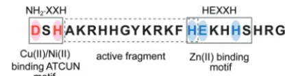
trans-port complex. Ultimately, it is transtrans-ported into the cell through the fungal polyamine transporters Dur3 and Dur31 in an energy-dependent process (38). Charge similarity between Hst 5 and polyamines is likely the reason why both of these cationic mole-cules can share a transporter. It has recently been shown that cel-lular uptake is enhanced by conjugation of the active fragment of Hst 5 (shown outlined by a broken line inFig. 1) with the poly-amine spermidine at either the N or C terminus (39). These con-jugates were highly effective as topical therapeutics in a murine model of oral candidiasis compared to the original peptide. In fact, lack of transporter-mediated intracellular transport is the basis for Hst 5 resistance inCandida glabrata, and expression ofC. albicans
Dur3 and Dur31 inC. glabrataresulted in both higher killing and uptake of Hst 5 (40), underscoring the key role of these transport-FIG 1Peptide sequence of Hst 5 with its functional domains. Hst 5 has a Cu/Ni binding ATCUN motif at the C terminus, while a Zn binding motif is present toward the N terminus. The active fragment of Hst 5 is shown outlined by a broken line.
on September 8, 2020 by guest
http://ec.asm.org/
ers in Hst 5 translocation. Hst 5 uptake and killing are lower inC. albicans ssa2⌬/⌬cells (35), showing that binding to the cell wall is an important step in allowing for localized retention before ener-gy-dependent uptake. However, surface binding, unlike uptake, is energy independent, since energy-depleted cells (cells treated with sodium azide, which reduces ATP synthesis) still had abundant Hst 5 binding but not intracellular uptake (29).
It is important to point out here that energy-dependent cyto-solic translocation is not the only mechanism that allows Hst 5 entry into the cells. Vacuolar localization of Hst 5 occurred in Hst 5-treatedC. albicanscells and this localization was reduced in endocytic mutants (28, 29), indicating a role for the endocytic pathway in uptake. However, endocytic uptake is insignificant in terms of Hst 5 toxicity, since cells defective in the endocytic path-way or cells lacking vacuoles had no differences in Hst 5 suscepti-bilities compared to the wild-type (WT) cells (28,29). In contrast,
C. albicanscells lacking genes encoding Rvs161 and Rvs167 Bin-Amphiphysin-Rvs homology domain proteins (that were shown to play a significant role in endocytosis) exhibited resistance to-ward Hst 5, leading the authors to conclude that endocytosis may play some role in the candidacidal mechanism of Hst 5 (41). Al-ternatively, this resistance may be an indirect result of changes in lipid composition or the physical properties of the plasma mem-brane in these mutants. Thus, uptake of Hst 5 may involve all three uptake pathways (transporter-mediated uptake, direct transfer across the membrane, and receptor-mediated endocytosis). The actual uptake potentially is a combination of one or more these processes, the choice of which is seemingly dependent on Hst 5 concentration (28,29).
CELL DEATH BY IONIC IMBALANCE AND VOLUME DYSREGULATION
Release of small molecules like potassium ions (42) and ATP (23) were among the first observations made by researchers trying to understand the mechanism of killing by Hst 5. K⫹release upon Hst 5 treatment offers one of the primary mechanisms by which Hst 5 interferes with cellular metabolism and ultimately leads to cell death. Cells lose volume, concomitantly with loss in viability, when treated with Hst 5 (22). Pharmacological inhibitors of anion transporters significantly prevented volume reduction and killing, suggesting that volume loss is a result of ion efflux (22). Subse-quently, Trk1 was identified as an ion transporter responsible for potassium loss, since cells lacking the protein were resistant to Hst 5 (43). Another observation, in line with such volume dysregula-tion, is that vacuolar expansion was also observed along with cy-tosolic translocation, but in either case, PI uptake (indicative of cell lysis) always followed but never preceded vacuolar expansion (29). Interestingly, extracellular NaCl (100 mM), but not sorbitol, prevented vacuolar expansion and PI entry in cells that already contained cytosolic Hst 5 (29), thus showing a critical role for ionic balance in Hst 5 toxicity.
ATP AND MITOCHONDRION-MEDIATED MECHANISM
Evidence also exists for the role of released cellular ATP in Hst 5-mediated killing. Treatment with Hst 5 in the presence of an ATP scavenger, apyrase, led to reduction in killing, suggesting a direct involvement for released ATP in cell death (44). Interest-ingly, anaerobically grown cells were less sensitive to Hst 5, show-ing that it is not entirely reduction in intracellular ATP content that is responsible for killing; rather, ATP efflux may present a
signal for cytotoxicity (45). It is known that released ATP signals cellular toxicity in higher eukaryotes by binding to membrane nucleotide P2X receptors (reviewed in reference44). The presence of such purinergic receptors in theC. albicanscell wall has been suggested by cross-reactivity with rat anti-P2X receptor antibody, so that released ATP itself may mediate Hst 5-induced killing (44). Because of the involvement of ATP in the Hst 5-mediated kill-ing, much attention was paid to the ATP-generating organelle, the mitochondrion, as a potential target. Treatment of cells with clas-sical mitochondrial inhibitors, prior to Hst 5 exposure, inhibited cell killing (32, 45, 46). However, similar results were obtained with generalized metabolic inhibitors and with high-salt condi-tions that disrupt ionic interaccondi-tions, such as those potentially oc-curring between Hst 5 and the cell wall (32,37). Thus, mitochon-drial involvement is likely to be primarily due to its role in maintenance of membrane potential that is important for Hst 5 internalization. Fluorescein isothiocyanate (FITC)-labeled Hst 5 was taken up by fungal cells only in metabolically active states, whereas metabolic inhibition of any sort, whether induced by per-turbing agents or due to slowing down of active respiration, re-sulted in loss of peptide uptake and killing (45,46). However, inhibition of mitochondrial transmembrane potential (47) along with localization of Hst 5 in the mitochondria (32) made it tempt-ing to conclude that Hst 5-mediated mitochondrial dysfunction is an important mechanism in killing. Furthermore, Hst 5 inhibited respiration in isolated mitochondria (47) as well as in intact yeast cells, and this inhibition correlated with cell killing (32). Interest-ingly, energy depletion itself induces changes in the physical state of the lipid bilayer, and this was offered as an alternative explana-tion for Hst 5 resistance of nonrespiring cells, independent of the involvement of mitochondria (48).
Interference with mitochondrial respiratory machinery can lead to generation of reactive oxygen species (ROS), and their role in Hst 5-mediated killing was also studied. Use of the oxygen radical-sensitive probe dihydroethidium (HEt) indeed revealed ROS generation in Hst 5-treatedC. albicanscells (49). In the pres-ence of the ROS scavengerL-cysteine, both ROS production and cell killing were prevented (49), establishing ROS generation as a mechanism for Hst 5-mediated killing. It is, however, imperative to point out that no direct interaction between Hst 5 and the respiratory chain enzymes was shown. Nonetheless, changes in the mitochondrial proteome have been shown in Hst 5-treatedC. albicanscells (50), including reduction of ubiquinol cytochromec
(cyt-c) reductase core protein 2, ATP synthase gamma chain, and malate dehydrogenase proteins, indicative of mitochondrial dys-function.
Hst 5-induced ROS generation as a cause of cell death has since been challenged. First, none of the classical markers of ROS-me-diated oxidative damage, including protein carbonylation and chromosome laddering, were observed as a result of Hst 5 treatment ofC. albicans(51). Second, Veerman et al. showed that TEMPO (2,2,66,6-tetramethylpiperidine-N-oxyl), a cell-permeant ROS scavenger, did not inhibit cell killing nor reduce the fluorescence of HEt (52). Furthermore, these authors showed that HEt-in-duced fluorescence upon Hst 5 treatment was in fact caused by redistribution of preexisting ethidium over the whole cell and not a result of ROS generation in the mitochondria. However, Hst 5-induced ROS generation can potentially be both transient and localized (the possibility of which is proposed and discussed in the following sections) and therefore more difficult to detect.
on September 8, 2020 by guest
http://ec.asm.org/
INVOLVEMENT OF MAPK SIGNALING
Mitogen-activated protein kinase (MAPK) pathways are signaling pathways that respond to environmental signals, and antifungal-mediated insult to the cell is no exception (reviewed in reference
53). Many antifungal drugs are known to activate these pathways inC. albicans. Hst 5-mediated oxidative stress as well as osmotic stress (as caused by ion loss) can therefore explain the induction of stress MAPK Hog1 in Hst 5-treatedC. albicanscells. Hst 5 treat-ment ofC. albicanscaused Hog1 phosphorylation, andhog1⌬/⌬ mutants were found to be more susceptible to Hst 5 (54). Hog1-induced glycerol production was enhanced in cells exposed to Hst 5 (54). All this corroborated with the proposed induction of os-motic stress as an important mechanistic mediator in Hst 5 can-didacidal activity. As mentioned above, the Hog1 pathway is also activated in response to oxidative stress, and therefore, Hst 5-in-duced oxidative stress cannot be completely ruled out.
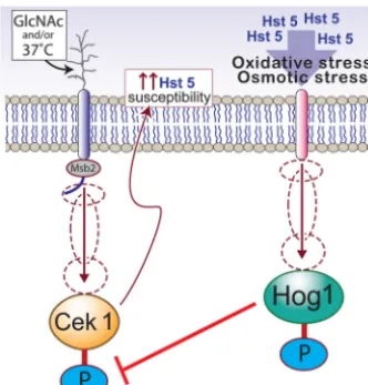
Recently, we found that the cell wall integrity MAPK Cek1 also plays a role in the fungicidal mechanism of Hst 5 (55). We ob-served that cells are more susceptible to Hst 5 under environmen-tal conditions that activate Cek1 phosphorylation, such as tem-perature shift to 37°C and use ofN-acetylglucosamine (GlcNAc) as the sole carbon source. This is not surprising, since Hst 5 binds toCandidacell wall components, including-glucans, whose lev-els are maintained by the Cek1 pathway (56). Interestingly, Ssa2, the cell wall binding partner of Hst 5, is a heat shock protein whose expression increases with a temperature shift from 30°C to 37°C (35), which incidentally is also a cue for Cek1 induction. Further-more, Hog1 represses activation of Cek1 MAPK (57); this poten-tially allows for the integration of various environmental signals with the outcome of Hst 5 treatment ofC. albicans. This interplay of MAPK activity inC. albicansin relationship to Hst 5 activity is depicted inFig. 2.
However,C. albicansresponse to environmental conditions may also reduce Hst 5 activity. Two such examples involve the
Cek1 MAPK pathway: (i)C. albicanssecreted aspartyl proteases (Saps) with a role in activation of the pathway (58) potentially degrade Hst 5 (59) and (ii) the interaction of the shed domain of Cek1 head sensor Msb2 with Hst 5 potentially offers protection against Hst 5 (60), although it still remains elusive whether the exact concentrations of shed Msb2in vivocan functionally inhibit Hst 5 activity in the oral cavity. The roles of shed Msb2 and Saps in Hst 5 activity are discussed in detail in theaccompanying minire-view(by Marc Swidergall and Joachim F. Ernst [61]).
Hst 5 METAL BINDING ABILITIES AS A POTENTIAL CANDIDACIDAL MECHANISM
Perplexingly, purified Hst 5 exhibits only 10 to 15% of itsin vitro
fungicidal activity when added into whole saliva (62, 63). Two major processes are likely to account for these differences: (i) the dynamic turnover of salivary proteins balancing secretion with proteolytic degradation (64, 65) and (ii) the phenomenon first described as “masking” (62) that is likely due to Hsts binding with salts and possibly with various metals, including Ca (66), Cu, and Zn in saliva. A similar phenomenon was also observed for the candidacidal activity of another antimicrobial salivary protein, lactoferrin (67). Studies have established that various transitional metals, such as Zn, Ni, Cu, and Fe, are intrinsically present in the saliva (68–70). However, the metal content of healthy or control subjects showed significant variation between individuals and ex-perimental conditions. Because of the potential peptide-metal in-teractions in saliva, the metal binding abilities of Hst 5 have been extensively studied. Hst 5 binds Zn and Cu (71) and possesses definitive metal binding motifs (Fig. 1) for Cu and Ni (ACTUN motif, encompassing the first 3 N-terminal amino acids, Asp-Ser-His) as well as for Zn (HEXXH motif at the C terminus [72,73]). Multiple strong binding sites within the binding motif are believed to occur for Cu, while Zn has only one strong putative metal binding site (74).
Although binding to Cu, Zn, or Ni did not stabilize the␣ -he-lical structure of Hst 5 (74), important consequences of metal binding have been observed. Zinc binding was shown to allow Hst 5 to catalyze fusion of lipid vesicles (73), and some increase in the bactericidal activity for Hst 5 has been reported for Zn-Hst 5 (75). However, effects of Cu binding provide unique mechanistic in-sights into novel potential candidacidal mechanisms for Hst 5. A high level of hydrogen peroxide production was observed in solu-tions with Hst 5 and Cu, in the presence of a reductant such as ascorbate (76–78). This raises an interesting possibility for ROS generation by a Cu-Hst 5 complex. Yeast mitochondria have been shown to contain detectable levels of nonprotein copper or “free” copper (79), and this, together with the fact that Hst 5 is shown to localize to the mitochondria, again raises ROS production as a possible mechanism involved in killing. The lack of global effects of ROS production in Hst 5-treated cells could be explained if production of ROS by Hst 5 is a transient effect that is localized only to the mitochondria. Interestingly,C. albicansmitochondrial superoxide dismutase (Sod5) is a Cu/Zn-containing protein that is upregulated during the yeast-to-hypha transition and induced during oxidative stress, while Hst 5 inhibits germination and Hst 5-mediated chelation of Cu and Zn could potentially interfere with the ability of Sod5 function.
Surprisingly, Hst 5 interactions with Fe, an important compo-nent of human diet and saliva, have not been investigated previ-ously. Iron is known to effectively coordinate with histidine resi-FIG 2Interplay of Hst 5 treatment with MAPK pathways ofC. albicans.
Ac-tivation of Cek1 MAPK by environmental conditions such as GlcNAc as a carbon source or growth temperature of 37°C may potentially enhance Hst 5 susceptibility by Cek1-mediated cell wall modifications (left). On the other hand, Hst 5 treatment itself causes oxidative stress, leading to the activation of Hog1 stress MAPK inC. albicans(right). Inhibition of Cek1 pathway by Hog1 activation offers another layer of complexity to this interplay between Hst 5 treatment and cellular MAPK pathways. Proteins that are phosphorylated are indicated by a blue P.
on September 8, 2020 by guest
http://ec.asm.org/
dues that are abundantly found in Hst 5. Our recent data show that iron does indeed bind to Hst 5 as evidenced by a change in Hst 5 structure when measured by circular dichroism (CD) upon titration of Hst 5 with iron (63). Although the most immediate effect of iron binding was a decrease in thein vitrocandidacidal activity of Hst 5, we found altered expression levels in large num-bers ofC. albicansiron uptake genes in response to Hst 5 treat-ment (63). This suggests that iron chelation, and metal chelation in general (as depicted inFig. 3), may be one of the primary and yet unexplored candidacidal mechanisms of Hst 5. Iron is an es-sential nutrient for all living organisms, since it serves as a cofactor in proteins involved in electron transfer and is a component of iron-sulfur cluster proteins or other iron-containing proteins that function as enzymes in major metabolic processes in the cell. Che-lation of iron and other important metals that serve as micronu-trients can create nutrient limiting conditions, negatively affecting pathogen growth. On the other hand, intracellular metal chelation by Hst 5 can potentially disrupt important fungal cellular func-tions. For example, mitochondria are the most susceptible to changes in cellular iron levels, owing to various iron-containing proteins in their organellar protein machinery. This suggests that the previously proposed mitochondrial dysfunction may instead be a response to Hst 5-induced disruption of cellular iron homeo-stasis that can be an important potential mechanism for Hst 5-me-diated killing ofC. albicans. Lastly, like Cu, Hst 5-Fe interactions can very well present a source for generation of ROS that could oxidize important proteins in the mitochondria, leading to cell death.
SUMMARY
Much progress has been made in our understanding of the mech-anism of Hst 5. It is clear that Hst 5 affects multiple intracellular targets. However, the ultimate cause of cell death seems to be counterintuitively simple, the loss of potassium ions leading to osmotic imbalance. Nevertheless, there is a possibility that addi-tional mechanisms are either simultaneously in play within the cell or that certain environmental conditions may favor one mechanism over the other. For example, metal binding abilities of Hst 5 would assume greater significance under nutrient-limited conditions, since metal binding by Hst 5 could potentially further
limit the essential metals needed for growth. However, metal binding would not be of major significance if nutrients were abun-dant in the environment. Similarly, environmental conditions leading to Cek1 MAPK activation inC. albicanswould make cells more susceptible to Hst 5, while MAPK Hog1 activation would potentially mitigate Hst 5-mediated oxidative and osmotic stress. Thus, Hst 5 may have evolved to utilize multiple environmentally induced physiological states ofC. albicansto maximize its killing potential.
The past 2 decades have highlighted the candidacidal potential of Hst 5. With all the considerable knowledge gained about the mechanism of action of this peptide, we are at an interesting junc-ture with regard to bench-to-bed translational breakthrough. This breakthrough will most likely involve either an intelligent design of novel Hst 5 derivatives for enhanced killing potential (as ob-served with the spermidine conjugates) or use of Hst 5 in conjunc-tion with other drugs or compounds that would potentiate Hst 5 activity. Examples of this are coadministration of Hst 5 with drugs like caspofungin that activate Cek1 MAPK to makeC. albicans
cells more susceptible, or coadministration with iron chelator drugs that can potentially “protect” Hst 5 activity from iron bind-ing-mediated loss in the killing potential of Hst 5. However, effec-tive routes of administration and treatment efficacies will need to be determined in appropriate experimental models for various forms of candidiasis, including topical use against mucosal candi-diasis (oral and dermal) and systemic use for more widespread disease.
ACKNOWLEDGMENT
We thank John Nyquist for the illustrations in this article.
REFERENCES
1.Oppenheim FG, Xu T, McMillian FM, Levitz SM, Diamond RD, Offner GD, Troxler RF.1988. Histatins, a novel family of histidine-rich proteins in human parotid secretion. Isolation, characterization, primary struc-ture, and fungistatic effects on Candida albicans. J. Biol. Chem.263:7472– 7477.
2.Johnson DA, Yeh CK, Dodds MW.2000. Effect of donor age on the concentrations of histatins in human parotid and submandibular/ sublingual saliva. Arch. Oral Biol.45:731–740.http://dx.doi.org/10.1016 /S0003-9969(00)00047-9.
3.Campese M, Sun X, Bosch JA, Oppenheim FG, Helmerhorst EJ.2009. Concentration and fate of histatins and acidic proline-rich proteins in the oral environment. Arch. Oral Biol.54:345–353.http://dx.doi.org/10.1016 /j.archoralbio.2008.11.010.
4.Sabatini LM, Azen EA.1989. Histatins, a family of salivary histidine-rich proteins, are encoded by at least two loci (HIS1 and HIS2). Biochem. Biophys. Res. Commun. 160:495–502. http://dx.doi.org/10.1016/0006 -291X(89)92460-1.
5.Castagnola M, Inzitari R, Rossetti DV, Olmi C, Cabras T, Piras V, Nicolussi P, Sanna MT, Pellegrini M, Giardina B, Messana I.2004. A cascade of 24 histatins (histatin 3 fragments) in human saliva. Suggestions for a pre-secretory sequential cleavage pathway. J. Biol. Chem. 279:
41436 – 41443.
6.Edgerton M, Koshlukova SE, Lo TE, Chrzan BG, Straubinger RM, Raj PA.1998. Candidacidal activity of salivary histatins. Identification of a histatin 5-binding protein on Candida albicans. J. Biol. Chem.273:
20438 –20447.
7.Flora B, Gusman H, Helmerhorst EJ, Troxler RF, Oppenheim FG.2001. A new method for the isolation of histatins 1, 3, and 5 from parotid secre-tion using zinc precipitasecre-tion. Protein Expr. Purif.23:198 –206.http://dx .doi.org/10.1006/prep.2001.1493.
8.Khan SA, Fidel PL, Jr, Thunayyan AA, Varlotta S, Meiller TF, Jabra-Rizk MA.2013. Impaired histatin-5 levels and salivary antimicrobial ac-tivity against in HIV infected individuals. J. AIDS Clin. Res.
4:pii⫽1000193.http://dx.doi.org/10.4172/2155-6113.1000193.
FIG 3Hst 5-mediated metal chelation as a potential candidacidal mechanism. Both extracellular and intracellular metal chelation can potentially contribute toward cell death, either by scavenging away important transition metals needed for growth (left), or by dysregulation of metal homeostasis that affects cell organellar functions such as mitochondrial functions (right).
on September 8, 2020 by guest
http://ec.asm.org/
9.Jensen JL, Lamkin MS, Oppenheim FG. 1992. Adsorption of human salivary proteins to hydroxyapatite: a comparison between whole saliva and glandular salivary secretions. J. Dent. Res.71:1569 –1576.http://dx .doi.org/10.1177/00220345920710090501.
10. Siqueira WL, Margolis HC, Helmerhorst EJ, Mendes FM, Oppenheim FG.
2010. Evidence of intact histatins in the in vivo acquired enamel pellicle. J. Dent. Res.89:626–630.http://dx.doi.org/10.1177/0022034510363384.
11. Oudhoff MJ, Blaauboer ME, Nazmi K, Scheres N, Bolscher JG, Veer-man EC.2010. The role of salivary histatin and the human cathelicidin LL-37 in wound healing and innate immunity. Biol. Chem.391:541–548. 12. Oudhoff MJ, Bolscher JG, Nazmi K, Kalay H, van’t Hof W, Amerongen AV, Veerman EC.2008. Histatins are the major wound-closure stimulat-ing factors in human saliva as identified in a cell culture assay. FASEB J.
22:3805–3812.http://dx.doi.org/10.1096/fj.08-112003.
13. Xu T, Levitz SM, Diamond RD, Oppenheim FG.1991. Anticandidal activity of major human salivary histatins. Infect. Immun.59:2549 –2554. 14. Helmerhorst EJ, Reijnders IM, van’t Hof W, Simoons-Smit I, Veerman EC, Amerongen AV.1999. Amphotericin B- and fluconazole-resistant Candida spp., Aspergillus fumigatus, and other newly emerging patho-genic fungi are susceptible to basic antifungal peptides. Antimicrob. Agents Chemother.43:702–704.
15. Lin AL, Shi Q, Johnson DA, Patterson TF, Rinaldi MG, Yeh CK.1999. Further characterization of human salivary anticandidal activities in a hu-man immunodeficiency virus-positive cohort by use of microassays. Clin. Diagn. Lab. Immun.6:851– 855.
16. Raj PA, Edgerton M, Levine MJ.1990. Salivary histatin 5: dependence of sequence, chain length, and helical conformation for candidacidal activ-ity. J. Biol. Chem.265:3898 –3905.
17. Driscoll J, Duan C, Zuo Y, Xu T, Troxler R, Oppenheim FG.1996. Candidacidal activity of human salivary histatin recombinant variants produced by site-directed mutagenesis. Gene177:29 –34. http://dx.doi .org/10.1016/0378-1119(96)00265-X.
18. Tsai H, Raj PA, Bobek LA.1996. Candidacidal activity of recombinant human salivary histatin-5 and variants. Infect. Immun.64:5000 –5007. 19. Raj PA, Soni SD, Levine MJ.1994. Membrane-induced helical
confor-mation of an active candidacidal fragment of salivary histatins. J. Biol. Chem.269:9610 –9619.
20. Brewer D, Hunter H, Lajoie G.1998. NMR studies of the antimicrobial salivary peptides histatin 3 and histatin 5 in aqueous and nonaqueous solutions. Biochem. Cell Biol.76:247–256.http://dx.doi.org/10.1139/o98 -066.
21. Raj PA, Marcus E, Sukumaran DK.1998. Structure of human salivary histatin 5 in aqueous and nonaqueous solutions. Biopolymers45:
51– 67.http://dx.doi.org/10.1002/(SICI)1097-0282(199801)45:1⬍51:: AID-BIP5⬎3.0.CO;2-Y.
22. Baev D, Li XS, Dong J, Keng P, Edgerton M. 2002. Human salivary histatin 5 causes disordered volume regulation and cell cycle arrest in Candida albicans. Infect. Immun. 70:4777– 4784. http://dx.doi.org/10 .1128/IAI.70.9.4777-4784.2002.
23. Koshlukova SE, Lloyd TL, Araujo MW, Edgerton M. 1999. Salivary histatin 5 induces non-lytic release of ATP from Candida albicans leading to cell death. J. Biol. Chem.274:18872–18879.http://dx.doi.org/10.1074 /jbc.274.27.18872.
24. Helmerhorst EJ, van’t Hof W, Breeuwer P, Veerman EC, Abee T, Troxler RF, Amerongen AV, Oppenheim FG.2001. Characterization of histatin 5 with respect to amphipathicity, hydrophobicity, and effects on cell and mitochondrial membrane integrity excludes a candidacidal mech-anism of pore formation. J. Biol. Chem.276:5643–5649.http://dx.doi.org /10.1074/jbc.M008229200.
25. Helmerhorst EJ, Van’t Hof W, Veerman EC, Simoons-Smit I, Nieuw Amerongen AV.1997. Synthetic histatin analogues with broad-spectrum antimicrobial activity. Biochem. J.326(Part 1):39 – 45.
26. Situ H, Balasubramanian SV, Bobek LA.2000. Role of alpha-helical conformation of histatin-5 in candidacidal activity examined by proline variants. Biochim. Biophys. Acta1475:377–382.http://dx.doi.org/10.1016 /S0304-4165(00)00096-9.
27. Baev D, Lil XW, Edgerton M. 2001. Genetically engineered human salivary histatin genes are functional in Candida albicans: development of a new system for studying histatin candidacidal activity. Microbiology
147:3323–3334.
28. Mochon AB, Liu H.2008. The antimicrobial peptide histatin-5 causes a spatially restricted disruption on the Candida albicans surface, allowing
rapid entry of the peptide into the cytoplasm. PLoS Pathog.4:e1000190.
http://dx.doi.org/10.1371/journal.ppat.1000190.
29. Jang WS, Bajwa JS, Sun JN, Edgerton M. 2010. Salivary histatin 5 internalization by translocation, but not endocytosis, is required for fun-gicidal activity in Candida albicans. Mol. Microbiol.77:354 –370.http: //dx.doi.org/10.1111/j.1365-2958.2010.07210.x.
30. Ruissen AL, Groenink J, Van’t Hof W, Walgreen-Weterings E, van Marle J, van Veen HA, Voorhout WF, Veerman EC, Nieuw Amerongen AV.2002. Histatin 5 and derivatives. Their localization and effects on the ultra-structural level. Peptides23:1391–1399.http://dx.doi.org/10.1016 /S0196-9781(02)00076-1.
31. den Hertog AL, van Marle J, van Veen HA, Van’t Hof W, Bolscher JG, Veerman EC, Nieuw Amerongen AV.2005. Candidacidal effects of two antimicrobial peptides: histatin 5 causes small membrane defects, but LL-37 causes massive disruption of the cell membrane. Biochem. J.388:
689 – 695.http://dx.doi.org/10.1042/BJ20042099.
32. Helmerhorst EJ, Breeuwer P, van’t Hof W, Walgreen-Weterings E, Oomen LC, Veerman EC, Amerongen AV, Abee T.1999. The cellular target of histatin 5 on Candida albicans is the energized mitochondrion. J. Biol. Chem.274:7286 –7291.http://dx.doi.org/10.1074/jbc.274.11.7286. 33. Chaffin WL, Lopez-Ribot JL, Casanova M, Gozalbo D, Martinez JP.
1998. Cell wall and secreted proteins of Candida albicans: identification, function, and expression. Microbiol. Mol. Biol. Rev.62:130 –180. 34. Li XS, Reddy MS, Baev D, Edgerton M.2003. Candida albicans Ssa1/2p
is the cell envelope binding protein for human salivary histatin 5. J. Biol. Chem.278:28553–28561.http://dx.doi.org/10.1074/jbc.M300680200. 35. Li XS, Sun JN, Okamoto-Shibayama K, Edgerton M.2006. Candida
albicans cell wall ssa proteins bind and facilitate import of salivary histatin 5 required for toxicity. J. Biol. Chem.281:22453–22463.http://dx.doi.org /10.1074/jbc.M604064200.
36. Sun JN, Li W, Jang WS, Nayyar N, Sutton MD, Edgerton M.2008. Uptake of the antifungal cationic peptide histatin 5 by Candida albicans Ssa2p requires binding to non-conventional sites within the ATPase do-main. Mol. Microbiol. 70:1246 –1260. http://dx.doi.org/10.1111/j.1365 -2958.2008.06480.x.
37. Jang WS, Li XS, Sun JN, Edgerton M.2008. The P-113 fragment of histatin 5 requires a specific peptide sequence for intracellular transloca-tion in Candida albicans, which is independent of cell wall binding. Anti-microb. Agents Chemother.52:497–504.http://dx.doi.org/10.1128/AAC .01199-07.
38. Kumar R, Chadha S, Saraswat D, Bajwa JS, Li RA, Conti HR, Edgerton M.2011. Histatin 5 uptake by Candida albicans utilizes polyamine trans-porters Dur3 and Dur31 proteins. J. Biol. Chem.286:43748 – 43758.http: //dx.doi.org/10.1074/jbc.M111.311175.
39. Tati S, Li R, Puri S, Kumar R, Davidow P, Edgerton M.2014. Histatin 5-spermidine conjugates have enhanced fungicidal activity and efficacy as a topical therapeutic for oral candidiasis. Antimicrob. Agents Chemother.
58:756 –766.http://dx.doi.org/10.1128/AAC.01851-13.
40. Tati S, Jang WS, Li R, Kumar R, Puri S, Edgerton M.2013. Histatin 5 resistance of Candida glabrata can be reversed by insertion of Candida albicans polyamine transporter-encoding genes DUR3 and DUR31. PLoS One8:e61480.http://dx.doi.org/10.1371/journal.pone.0061480. 41. Douglas LM, Martin SW, Konopka JB. 2009. BAR domain proteins
Rvs161 and Rvs167 contribute to Candida albicans endocytosis, morpho-genesis, and virulence. Infect. Immun.77:4150 – 4160.http://dx.doi.org /10.1128/IAI.00683-09.
42. Pollock JJ, Denepitiya L, MacKay BJ, Iacono VJ.1984. Fungistatic and fungicidal activity of human parotid salivary histidine-rich polypeptides on Candida albicans. Infect. Immun.44:702–707.
43. Baev D, Rivetta A, Vylkova S, Sun JNN, Zeng GF, Slayman CL, Edgerton M.2004. The TRK1 potassium transporter is the critical effector for killing of Candida albicans by the cationic protein, histatin 5. J. Biol. Chem.279:55060 –55072.http://dx.doi.org/10.1074/jbc.M411031200. 44. Koshlukova SE, Araujo MW, Baev D, Edgerton M.2000. Released ATP
is an extracellular cytotoxic mediator in salivary histatin 5-induced killing of Candida albicans. Infect. Immun.68:6848 – 6856.http://dx.doi.org/10 .1128/IAI.68.12.6848-6856.2000.
45. Gyurko C, Lendenmann U, Troxler RF, Oppenheim FG.2000. Candida albicans mutants deficient in respiration are resistant to the small cationic salivary antimicrobial peptide histatin 5. Antimicrob. Agents Chemother.
44:348 –354.http://dx.doi.org/10.1128/AAC.44.2.348-354.2000. 46. Gyurko C, Lendenmann U, Helmerhorst EJ, Troxler RF, Oppenheim
FG.2001. Killing of Candida albicans by histatin 5: cellular uptake and
on September 8, 2020 by guest
http://ec.asm.org/
energy requirement. Antonie Van Leeuwenhoek79:297–309.http://dx .doi.org/10.1023/A:1012070600340.
47. Petruzzelli R, Clementi ME, Marini S, Coletta M, Di Stasio E, Giardina B, Misiti F.2003. Respiratory inhibition of isolated mammalian mito-chondria by salivary antifungal peptide histatin-5. Biochem. Biophys. Res. Commun.311:1034 –1040.http://dx.doi.org/10.1016/j.bbrc.2003.10.104. 48. Veerman EC, Valentijn-Benz M, Nazmi K, Ruissen AL, Walgreen-Weterings E, van Marle J, Doust AB, van’t Hof W, Bolscher JG, Amerongen AV. 2007. Energy depletion protects Candida albicans against antimicrobial peptides by rigidifying its cell membrane. J. Biol. Chem.282:18831–18841.http://dx.doi.org/10.1074/jbc.M610555200. 49. Helmerhorst EJ, Troxler RF, Oppenheim FG.2001. The human salivary
peptide histatin 5 exerts its antifungal activity through the formation of reactive oxygen species. Proc. Natl. Acad. Sci. U. S. A.98:14637–14642.
http://dx.doi.org/10.1073/pnas.141366998.
50. Komatsu T, Salih E, Helmerhorst EJ, Offner GD, Oppenheim FG.2011. Influence of histatin 5 on Candida albicans mitochondrial protein expres-sion assessed by quantitative mass spectrometry. J. Proteome Res.10:646 – 655.http://dx.doi.org/10.1021/pr100861k.
51. Wunder D, Dong J, Baev D, Edgerton M.2004. Human salivary histatin 5 fungicidal action does not induce programmed cell death pathways in Candida albicans. Antimicrob. Agents Chemother.48:110 –115.http://dx .doi.org/10.1128/AAC.48.1.110-115.2004.
52. Veerman EC, Nazmi K, Van’t Hof W, Bolscher JG, Den Hertog AL, Nieuw Amerongen AV.2004. Reactive oxygen species play no role in the candidacidal activity of the salivary antimicrobial peptide histatin 5. Biochem. J.381:447– 452.http://dx.doi.org/10.1042/BJ20040208. 53. Hayes BM, Anderson MA, Traven A, van der Weerden NL, Bleackley
MR.14 February 2014. Activation of stress signalling pathways enhances tolerance of fungi to chemical fungicides and antifungal proteins. Cell. Mol. Life Sci.http://dx.doi.org/10.1007/s00018-014-1573-8.
54. Vylkova S, Jang WS, Li W, Nayyar N, Edgerton M.2007. Histatin 5 initiates osmotic stress response in Candida albicans via activation of the Hog1 mitogen-activated protein kinase pathway. Eukaryot. Cell6:1876 – 1888.http://dx.doi.org/10.1128/EC.00039-07.
55. Li R, Puri S, Tati S, Edgerton M.2014. Cek1 phosphorylation facilitates antifungal activity of Hst 5, poster 97A, p 146. Final Prog. Abstr. 12th ASM Conf. Candida candidiasis. American Society for Microbiology, Washing-ton, DC.
56. Galan-Diez M, Arana DM, Serrano-Gomez D, Kremer L, Casasnovas JM, Ortega M, Cuesta-Dominguez A, Corbi AL, Pla J, Fernandez-Ruiz E.2010. Candida albicans beta-glucan exposure is controlled by the fungal CEK1-mediated mitogen-activated protein kinase pathway that modu-lates immune responses triggered through dectin-1. Infect. Immun.78:
1426 –1436.http://dx.doi.org/10.1128/IAI.00989-09.
57. Roman E, Nombela C, Pla J. 2005. The Sho1 adaptor protein links oxidative stress to morphogenesis and cell wall biosynthesis in the fungal pathogen Candida albicans. Mol. Cell. Biol.25:10611–10627.http://dx .doi.org/10.1128/MCB.25.23.10611-10627.2005.
58. Puri S, Kumar R, Chadha S, Tati S, Conti HR, Hube B, Cullen PJ, Edgerton M.2012. Secreted aspartic protease cleavage of Candida albi-cans Msb2 activates Cek1 MAPK signaling affecting biofilm formation and oropharyngeal candidiasis. PLoS One7:e46020.http://dx.doi.org/10 .1371/journal.pone.0046020.
59. Meiller TF, Hube B, Schild L, Shirtliff ME, Scheper MA, Winkler R, Ton A, Jabra-Rizk MA.2009. A novel immune evasion strategy of Can-dida albicans: proteolytic cleavage of a salivary antimicrobial peptide. PLoS One4:e5039.http://dx.doi.org/10.1371/journal.pone.0005039. 60. Szafranski-Schneider E, Swidergall M, Cottier F, Tielker D, Roman E,
Pla J, Ernst JF.2012. Msb2 shedding protects Candida albicans against antimicrobial peptides. PLoS Pathog.8:e1002501.http://dx.doi.org/10 .1371/journal.ppat.1002501.
61. Swidergall M, Ernst JF.2014. Interplay betweenCandida albicansand the antimicrobial peptide armory. Eukaryot. Cell13:950 –957.
62. Helmerhorst EJ, Flora B, Troxler RF, Oppenheim FG.2004. Dialysis
un-masks the fungicidal properties of glandular salivary secretions. Infect. Im-mun.72:2703–2709.http://dx.doi.org/10.1128/IAI.72.5.2703-2709.2004. 63. Puri S, Li R, Edgerton M.2014. Iron binding negatively impacts the
fungicidal activity of salivary protein histatin 5 againstCandida albicans, abstr S10C:6, p 77. Final Prog. 12th ASM Conf. Candida candidiasis. American Society for Microbiology, Washington, DC.
64. Helmerhorst EJ, Alagl AS, Siqueira WL, Oppenheim FG.2006. Oral fluid proteolytic effects on histatin 5 structure and function. Arch. Oral Biol.51:1061–1070.http://dx.doi.org/10.1016/j.archoralbio.2006.06.005. 65. Xu L, Lal K, Santarpia RP, III, Pollock JJ.1993. Salivary proteolysis of histidine-rich polypeptides and the antifungal activity of peptide degrada-tion products. Arch. Oral Biol. 38:277–283. http://dx.doi.org/10.1016 /0003-9969(93)90133-7.
66. Dong J, Vylkova S, Li XS, Edgerton M.2003. Calcium blocks fungicidal activity of human salivary histatin 5 through disruption of binding with Candida albicans. J. Dent. Res. 82:748 –752.http://dx.doi.org/10.1177 /154405910308200917.
67. Viejo-Diaz M, Andres MT, Fierro JF. 2004. Modulation of in vitro fungicidal activity of human lactoferrin against Candida albicans by ex-tracellular cation concentration and target cell metabolic activity. Antimi-crob. Agents Chemother.48:1242–1248.http://dx.doi.org/10.1128/AAC .48.4.1242-1248.2004.
68. Garhammer P, Hiller KA, Reitinger T, Schmalz G.2004. Metal content of saliva of patients with and without metal restorations. Clin. Oral Invest.
8:238 –242.http://dx.doi.org/10.1007/s00784-004-0281-4.
69. Matos de Souza R, Macedo de Menezes L.2008. Nickel, chromium and iron levels in the saliva of patients with simulated fixed orthodon-tic appliances. Angle Orthod.78:345–350.http://dx.doi.org/10.2319 /111806-466.1.
70. Mikulewicz M, Chojnacka K, Wozniak B, Downarowicz P.2012. Release of metal ions from orthodontic appliances: an in vitro study. Biol. Trace Elem. Res.146:272–280.http://dx.doi.org/10.1007/s12011-011-9233-4. 71. Gusman H, Lendenmann U, Grogan J, Troxler RF, Oppenheim FG.
2001. Is salivary histatin 5 a metallopeptide? Biochim. Biophys. Acta1545:
86 –95.http://dx.doi.org/10.1016/S0167-4838(00)00265-X.
72. Grogan J, McKnight CJ, Troxler RF, Oppenheim FG.2001. Zinc and copper bind to unique sites of histatin 5. FEBS Lett.491:76 – 80.http://dx .doi.org/10.1016/S0014-5793(01)02157-3.
73. Melino S, Rufini S, Sette M, Morero R, Grottesi A, Paci M, Petruzzelli R.1999. Zn(2⫹) ions selectively induce antimicrobial salivary peptide histatin-5 to fuse negatively charged vesicles. Identification and character-ization of a zinc-binding motif present in the functional domain. Bio-chemistry38:9626 –9633.
74. Brewer D, Lajoie G.2000. Evaluation of the metal binding properties of the histidine-rich antimicrobial peptides histatin 3 and 5 by electro-spray ionization mass spectrometry. Rapid Commun. Mass. Spectrom.
14:1736 –1745.http://dx.doi.org/10.1002/1097-0231(20001015)14:19
⬍1736::AID-RCM86⬎3.0.CO;2-2.
75. Rydengard V, Andersson Nordahl E, Schmidtchen A.2006. Zinc poten-tiates the antibacterial effects of histidine-rich peptides against Enterococ-cus faecalis. FEBS J. 273:2399 –2406. http://dx.doi.org/10.1111/j.1742 -4658.2006.05246.x.
76. Cabras T, Patamia M, Melino S, Inzitari R, Messana I, Castagnola M, Petruzzelli R.2007. Pro-oxidant activity of histatin 5 related Cu(II)-model peptide probed by mass spectrometry. Biochem. Biophys. Res. Commun.358:277–284.http://dx.doi.org/10.1016/j.bbrc.2007.04.121. 77. Houghton EA, Nicholas KM.2009. In vitro reactive oxygen species
pro-duction by histatins and copper(I,II). J. Biol. Inorg. Chem.14:243–251.
http://dx.doi.org/10.1007/s00775-008-0444-x.
78. Tay WM, Hanafy AI, Angerhofer A, Ming LJ.2009. A plausible role of salivary copper in antimicrobial activity of histatin-5–metal binding and oxidative activity of its copper complex. Bioorg. Med. Chem. Lett.19:
6709 – 6712.http://dx.doi.org/10.1016/j.bmcl.2009.09.119.
79. Cobine PA, Ojeda LD, Rigby KM, Winge DR.2004. Yeast contain a non-proteinaceous pool of copper in the mitochondrial matrix. J. Biol. Chem.279:14447–14455.http://dx.doi.org/10.1074/jbc.M312693200.