DRCMR Annual Report 2009-2010
64
0
0
Full text
(2) Edited by: Karam Sidaros, Olaf B. Paulson, Majken L. Møller and Hartwig R. Siebner Layout by: Majken L. Møller and Karam Sidaros.
(3) CONTENTS Introduction. 4. Dansk resumé. 5. Flashback on the last 15 years at DRCMR. 6. Ultra-high field MRI. 7. Staff at DRCMR. 8. Clinical work at DRCMR. 10. Research at DRCMR. 13. Collaborative Research Projects. 48. Reader Centre. 49. Collaboration. 51. Publications. 53. Acknowledgements. 61. DRCMR retreat at the Helene Elsass Center in Charlottenlund, April 2010. 3.
(4) INTRODUCTION The years 2009 and 2010 have been some of the most exciting in the history of the Danish Research Centre for Magnetic Resonance (DRCMR). Public funding had already been obtained in 2008 for two new scanners for clinical use, a 3 and a 1.5 Tesla scanner to replace two older scanners dating back to 1994. In connection with the installation of the new scanners the department was completely reconstructed and is now a modern user-friendly department with good facilities for the patients. We would like to take the opportunity to thank all who were involved in the installation of the new scanners and the rebuilding of the MRI-facilities. Our special thanks go to the clinical team who showed great commitment and flexibility during the period of reconstruction. The ‘new’ department with its new scanners was officially inaugurated at the end of 2010. The reconstruction of the department further involved that the Department of Radiology’s MR scanner was integrated in the MR Department and a closer collaboration between the two departments was established. The clinical scanners at the MR department now consist of two 3 Tesla and two 1.5 Tesla whole-body scanners. Funding was also obtained in 2008 for upgrading of the department’s small bore 4.7 Tesla MR-scanner used for experimental studies. This upgrade took place in 2009. Although the magnet is two decades old the scanner is now a modern completely up to date scanner. The upgrade of the scanner is also of utmost importance for the department’s research activities with hyperpolarized Carbon-13.. Olaf B. Paulson. Hartwig R. Siebner. The excitement in the department’s history reached a new milestone in December 2009 where the Danish Agency for Science, Technology and Innovation granted 40% of the budget to establish a national 7 Tesla centre for human studies at the MR department. In June 2010 The John & Birthe Meyer Foundation gave a substantial grant covering the rest of the budget. The funding for the 7 Tesla centre now became a reality. The 7 Tesla scanner is expected to be installed in 2013. In October 2010 the DRCMR received a major research grant. A “grant of excellence” was awarded by the Lundbeck Foundation to Professor Hartwig R. Siebner for a project entitled "control of actions" (ContAct). The mission of the ContAct group is to investigate how the human brain flexibly integrates relevant contextual dimensions into appropriate actions. Further the Lundbeck Foundation prolonged the Center for Integrated Molecular Imaging (CIMBI) headed by Prof. Gitte Moos Knudsen (Neurobiological Research Unit, Department of Neurology, Rigshospitalet) with major funding for additional five years in which the DRCMR is a major partner. 2009 and 2010 were years with new initiatives in the research organisation at DRCMR. In the fall 2010 a change in the leadership took place. Hartwig R. Siebner took over as Head of the Department after Olaf B. Paulson. Moreover, the previous structure with a research coordinating group was replaced in 2010 by twelve research groups. Each group focuses on a specific research theme and is headed by a research group leader (RGL). The motivation behind establishing a new organisational structure is to sharpen the neuroscientific and methodological profile of the DRCMR and to give the senior researchers more responsibility. The activities of the research groups and their interactions are highlighted in this rapport. Some overlap obviously takes place as many researchers have activities in more than one group. The last two years have also witnessed a boost in the scientific output of the DRCMR with a marked increase in the number of published scientific papers of more than 70% as compared to the two preceding years (2007-2008). We would like to express our gratitude towards the foundations and institutions whose support and collaboration over the years has enabled the Danish Research Centre for Magnetic Resonance to advance the use of magnetic resonance imaging as an investigative tool in biomedical science and to secure the DRCMR’s frontline position in MR research. Olaf B. Paulson and Hartwig R. Siebner 4. The magnet of one of the new MR scanners being lowered into the hospital building in March 2010..
(5) DANSK RESUMÉ Denne rapport giver et indblik i målene, visionerne og organisationen af MR afdelingen på Hvidovre Hospital og beskriver afdelingens aktiviteter i 2009-2010. MR afdelingen har i denne periode gennemgået en voldsom udvikling, som afspejles i flotte resultater i flere forskellige af afdelingens produktionskanaler og som ligeledes kan ses på afdelingens nye struktur. En bevilling af to nye skannere i 2008 førte til en større ombygning af den kliniske sektion, som stod klar til indvielse i sensommeren 2010. I forbindelse med ombygningen overgik ansvaret for Røntgenafdelingens MR skanner til MR afdelingen. MR afdelingen råder således nu over to stk. 3 Tesla og to stk. 1,5 Tesla MR skannere, der sammen med forbedrede vente- og patientarealer har skabt bedre forhold ikke kun for patienterne, men også for personalet på afdelingen. Yderligere råder afdelingen over en 4,7 Tesla eksperimentel skanner, der netop er blevet opgraderet, hvilket har haft positiv betydning for alle studier, der køres på denne. På det organisatoriske plan er der også sket markante ændringer. I efteråret 2010 overtog Professor Hartwig R. Siebner ledelsen af afdelingen efter Olaf B. Paulson. En ny forskningsgruppestruktur har ligeledes set dagens lys, hvor de 11 forskningsgrupper fordeler sig i to kategorier: Metodologisk forskning og Anvendt forskning. Hver gruppe har en eller flere forskningsgruppeleder(e), som har hovedansvaret for at gruppens forskning er med til at styrke afdelingens kerneforskningsområder. Ligeledes har det været en pointe med den nye struktur at gøre forskningsgruppernes profil endnu skarpere. Grupperne samt deres forskningsaktiviteter er beskrevet her i rapporten. 2009-2010 har ligeledes vist en markant stigning i publikationer på mere end 70% i forhold til (2007-2008), samtidig har centrets forskere deltaget og bidraget til flere internationale konferencer end foregående år. Opnåelsen af ekstern finansiering nåede ligeledes usete højder, da Forsknings- og Innovationsstyrelsen (40%) og John & Birthe Meyer Fonden (60%) tilsammen donerede 66 millioner kr. til et nationalt 7 Tesla forskningscenter på afdelingen. Installationen af den nye 7 Tesla skanner forventes at ske i 2012. Derudover har Hartwig R. Siebner modtaget Lundbeckfondens Grant of Excellence i 2010 til et projekt, der vedrører hjernens kontrol af handlinger. Afdelingen forventer meget af disse store projekter og ser frem til spændende forskningsaktiviteter og resultater i de kommende år, hvilket vil være med til at sikre afdelingen som et af de førende kliniske og forsknings MR-centre i denne del af Europa.. The organizational structure of DRCMR with a clinical and a research section and a number of research groups.. 5.
(6) Flashback on the last 15 years at DRCMR. Now that I have decided to step down as chairman of the department, but to continue as active scientist, it is worthwhile to give a flashback over the last 15 years during which I chaired the department. The department was established in 1985 and the first chairman of the department was Ole Henriksen, who successfully developed the department to an international level with international cooperation and funding. Unfortunately, Ole became suddenly disabled in 1995 at a much too young age and could not continue with his professional activities. The department suddenly lacked leadership which was problematic for its many ongoing research activities. Then in the beginning of August 1995, Erik Juhl, the chairman of the Copenhagen hospital co-operation called and asked me to take over the chairmanship of DRCMR (simultaneous to my duties as professor of neurology at Rigshospitalet). I couldn’t resist and started the following day. The department that I took over after Ole was in very good shape, equipped with 3 scanners for clinical studies – two of them being only one year old as well as one scanner for small animal studies. For the first year the task seemed rather easy, just to guide and continue the research in Ole’s spirit. But, as time passed new activities had to be initiated and new hardware became necessary for keeping the departments research on the cutting edge. Hardware has also been renewed. The first 3 Tesla MR scanner in Denmark was installed at DRCMR in 2002 (Simon Spies Foundation). In 2008, national funding was obtained to replace the department’s two older scanners (dating back to 1994) with two new scanners. Also in 2008, funding (Simon Spies Foundation) was obtained to establish hyperpolarised carbon-13 research; an initiative taken by Per Åkeson. The department’s scanner for small animal studies has continuously been updated but a major and complete update took place in 2009 and the scanner is now a brand new scanner just with the exception of the well functioning magnet, which dates back to 1989. The newest major funding obtained during my leadership of the department was in 2009 and 2010 for a 7 Tesla scanner for human studies (The Danish Agency for Science, Technology and Innovation and the John and Birthe Meyer foundation). This is the first funding for the 7 Tesla scanner in the Nordic countries. It is worthwhile to mention that the establishment for the 7 Tesla centre was a national initiative, and especially Århus from the western of Denmark and Rigshospitalet in Copenhagen were, in a very early stage, key players in this initiative. The research kept growing steadily during the years and many talented young people came to write their PhD theses. Many of them were so tal6. ented that it became possible to get funding in order to keep them in the department as PostDocs or senior researchers securing the long term research of the department. Six of them are now among the 12 research principals in the department and of these four have finished their PhD theOlaf B. Paulson ses within the last four years. Other research principals have been headhunted either from the Niels Bohr Institute, USA, Sweden, or Germany. In 2006 a new clinical team was established and Per Åkeson was appointed as leader of the clinical section. New initiatives were now taken in clinical MR. With strengthening of the senior research leadership in 2009 and 2010 with the appointment of Hartwig R. Siebner it became possible to expand the existing broad collaboration with other institutes and new collaborators have been added. The future is challenging with major responsibilities for Hvidovre Hospital and the commitment to secure a true national strategy and use of the 7 Tesla facilities. In conclusion I would say that I am proud of what has been achieved during these 15 years in cooperation with many good co-workers at Hvidovre Hospital and not to forget with many good coworkers in other Danish institutions. They have supported the concept that joining forces leads to the best result as reflected in the present annual report. When I became chairman of the Department of Neurology at Rigshospitalet 3 decades ago I promised myself that when I stepped down as chairman of a department it should be at a top level and preferentially still on an upwards slope. I feel that I have fulfilled this promise at DRCMR. Olaf B. Paulson.
(7) Ultra-high field MRI As mentioned earlier funding for a 7 Tesla scanner for human use has been obtained by the hospital during the last 2 years, The Danish Agency for Science, Technology and Innovation funded 40% of the project in December 2009 and The John and Birthe Meyer Foundation funded the remaining 60% in June 2010. This is the first funding in the Nordic countries for a 7 Tesla MR-scanner for human use. The initiative to the 7 Tesla project was taken up by Olaf B. Paulson who gathered Danish neuroscience researchers from both eastern and western Denmark in a national effort. Olaf B. Paulson has been instrumental throughout this process both by coordinating the efforts to obtain funding and leading the 7 Tesla project. According to the national frame of the project, the scientific work will be guided by a Steering group representing a broad range of institutions that have participated in the application and that will be involved in the coming research. The timing seems excellent as a new generation of 7 Tesla scanners has been developed recently by all major vendors. Most importantly these scanners are now actively shielded comparable to ordinary MR scanners in clinical settings. This allows more flexible installation of a 7 Tesla scanner within a hospital environment without shielding with 300400 tons of steel. After the summer 2010 evaluation, the specification of requirements and possibilities for installation took its start. A tender call will be ready in the spring 2011, and following site visit to major international centres a decision and contract negotiation is expected to take place in the fall 2011. The installation is expected in the beginning of 2013 and research activities are expected to start in summer 2013.. International 7 Tesla meeting in Copenhagen. An international workshop on Applications of Human Ultra-High Field MRI held at the Helene Elsass Centre in May 2010, just after the annual meeting of The International Society for Magnetic Resonance in Medicine in Stockholm. The meeting was organized by Olaf B. Paulson with Lars G. Hanson (program chair), Hartwig R. Siebner, Freddy Ståhlberg, Carsten Thomsen, Leif Østergaard, and Per Åkeson as co-organizers.. Thanks to generous donations the DRCMR can soon acquire extremely detailed images of the brain using ultrahigh field technology. (Image courtesy of Prof. Oliver Speck). The meeting took place after the first part of the funding for the Danish 7 Tesla MRI centre had been obtained. The scientific programme included a wide range of lectures given by 11 highly profiled invited speakers. The speakers provided an impressive update of the ongoing research at the frontiers of ultra-high field MRI. The meeting attracted a great deal of interest with more than 80 national and international participants. It also strengthened the Danish desire to establish a 7 Tesla MRI centre. Six weeks later we obtained the final funding. A milestone was reached in Danish MR research.. 7.
(8) STAFF AT DRCMR. Daily life in the clinical section at DRCMR. Staff - Clinical Section In 2009-2010 the clinical section of DRCMR included the following staff members: Senior staff: Marianne Dalsgaard, Head Technologist Stefán Kristjánsson, MD, Senior Physician Erland Magnussen, MD, Senior Physician Per Åkeson, MD, DMSc, Senior Physician and Head of Clinical MRI Junior medical staff: Bodil Damgaard, MD Camilla Gøbel Madsen, MD. DRCMR staff at the DHL relay race 2010. 8. Technologists: Ravanbakhsh Ahmadnija, Radiographer Yalda Ansari, Radiographer Marion Berge, Radiographer Siri Eggum, Radiographer Jane Næsby, Radiographer Pia Olsen, Radiographer Ann-Sofi Sjöqvist, Radiographer Secretarial and Administrative staff: Lena Bech, Clinical Secretary Kia Iben Hjølund, Clinical Secretary Trainee Joan Husted, Clinical Secretary Hanne Isgaard, Clinical Secretary Tina Oppermann, Clinical Secretary Nana Siggard-Andersen, Senior Clinical Secretary Ruth Kielstrup, Hospital Assistant.
(9) Staff - Research Section In 2009-2010 the research section of DRCMR included the following staff members: Senior Staff William Baaré, PhD, Psychologist Daniela Balslev, PhD, MD Mark Schram Christensen*, PhD, Engineer Tim B. Dyrby, PhD, Engineer Bjørn H. Ebdrup*, PhD, MD Ellen Garde, PhD, MD Lars G. Hanson*, PhD, Physicist Susanne Henningsson, PhD, Engineer Pernille Iversen, PhD, Biophysicist Terry L. Jernigan*, Professor, PhD, Psychologist Julian Macoveanu, PhD, Engineer Kristoffer H. Madsen*, PhD, Engineer Peter Magnusson, PhD, Physicist Olaf B. Paulson*, Professor Maurice Ptito*, Guest Professor, PhD, DMSc Thomas Z. Ramsøy*, PhD, Psychologist Poul Ring, MSc, Engineer Karam Sidaros*, PhD, Engineer Hartwig R. Siebner, Professor & Head of Department Arnold Skimminge, PhD, Physicist Lise Vejby Søgaard, PhD, Physicist Xingchen Wu, PhD, MD Technologists Sascha Gude. Laboratory Technician Sussi Larsen, Research Technician Hanne Schmidt, Radiographer Secretarial and Administrative Staff Elsebeth Nielsen, Hospital Assistant Ina Tech Andersen, Research Secretary Susanne Steffensen, BSc, Research Secretary Torkil Svensgaard, Information Technologist Junior Staff PhD students Christian T. Brandt*, MD Brian V. Broberg*, MSc, Pharmacologist Sadia Asghar Butt, MSc, Biochemist Anne-Marie Dogonowski, MD. Torsten Dorniok, MSc, Physicist Kristian S. Frederiksen*, MD Morten Friis-Olivarius*, MSc, Biologist Sofie V. Gelskov*, MSc, Biologist Mette H. Lauritzen, MSc, Human Biologist Trine Bjørg Hammer*, MD Frederik Hengstenberg*, MD Damian Herz, MD Jens Hjortkjær*, MA, Music Psychologist Bettina Hornbøll, MSc, Biologist Betina V. Jensen*, MSc Kasper W. Jørgensen*, MSc Tanja Kassuba*, Msc, Psychologist Helle R. Laursen, MSc, Psychologist Christoffer Laustsen*, MSc Matthew G. Liptrot, MSc, Engineer Astrid R. Lou*, MD Henrik Lund, MSc, Human Biologist Henrik Lundell*, MSc, Engineer Mark Lyksborg*, MSc, Engineer Kathrine Skak Madsen, MSc, Biologist David Meder, MSc, Psychologist Ayna Baladi Nejad*, MSc, Psychologist Robin de Nijs, MSc, PDEng, Medical Physicist Nina L. Reislev, MSc, Engineer Charlotte Ryberg*, MSc, Biologist Annette Sidaros*, MD Anders A. Skjolding*, MD Martin Skov*, MA, Nordic Languages and Literature Joyce van der Vegt*, MD Martin Vestergaard, MSc, Psychologist Jon Wegener*, MSc, Life Sciences and Chemistry Junior Researchers Vibe Nordahn Bredsdorff, MSc Student Eline Bruun Ofei, MSc Psychology Student Sajjad Ahmad Chughtai, Student Assistant Emil Enemærke, MSc Physics Student Julie Hagstrøm, MSc Psychology Student Louise B. Johansen, MSc Engineer Student Brith Klarborg, MSc, Psychologist Martin Kristensson, MSc Engineer Student Yngve Munck-Lindblom, MSc Physics Student Conscientious Objectors Theis Groth. * DRCMR staff with dual affiliations — see more details in the text describing research activities.. 9.
(10) CLINICAL WORK AT DRCMR During the last couple of years the organisation has undergone some changes as two radiologists have moved on to greater challenges. One resident has moved to a consultant position at Aalborg Hospital and one consultant has become Head of Radiology at Herlev Hospital. This, together with the fact that the clinical section has described all orthopaedic MRI cases from the Department of Radiology at Copenhagen University Hospital Hvidovre (HH), has led to a lack of radiologists hampering the production during 2009. However, it was possible to maintain the patient throughput at around 3500 examinations in 2009. In September 2009, two new radiologists were employed, one resident and one consultant. The consultant was an experienced radiologist with a Swedish specialty in neuroradiology. He was unfortunately (for us) recruited to a position as a consultant at a hospital in Jeddah, Saudi Arabia and left in June 2010. However, during 2010 he also managed to work as a part-time consultant for DRCMR holding a 20% position describing scans from home. Two new MR-scanners, a 3 Tesla and a 1.5 Tesla scanner, ordered during 2008 were planned to be installed during 2009. This was not achieved as the building plans were changed and the rebuild became larger, but also better, allowing for a more effective patient flow. The rebuilding started in 2010 and this implied that there was only one MR scanner available during daytime. In addition, the MR-scanner at the Department of Radiology (HH) was used during evenings and on some weekends to cope with the clinical workload. After a long period of working in a building site, the new department was finally (almost) ready on June 14, 2010. Both of the new scanners were started and the first patients were scanned during the afternoon. The leap in image quality was huge. The workflow was also very much improved with cabins for the patients to change clothes in, and a more patient friendly. Discussion at the clinic. 10. Patient with epileptic seizures and heterotopia. The arrow is pointing towards the grey matter, located in the wrong part of the brain.. waiting room. In the autumn the Department of Radiology’s scanner was also integrated in the MRdepartment’s organisation and the department now runs two 3 Tesla and two 1.5 Tesla MR-scanners. Due to the increased speed of the new scanners the number of patients that could be scanned per day was also increased. The majority of the examinations are referrals from HH, although about half of these are referred from other hospitals or specialists inside or outside the Copenhagen area. Investigations of neurological diseases, e.g. suspicion of stroke, multiple sclerosis, intracranial tumours, intracranial haemorrhage, dementia and epilepsy are an important part of daily clinical radiology and still dominate the panorama of examinations together with spinal examinations. Diffusion tensor imaging is now developing more into a clinical tool although still not used extensively. The same situation is true for functional MRI, although both methods are increasingly used to aid neurosurgeons. The clinical section is represented in the ‘EPI-KIR’ group, a group responsible for national epilepsy patient management as well as postoperative patient management, thus selecting patients suitable for surgical intervention. Consequently, many patients with epilepsy have been imaged for the presence of structural brain abnormalities causing seizures. Patients are received from all over Denmark for these examinations. An image of a patient with epileptic seizures starting with optical sensations is shown on the previous page..
(11) Patient with Moya-Moya Syndrome, i.e. inflammatory changes in the Intracranial arteries.. Patients with suspected intracranial vascular diseases such as arteriovenous malformations and aneurysms are regularly referred to the department for investigation with MRI and MR angiography. MR imaging and angiography are performed both without and with contrast agents. The use of the 3 Tesla scanner for these examinations has further improved the results due to the very high resolution that can be achieved and has become routine. Above to the right is a picture of a patient with inflammatory changes in the intracranial arteries, so-called Moya-Moya syndrome. Infectious diseases like encephalitis of different origins, isolated affections of one cranial nerve or central nervous system tuberculosis and different other more or less seldom infections have also continued to increase in numbers. Paediatric radiology, especially paediatric neuroradiology is still increasing both in neonates and older children with different neurological diseases such as hypoxic complications occurring around delivery and seizures in the postnatal period. The pictures below show diffusion imaging as well as anatomical imaging of a four-day-old infant, which has suffered an infarction, a stroke, at birth. For. the investigation of diseases such as congenital malformations, cerebral and spinal as well as metabolic diseases, MRI is the method of choice readily visualizing most diseases. DRCMR is an active member of the Copenhagen network meeting regularly to evaluate difficult cases of neurological malformations and diseases. Many examinations of children in particular are performed in general anaesthesia and the department is now with the help of the Department of Anaesthesiology (HH) performing MR-scanning under general anaesthesia two days per week. Patients with suspected cervical spinal stenoses or suspected cervical disc herniation are also preferentially investigated with MRI. Again, when there is suspicion of lumbar disc herniation, spinal stenosis, post-operative recurrent disc herniation, or infection, MRI is the preferred diagnostic method. Also, intradural pathology such as tumours of the spinal cord, intradural meningeomas and neurinomas are well characterised by MRI. Below are pictures of a patient with a tumour of the spinal cord. Investigations of musculoskeletal cases, e.g. intraarticular diseases such as meniscal tears, osteoarthritis or infections, extraarticular diseases. Diffusion (left) and anatomical imaging (middle and right) of 4day old infant who suffered an infarction at birth. 11.
(12) fractures as well as humps. MR-arthrography of hip joints has improved the diagnostic results especially concerning labral lesions. Other areas where MRI is used are tendon tears around the ankle, different diseases in the foot and inflammatory diseases in the spine and the sacro-iliac joints. Infection and sift tissue tumours are another area where MRI is useful both for diagnosis and treatment planning. The number of studies with these indications has increased substantially during 2009. The figure below shows a patient with a tumour in the left side of the pelvis, which turned out to be a lymphoma, a tumour originating from lymphocytes in blood.. Patient with a tumour (blue arrow) in the spinal cord resulting in cystic changes inside the cord (red arrow).. such as tendinitis and soft tissue diseases as well as soft tissue tumours have continued to increase in numbers. The clinical section at DRCMR has also described the orthopaedic MRI-scans from the Department of Radiology (HH) since June 2009. The clinical section has also continued to help this department during vacation and illness concerning abdominal cases. Three-dimensional imaging has become routine and is showing much promise. MRI is used extensively in the evaluation of meniscal lesions, lesions in the cruciate ligaments, collateral ligaments and damage to the cartilage in the knees. In the shoulder, MRI is used in diagnosing labral lesions, rupture of the rotator cuff and so forth. MR-arthrography of the shoulder has increased in numbers with good diagnostic results. In the hip, MRI is used to diagnose labral lesions, cartilage diseases and sometimes to find difficult hip. Patient with a tumour in the pelvis, that originated from lymphocytes in the blood, i.e. lymphoma.. 12. Due to the inclusion of the Department of Radiology’s MR-scanner into the organisation in the end of 2010, the scope of examinations has widened and a variety of abdominal MR-examinations are now performed including rectal cancer examinations, small bowel examinations, MRCPs etc. These are described in co-operation with the Department of Radiology. The new scanners have also allowed for good quality cardiac scanning and this has been implemented during 2010 together with cardiologists from the Department of Cardiology and Pulmonology at HH..
(13) RESEARCH AT DRCMR A unique strength of the DRCMR is the multidisciplinary nature of its research activities. The centre has for many years had a vigorous basic research programme alongside both clinical and preclinical research programmes. The centre’s research activities thus range from the development of new hardware, software and analyses methods through research that leads to a better biological understanding of the healthy human body all the way to clinical research that provides unique insight into the progression of disease. The DRCMR has thus implicitly had a large number of research groups throughout the years. During 2009, the previous research coordinating group was transformed into more structured research groups each headed by a research group leader. The motivation for this new research structure was to sharpen the profile of these research groups and to ensure that the focus on the centre’s core research areas is maintained.. neuroscientific questions. At DRCMR, postdoc Arnold Skimminge is leading the work on exploiting the complementarities between different modalities, in order to emphasise each modality’s qualities. Furthermore, work is focused on selecting the appropriate modality to the specific purpose as well as developing computational methods that integrate MRI and non-MRI methodologies to construct synergic value. Apart from these research groups, DRCMR also participates in major collaborative projects, as a part of a multicentre effort, e.g. in the Center for Clinical Intervention and Neuropsychiatric Schizophrenia Research (CINS). In the following, these research groups and the research they conduct, will be presented.. Each group is headed by one or multiple research group leaders. The new structure helps give the senior researchers at DRCMR a sharper profile and clarifies the role and expectations of being a research group leader. The responsibilities of the group leaders include developing a strategy and infrastructure within their research areas as well as project management and fundraising. The groups have a wide range of collaborators, both nationally and internationally, and the research group leaders constitute the centre’s points of contact with these collaborators. Initially, twelve research groups were defined, five covering methodological research areas and seven covering applied and clinical research areas, an overview of these is provided below. Across different research areas, we have become increasingly successful in combining multiple imaging modalities and computational methods to address. Senior Researcher William Baaré at work. Applied research areas. Aging & Dementia research group (led by Ellen Garde). Brain Maturation research group (led by William Baaré and Terry L. Jernigan). Methodological research areas. MR Physics & Acquisition research group (led by Lars G. Hanson). Computational Analysis & Modelling research group (led by Kristoffer H. Madsen). Diffusion Imaging research group (led by Tim B. Dyrby). Preclinical research group (led by Lise Vejby Søgaard). Hyperpolarized MRI research group (led by. Peter Magnusson, Lise Vejby Søgaard and Per Åkeson). Decision Neuroscience research group (led by Thomas Z. Ramsøy and Hartwig R. Siebner). Multiple Sclerosis research group (led by Hartwig R. Siebner). Plasticity in the Visual System research. group (led by Maurice Ptito, Olaf B. Paulson and Hartwig R. Siebner). Sensorimotor Integration research group (led by Mark Schram Christensen and Hartwig R. Siebner). Cimbi (led by Terry L. Jernigan and Hartwig Siebner) 13.
(14) MR Physics & Acquisition The ‘MR physics and acquisition methodology group’ at the DRCMR conducts research and development aimed at improving MR scanning, e.g. with respect to speed, sensitivity or specificity. The developed methods are used in a range of projects.. DRCMR members. Lars G. Hanson (group leader), Lise Vejby Søgaard, Peter Magnusson, Kristoffer H. Madsen, Arnold Skimminge, Karam Sidaros, Henrik Lundell, Torsten Dorniok, Emil Enemærke, Yngve Munck-Lindblom, Robin de Nijs and Martin Kristensson.. External collaborators. Associate Professor Luke Haseler and Professor. . . Bengt Saltin, Griffith University, Australia, and Copenhagen Muscle Research Centre, University of Copenhagen, Denmark Professor Lars Kai Hansen, Professor Jens E. Wilhjelm and PhD student Jonas DuunHenriksen, Technical University of Denmark, Denmark Dr Christine i Dali, Copenhagen University Hospital Rigshospitalet, Denmark Dr Norman Barton, Shire Plc, Jersey, USA Dr Jessica Schulz and Professor Robert Turner, Max Planck Institute for Human Cognitive and Brain Sciences, Leipzig, Germany. Research projects. Real-time motion correction. Evaluation of treatment efficacy by spectroscopic and diffusion techniques.. Visualisation of Magnetic Resonance techniques. Rapid transmit field mapping.. Research methods. The group members have technical backgrounds, e.g. in physics, mathematics and engineering. The employed methods range from fundamental physics to advanced data processing techniques Research Group needed to extract important Leader: Lars G. physiological parameters from Hanson the measurements. Targets of the development include imaging, spectroscopy, and acquisition combined with other non-MR techniques. The group also provides education in MRI physics and techniques at the DRCMR, the Technical University of Denmark (DTU) and elsewhere. Educational material and software developed for this is made freely available, and is used internationally. Group activities are now being directed towards techniques of particular relevance for high field, e.g. multi-channel techniques, motion correction and field inhomogeneity handling.. Research activities. A major achievement in 2009 was the appointment of Lars G. Hanson as an Associate Professor in the Biomedical Engineering group at the Biomedical Engineering / Medicine & Technology at the Department of Electrical Engineering, DTU. Independently, Lars stayed in his former position as Senior Researcher and group leader at the DRCMR. It has been a mutual wish to strengthen the MR research and education at the DTU, in particular for the Medicine & Technology programme that is a collaboration between the DTU and the Faculty of. Accelerated lung imaging by ‘compressed sensing’ for healthy volunteer (top) and patient with Chronic Obstructive Pulmonary Disease (bottom). The images here show diffusion coefficients (ADCs) of magnetized 3He gas in the lungs. Only a fraction of the data normally required to calculate images were used (left to right: 100%, 60%, 39%, 19%). Good image quality is found even for significant acceleration factors (Søgaard et al. 2009).. 14.
(15) Only local contrast variation is attributed any significance in ‘raw’ MR images. One reason is evident in this graph showing how the image intensity varies with sensitivity of the coil (antenna) used for rotating the body magnetization. There is pronounced signal variation even within each particular type of tissue (Each voxel appears as a colored dot. Red: white matter. Black: grey matter. Green: cerebrospinal fluid). Methods were developed to remove this variation when multichannel coils are used. (Graph from the master’s project of Emil Enemærke). Medicine at the University of Copenhagen. Similarly, technical expertise at DTU now benefits patients and research via improved contacts to MRI sites. The double appointment was an important step in that direction.. ducted Master’s degree projects at the DRCMR on radio wave field mapping methods for quantitative imaging and on hippocampal spectroscopy. Martin Kristensson from DTU initiated his Masters project on rapid field mapping.. Some of the group activities described briefly here cover topics that are implemented in other groups’ research activities. Peter Magnusson and Lise Vejby Søgaard continued their work to optimise fast and efficient data acquisition methods that are crucial for hyperpolarisation studies. Lise presented results on the use of compressed sensing for hyperpolarised 3He lung imaging at the international MR conference ISMRM. Torsten Dorniok intensified his work in non-linear image registration of hyperpolarised 3He lung MRI data and the computation of ventilation maps.. Physicist Robin de Nijs finished his PhD thesis, financed by The Danish Medical Research Council, and published a paper on motion correction methods for MR-spectroscopy.. Physics students Emil Enemærke and Yngve MunckLindblom from the University of Copenhagen con-. Lars G. Hanson continued analysis of data from metabolic and diffusion measurements in a major study of a rare genetic disease, metachromatic leukodystrophy. Remarkable correlations between function and metabolite measures were found. Also analysis of phosphorous muscle spectroscopy data was conducted. These data acquired in collaboration with the Copenhagen Muscle Research Center and Griffith University, Australia, fits earlier remarkable findings of increased muscle efficiency 15.
(16) when the blood supply is blocked pharmacologically. Several teams of DTU students conducted projects on EEG-fMRI, polarisation measurements, magnetisation transfer MRI and more. Much effort was devoted to educational activities, and it paid off in terms of international recognition. Educational software by Lars G. Hanson for simulating MRI physics was awarded the ‘InfoRESO award’ for best use of information technology by the European Society for Magnetic Resonance in Medicine and Biology (ESMRMB). Following a donation from ‘Tips and Lotto Midlerne’, the developed ‘Bloch Simulator’ was later ported to run directly in a browser. Widely used Danish notes on MR, written for a broad audience, were revised and also translated to English in collaboration with Theis Groth under the name ‘Introduction to Magnetic Resonance Imaging Techniques’. This text and other DRCMR educational material has since been referenced internationally in blogs and on numerous websites. A new ‘java applet’ for illustrating basic magnetic resonance was published via the DRCMR homepage. Other educational software was developed in collaboration with Jonas Duun-Henriksen and Jens E. Wilhjelm at the DTU, and is used in the DTU course ‘Introduction to Medical Imaging’. In addition, Lars G. Hansen offered a well-attended course on MR imaging in collaboration with radiological departments at Copenhagen University Hospitals as a consequence of his DTU appointment. This newly established course was awarded for obtaining the best student evaluations at the Department of Electrical Engineering, DTU, spring 2010. Locally, the annual open and free DRCMR courses on MRI techniques coordinated by Arnold Skimminge and Lars G. Hanson attracted many external participants in addition to local students.. Selected publications. i Dali C; Hanson LG; Barton NW; Fogh J; Nair N; Lund AM: Brain N-acetylaspartate levels correlate with motor function in metachromatic leukodystrophy. Neurology. 2010;75(21):1896-1903. de Nijs R; Miranda MJ; Hansen LK; Hanson LG: Motion correction of single-voxel spectroscopy by independent component analysis applied to spectra from nonanesthetized pediatric subjects. Magn Reson Med. 2009;62(5):1147-1154.. 16. A simple ‘java applet’ for demonstrating basic magnetic resonance was made available at the DRCMR web page. The applet allows users to interactively experiment with the basic ingredients of MR. Most will discover the resonance phenomenon by themselves while doing so.. de Nijs R: Corrections in clinical Magnetic Resonance Spectroscopy and SPECT. PhD thesis, Informatics and Mathematical Modeling, Technical University of Denmark, 2010. Hanson LG: MR-skanning ved 7 tesla feltstyrke etableres i Danmark. Medicinsk Teknologi og Informatik. 2010;7(4):20-22. Sardanelli F; Fausto A; Di Leo G; de Nijs R; Vorbuchner M and Podo F: In vivo proton MR spectroscopy of the breast using the total choline peak integral as a marker of malignancy. AJR Am J Roentgenol. 2009; 192(6):1608-1617..
(17) Computational Analysis & Modelling The computational analysis group at DRCMR is engaged in research, development and application of advanced computational data analysis techniques for the analysis of brain imaging data. The efforts within this group strive to improve sensitivity and interpretability of the vast amounts of data that are acquired using modern neuroimaging techniques.. Members. The computational analysis group is headed by Kristoffer H. Madsen. In addition, the group consists of PhD students Kasper W. Jørgensen and Toke J. Hansen enrolled at the cognitive systems section at Informatics and Mathematical Modelling (IMM) at the Technical university of Denmark (DTU). The computational analysis group at DRCMR maintains a particular strong collaboration with the cognitive systems group at DTU headed by professor Lars Kai Hansen and is actively involved in collaboration and supervision of several MSc and PhD projects at DTU.. External collaborators. Assistant professor Morten Mørup, Cognitive . systems, Informatics and Mathematical Modelling, DTU Professor Lars Kai Hansen, Cognitive systems, Informatics and Mathematical Modelling, DTU PhD student Peter M. Rasmussen, Cognitive systems, Informatics and Mathematical Modelling, DTU Associate professor Torben E. Lund, Center of Functionally Integrative Neuroscience, Aarhus University Associate professor Bharat Biswal, Department of Radiology, University of Medicine and Dentistry New Jersey Professor Stephen Strother, Rotman Research Institute, Department of Medical Biophysics, University of Toronto. Research activities. In addition to providing infrastructure, education and support related to data analysis for the other DRCMR groups, the computational analysis group at DRCMR is involved in research and development of novel data analysis techniques useful in the analysis of neuroimaging data. These efforts are mainly. focused on multivariate modelling and predictive models (brain state decoding). Traditionally, analysis of neuroimaging data has relied on two very important assumptions; independence and linearity. The independence asResearch Group Leader: Kristofsumption is at the crux of the fer H. Madsen w e l l e st a b l i sh e d m a ssunivariate modelling methods often applied in brain mapping studies (activation localisation) where all locations in the brain (voxels) are essentially assumed independent. In contrast to these univariate approaches multivariate methods such as factor analysis, independent component analysis and extensions to multilinear decomposition methods are able to capture spatiotemporal patterns in the data. Such methodology was applied in the analysis of resting state scans collected from a large group of MS patients. The analysis revealed increased sub-cortical expression of the motor-network compared with a group of matched controls. Univariate approaches are essentially limited because they only consider individual voxels. Therefore, these methods are inappropriate to capture distributed changes in multiple voxels mediated by the connections and interactions between areas. Non-linear multivariate models can be employed in order to capture complex relations between several brain regions. However, these models are typically much more computationally demanding and can cause severe overfitting in cases where the model is too flexible or where the control parameters of the model have inappropriate values. In addition, multivariate and in particular non-linear models are often much harder to interpret and visualise due to the complex interactions they are able to capture. To help circumvent these shortcomings the computational analysis group is involved in efforts aiming to visualise and investigate the reproducibility of these methods together with PhD student Peter M. Rasmussen from DTU. In 2009 the group also initiated a project aimed at real-time analysis of functional MRI (fMRI) data, in this setting imaging data is analysed as it is acquired at the scanner. This project has many inter-. The motor network as indentified using independent component analysis.. 17.
(18) Visualisation of classification model using sensitivity map. The sensitivity map shows which areas are important for classification by quantifying each voxel’s contribution to the cost function. The sensitivity map is calculated based on a linear support vector machine classification between left and right hand in a simple finger tapping experiment. The figure displays axial brain slices with red colours indicating areas that are more expressed for right than for left finger tapping whereas blue indicate the opposite.. esting applications including using the analysed data for biofeedback and quality control of functional brain scans. Related to this topic PhD student Toke Jansen Hansen initiated his master project entitled “Real-time analysis of brain imaging data” after a successful defence he now continues the work in a PhD project granted by the Technical University of Denmark. During his master project Toke developed a high-performance real-time pipeline for the processing and analysis of real-time fMRI data. This pipeline enables running in the order of several hundreds of state-of-the-art classification models on real-time fMRI data and open possibilities of constructing classifiers that dynamically adapt to changes in the subjects strategy. The project already shows promising results using biofeedback in a simple fMRI motor task. MSc students Nina L. Reislev and Louise B. Johansen (IMM, DTU) performed a study on methodology related to follow-up analyses of reti-. notopic mapping fMRI data. Retinotopic mapping is used to determine the mapping of visual input to the visual cortex. In their master project Nina and Louise investigated how this mapping is affected by acute changes in visual acuity. Communication between different areas in the brain can be represented as functional interactions in a complex network. Functional connectivity as measured by fMRI enables us to peak into how communication is distributed in the brain and create a graph with links (connections) between nodes (spatial locations). However, graphs based on these techniques are usually very noisy, this together with the large size of the networks makes it extremely difficult to grasp the information that is contained in such graphs. Together with assistant professor Morten Mørup (IMM, DTU) the computational analysis group is developing techniques that enable complex network dynamics to be captured and represented in more accessible ways.. Illustration of the fMRI realtime processing pipeline.. 18.
(19) The figure illustrates how the infinite relational model can be used to capture interactions between functional units in the brain. First the mutual information between nodes (2x2x2 voxel groups) are used to form a graphs for each subject, then the IRM establishes functional units (shared between all subjects) which communicate in a consistent way with the remaining units. The relations between the functional units are encoded in the subject specific \rho, containing an estimate of the connectivity between the functional units in that specific subject.. The infinite relational model used on fMRI is an example of such a model. The model identifies functional units (groups of nodes) that interact with the rest of the network in a consistent way. Such methodology directly takes advantages of interactions in the network whereas must other methods (such as independent component analysis and graph cut techniques) attempt to divide the network into maximally disconnected regions. In 2009 the group was involved in a large multicenter study of resting-state fMRI data (the 1000 functional connectomes project). The study combined more than 1000 resting state fMRI scans from multiple centers around the world and showed how knowledge on the human connectome can be assessed using resting-state fMRI data. The work resulted in a joint publication in the prestigious journal PNAS.. Publications. M. Mørup, K. H. Madsen, A. M. Dogonowski, H. Siebner, L. K. Hansen, Infinite Relational Modeling of Functional Connectivity in Resting State fMRI, Neural Information Processing Systems 2010, 2010 R Kupers, DR Chebat, KH Madsen, OB Paulson, M Ptito; Neural correlates of virtual route recognition in congenital blindness, Proceedings of the National Academy of Sciences USA. Volume 107, 2010, number 28, pages 4734-4739 BB Biswal, et. al.; Toward discovery science of human brain function. Proceedings of the National Academy of Sciences USA, volume 107, 2010, number 10, pages 4734-4739. Toke J. Hansen; “Real-time Analysis of Brain Imaging Data : A High-performance Distributed Pipeline for Multivariate Spatio-temporal Brain State Classification” M.Sc. Thesis, DTU Informatics, 2010 Nina L. Reislev and Louise B. Johansen; Analysis of functional Magnetic Resonance Imaging: Activation Changes Related to Cataract Surgery, DTU Informatics, 2010. 19.
(20) Diffusion Imaging The Diffusion Imaging Group (DIG) was founded at the DRCMR with the dual roles of maintaining a high international level of expertise in this rapidly expanding research field, where the demand for method verification and validation continues, and also to lead basic research into new ways in which diffusion MRI can be tuned to reflect specific features of tissue microstructure, such as cell size and density, and even its organisation.. DRCMR members. The DIG includes: Tim B. Dyrby (group leader), Lise Vejby Søgaard, Henrik Lundell, Matthew G. Liptrot, Nina L. Reislev, Mark Lyksborg and Kristian S. Frederiksen.. External members. External members of the DIG group are: Professor Maurice Ptito (Montreal University, Montreal, Canada) and Assistant Professor Mark Burk (Howard University, Washington DC, USA).. External collaborators. Professor Daniel C. Alexander, University College London, United Kingdom. Professor Geoff G. Parker, University of Manchester, United kingdom. Dr Penny Hubbard, University of Manchester, United Kingdom. Professor Bente Pakkenberg, Copenhagen University Hospital Bispebjerg, Denmark. Professor Rasmus Larsen,. The Technical University of Denmark, Denmark Professor Jens Bo Nielsen, University of Copenhagen, Denmark. Research activities. Research Group Tim B. Dyrby MSc, Phd, is head Leader: Tim B. of DIG and co-supervisor for Dyrby Henrik Lundell, Matthew G. Liptrot, Nina L. Reislev, Mark Lyksborg and Kristian S. Frederiksen. Tim B. Dyrby has his main interest in mapping and in understanding the underlying microstructrual changes in brain plasticity (especially maturation) by combining diffusion MRI, tractography and mathematical models of tissue microstructure. High-quality data sets are crucial for the verification and method validation.. Along these lines, Tim B. Dyrby, Lise Vejby Søgaard and colleagues have over the past four years developed an ex vivo imaging pipeline on DRCMR's 4.7 Tesla preclinical MR scanner to meet this demand and have successfully employed it on post mortem pig brains and monkey brains. The Lundbeck Foundation supported Olaf B. Paulson, Tim B. Dyrby and collaborators in collecting a high-quality ex vivo monkey dataset to study brain maturation (see section on preclinical research).. Dr Thomas Knöesche, Max Planck Institute for Human Cognitive and Brain Science, Germany. Associate Professor Yaniv Assaf, Tel-Aviv University, Israel Dr Ron Kupers, University of Copenhagen, Denmark. What is Diffusion MRI? Diffusion MRI uses special scanner sequences to generate images that are sensitive to the molecular diffusion of water within each imaged voxel. Such diffusion occurs at a microscopic scale, but if the tissue microstructure such as the axons (diameter range: ½ - 20 μm) within a voxel is homogenous, then the diffusion signal acquired will be too. Luckily, this situation occurs frequently, especially within the brain's white matter where the fibre-bundles, comprised of thousands of parallel axons, traverse the brain connecting the processing centres to each other. Consequently, diffusion imaging is able to generate maps where each voxel contains information about the preferred water diffusion direction(s).. 20. What is Tractography? Tractography is the processing of the diffusion imaging data, using the directional information within each voxel to reconstruct a ‘road-map’ of the white matter fibre-bundles from which the data actually originated. The voxel data is first processed in order to generate a model of the fibre-direction(s) within it. Next, these direction ‘sign-posts’ are chained together to produce estimates of the fibre pathways. A layer of statistics and maths is then applied to help ascertain the validity of the pathways thus found.. In December 2009 Europe’s leading diffusion MRI researchers, including DRCMR's Tim B. Dyrby as a partner, joined forces in the ‘CONNECT’ consortium (Consortium for the Non-Invasive Exploration of Connectivity and Tracts) in a multi-disciplinary team from 12 laboratories across Europe with financial support of the Future and Emerging Technologies (FET) within the European Commission (FP7) under FET-Open. DRCMR's contribution to CONNECT will be in verification and validation of.
(21) microstructural imaging techniques towards defining the optimal white matter imaging protocol to include diffusion MRI microstructure measurements. In the CONNECT consortium, Tim B. Dyrby is Work Package leader of the Validation part.. Mapping tissue microstructures in the living human brain tissue using diffusion MRI. Data from which indices of axon diameter (right) and fibre density (left) can be mapped were acquired on a clinical MR scanner in less than one hour. The optimised imaging protocols facilitate mapping of tissue microstructure independent upon the orientation of the fibre bundles. We can therefore, as shown, map microstructure in both the corpus callosum with in-plane fibre orientation, and the corticospinal tract with up-down fibre direction. Axon indices follow the histology literature with a low-higher-lower trend starting from genu, and the opposite for fibredensity.. At an international conference in 2009, Tim B. Dyrby, Professor Daniel Alexander, Professor Geoff Parker, Dr Penny Hubbard and Professor Maurice Ptito presented the first microstructrual imaging results of a live human brain, acquired in less than an hour on a clinical MR scanner (see figure above). The in vivo results were supported by microstructrual imaging of an ex vivo, fixated monkey brain. The results included indices of axon diameter and fibre density estimated from very few measurements. Optimised imaging protocols were generated using the active imaging approach introduced in 2008 by Professor Daniel Alexander. Ongoing research on the power-full experimental 4.7 Tesla Varian MR scanner focused upon understanding how axon diameter index is influenced by constrains such as maximal gradient strength and sequence used. The first results were presented at an international conference in 2010. This work is a part of CONNECT. In a more clinical aspect, Tim B. Dyrby has, together with DRCMR's Per Åkeson initiated investigations into the future potential of using diffusion MRI (together with tractography) in pre-surgical planning for e.g. tumour patients (see previous. section on Clinical Work at DRCMR) and a PhD project is planned initiated in the spring 2011. Over the past year, Matthew G. Liptrot has been developing a methodological improvement to the conventional, yet increasingly popular, tractographic analysis approach used to assess brain connectivity. These conventional processing techniques are far from perfect, and their accuracy is often compromised by artefacts, which can give a false representation of the true connections. However, with a new approach, named ICE-T (Iterative Confidence Enhancement for Tractography), the DIG have succeeded in drastically reducing these artefacts, enabling tractography to produce more accurate connection maps of the brain (illustrated below). The work has been collected in a thesis with several conference papers, and journal articles are under preparation. In October 2010, Matthew defended his PhD at Imperial College, University of London, United Kingdom. Henrik Lundell, PhD student at DRCMR and Department of Exercise and Sport Sciences, University of Copenhagen, is working on DWI of the spinal cord. Two main branches are developed in his work. Firstly, human in vivo DWI of healthy controls and patients with spinal cord injury and secondly, post mortem DWI on excised spinal cord tissue.. An example of the improvement in the tractography results achieved by Matthew G. Liptrot's ICE-T Framework. The three slices show the brain sliced along the 3 orthogonal axes. The lower left image shows the axial (top-down) view with the seed region (the start of the tractography path-finding routine) in green. Conventional tractography manages to find the red fibre-tracts, whilst the new ICE-T method finds many more (shown in blue), all the way to the contra-lateral side, in agreement with a previous invasive tracer validation study on the same data. Note how ICE-T successfully traverses a problematic region in the mid-brain which was too difficult for conventional tractography to penetrate through.. 21.
(22) In vivo DWI of the spinal cord is complicated due to physiological motion and image distortions due to field variations around the lungs and the vertebras. Henrik has investigated distortion-mapping techniques for the spinal cord, giving better geometrical consistency - something which is crucial for tractography (see figure below).. Mark Lyksborg started his PhD project in the spring 2010 and has an Engineering background with a MSc in mathematical modelling from the Technical University of Denmark. The aim of his PhD project is to investigate and develop mathematical models applied to multimodal MRI as potential biomarkers in patients with Multiple Sclerosis (MS). The focus of the project is to identify regional changes in structural brain connectivity in the major fibre tracts in MS patients using diffusion MRI and link these to specific functional impairments. The project is conducted in collaboration with Finn Sellebjerg at the Multiple Sclerosis Clinic at Rigshospitalet, and Professor Rasmus Larsen, Technical University of Denmark. Kristian S. Frederiksen is an MD and will in his PhD project examine the effects of cortical deposits of beta-amyloid on white matter structure in AD patients by combining diffusion MRI and PET imaging. For more details see the section on Aging and Dementia.. Selected publications Uncorrected data is too distorted to be used for tractography in the spinal cord (left). By applying corrections we are able to follow the path of white matter in the full length of the cervical spinal cord (right).. Alexander DC; Hubbard PL; Hall MG; Moore EA; Ptito M; Parker GJ; Dyrby TB: Orientationally invariant indices of axon diameter and density from diffusion MRI. Neuroimage. 2010;52(4):1374-89.. By adopting methods developed at DRCMR for imaging and analysing brain data post mortem to the spinal cord it is possible to create golden standards for in vivo imaging (see figure below to the right). Henrik Lundell has submitted his PhD thesis and the defence will be held in the spring 2011. The PhD project has kindly been funded by the Elsass Foundation. After finishing her MSc project on analysis of functional MRI from the department of Medicine and Technology at the Technical University of Denmark and University of Copenhagen, Nina L. Reislev joined DIG as a Research Assistant in the summer 2010. Nina has been working on initiating a new project investigating the structural connectivity of the brain of blind people using diffusion MRI and fMRI. The overall aim of the project is to gain insights into the plasticity of the brain, thereby being able to map the reorganisation of the brain following vital sensory deprivations. The project is in close collaboration with experts within the field of visual neuroscience namely Dr Ron Kupers and Professor Maurice Ptito. The project has kindly been supported by FSS and the Lundbeck foundation.. 22. Left: A schematic illustration of the decending fibers in the lateral column of the spinal cord. Right: Axial DWI of the monkey spinal cord with the fiber direction in each voxel determined with two different techniques; the diffusion tensor (DT) and Persistent Angular Structures (PAS). By using multi fiber reconstruction techniques, like PAS in this case, we are able to detect the small compartment of fibers bending into the grey matter in the core of the spinal cord..
(23) Preclinical Research The preclinical research group aims for better understanding and characterisation of healthy and diseased tissue. Models of common major diseases are used for longitudinal investigations, including effects of treatment. The group represents a multidisciplinary team with members of various backgrounds from engineering, biochemistry, human biology, pharmacology and medicine.. DRCMR members. The group includes: Lise Vejby Søgaard (group leader), Sadia Asghar Butt, Mette H. Lauritzen, Tim B. Dyrby, Henrik Lundell, Peter Magnusson, Brian V. Broberg, Anders D. Skjolding, Ellen Garde and Sascha Gude.. External Collaborators. Dr Niels Plath, H. Lundbeck A/S, Denmark Dr Börje Bjelke, Geriatric Clinic, Blekingesjukhuset, Sweden. Professor Marianne Juhler, Department of Neu-. rosurgery, Copenhagen University Hospital Rigshospitalet, Denmark Dr Lars Engelholm, Finsen Laboratory, Copenhagen University Hospital Rigshospitalet, Denmark. ting hot’. This has had the important implication for the ex vivo studies that they can now be performed within a weekend instead of lasting ten days. In general, all studies on the Varian scanner have benefitted from the upgrade as the faster and stronger gradients allow for a faster generation of MR images.. Research Group Leader: Lise Vejby Søgaard. Thanks to a donation from Simon Fougner Hartmanns Familiefond the scanner was further upgraded in 2010, now to include four receive channels instead of one. This allows for the use of array coils and accelerated data acquisition using parallel imaging. A four channel 13C heart array is now used for the hyperpolarised 13C heart studies providing a better coverage of the heart. Next to the scanner room the preclinical group has a laboratory, which is used for preparing the animals for scanning. This laboratory was rebuilt during 2009 and a new LAF bench was installed. The resulting laboratory provides a nice working environment for laboratory technician Sascha Gude and other members of the group.. Research content and infrastructure. A 4.7 Tesla Varian MR scanner designed for small animal imaging and spectroscopy research is used for the preclinical group’s studies. Focus areas in 2009-2010 have been in vivo studies of cell metabolism using hyperpolarised 13C enriched substances in mammary cancer, ischemic heart disease and normal brain, and ex vivo studies of brain and spinal cord tissue microstructure and connectivity using diffusion imaging. In addition, pharmacologically induced brain activation in a schizophrenia model has been investigated to provide basis for development of new drugs for this disabling disorder. In 2009 the 4.7 Tesla scanner was upgraded with a new gradient coil as well as a new console, resulting in a greatly improved system with high performance. The new gradient coil can provide a gradient strength of 400mT/m – a considerable improvement compared to the old gradient coil (140 mT/m). A high gradient strength is particularly important for the group’s diffusion studies where gradients are used to reveal the diffusion properties of the water molecules in the tissue of interest, and the upgrade was made possible thanks to a grant from the Lundbeck Foundation based on an application focused on ex vivo diffusion MRI on fixed tissue. The new gradient coil also has a considerable higher duty cycle, in simple terms meaning that ‘it can run faster without get-. Research activities. Sadia Asghar Butt was responsible for a large study aiming at characterising the disease progression in a mammary cancer mouse model. The transgenic mouse model of breast cancer, expressing the oncoprotein polyoma middle T antigen (PymT) in the mammary epithelium, resembles human breast cancers in many ways. The PymT mice form spontaneous tumours and are well-suited to study cancer progression and treatment and for characterisation of metabolism by MR based methods. The conversion rate of hyperpolarised pyruvate to lactate catalysed by the enzyme lactate dehydogenase (LDH) was measured at different stages of the disease (different ages) and was correlated to 1) tumour volume based on delineation of tumour regions on 3D MR images, 2) LDH activity based on biochemical analysis, 3) cellular expression pattern of LDH based on immunohistochemistry 4) disease stage based on histology. Another study followed the progression of the disease longitudinally with multiple scans for each mouse to study the effect of treatment. Mette H. Lauritzen pursued a study using hyperpolarised pyruvate to evaluate heart ischemia where the pyruvate metabolism is known to be altered. The hypothesis is that the technique can be used as a tool to determine the degree of tissue damage after ischemia by characterising the metabolic 23.
(24) Anatomical coronal CINE images of a rat heart. The frames show different phases of the cardiac cycle from diastole (A) to systole (C). In order to obtain conclusive images of the heart, and avoid motion artefacts, the image acquisition is synchronised (gated) to both the respiration and the cardiac cycle. A new and improved gating technology was introduced in the preclinical research group in 2009, and has made it possible to perform more advanced heart research in small animals.. changes regionally in the heart muscle. Transient ischemia is induced in the rat heart by ligating the left descending coronary artery by a suture resulting in either a minor ischemic lesion or a more severe infarct depending on the ligation time. Chemical shift images show changed metabolism in the area of ischemia and were correlated to proton anatomical images, see figure above. More details on the hyperpolarisation studies can be found in the section on hyperpolarisation. Brian V. Broberg finished his work investigating brain activity in a putative rat disease model of schizophrenia in 2009 in a collaborative study involving DRCMR, University of Copenhagen and H. Lundbeck A/S. A method for evaluating neuronal brain activity in rats was set up. The method utilises cerebral blood volume (CBV) as a marker for neuronal activity and evaluates regional changes in brain activity following pharmacological stimulation. Specifically, the effect of acute stimulation with the drug phencyclidine (PCP), which induces psychotic symptoms in humans, was evaluated in a large study comparing responses between ‘schizophrenic’ and healthy rats. The aim was to further validate the disease model by evaluation of the results against findings in humans. Brian V. Broberg defended his PhD on the subject in July 2010. The PhD was co-financed by the University of Copenhagen, The Danish Medical Research Council and H. Lundbeck A/S. Tim B. Dyrby optimised the diffusion measurement procedures to study brain maturation, taking advantage of the improved performance of the new gradient coil. A protocol to probe the microstructure in more detail by providing a measure of axonal diameters was also implemented. These studies were carried out ex vivo on perfusion fixed brains from pigs and Vervet monkeys. Ex vivo imaging benefits over in vivo, due to the absence of physiological noise such as head motion, respiration and cardiac cycle. Additionally, scanning time Diffusion MRI of fixed brain tissue benefits from the absence of physiological noise as well as unlimited scanning time, higher image resolution and SNR. The three images show colour-coded diffusion tensor imaging (DTI) calculated from diffusion MRI of the major fibre direction in each voxel where green is frontalposterior, blue up-down and red right-left direction. Comparing DTI reconstructed from in vivo diffusion MRI of a human brain (left) with that of ex vivo monkey brain (middle) or pig brain (right), the image quality of ex vivo is much higher.. 24. is unlimited (in principle) and we can therefore go for higher image resolution and higher signal-tonoise (SNR). (See figure below). Henrik Lundell also used diffusion imaging to study the axonal structure in the spinal cord ex vivo. Similar techniques as used for the brain were implemented and the acquisition set-up was optimised for spinal cord. More details on the diffusion studies can be found in the diffusion section of this report. The scanner upgrade mentioned previously also made it possible to obtain good quality EPI images. This was used in a collaborative study with a group from University College London to acquire in vivo diffusion weighted EPI images for detailed microstructure modelling. In 2010 Anders D. Skjolding began optimising the protocol for studies of hydrocephalic rats. The studies, which will investigate treatment of hydrocephalus using vasopressin delivered using an osmotic pump, are a continuation of the hydrocephalus studies carried out in 2007, and financed by the University of Copenhagen.. Selected publications. Dyrby TB; Baare WFC; Alexander DC; Jelsing J; Garde E; Søgaard LV: An ex vivo imaging pipeline for producing high-quality and high-resolution diffusion-weighted imaging datasets. Human Brain Mapping (in press). Broberg B: Animal disease models of schizophrenia. PhD Thesis, Faculty of Health Sciences and Faculty of Life Sciences, University of Copenhagen, 2010..
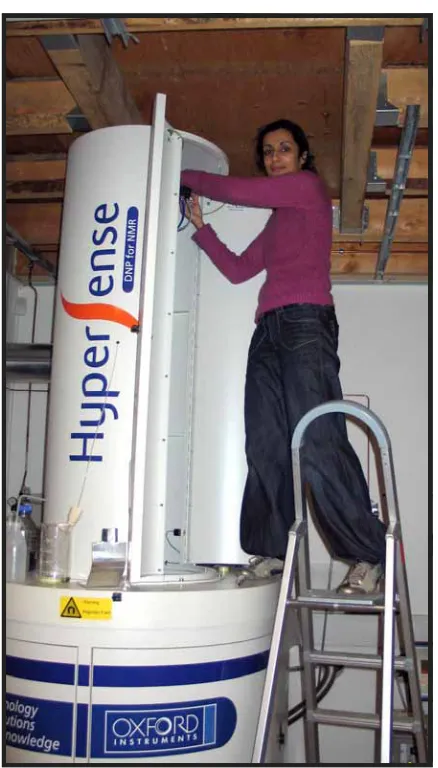
(25) Hyperpolarised MRI The hyperpolarisation research group aims for a better understanding and diagnosis of major chronic diseases by means of hyperpolarisation techniques. The group branches into two hyperpolarisation modalities: The use of hyperpolarised 3He-gas techniques (HP-3He) for studying chronic obstructive pulmonary disease (COPD) The use of hyperpolarised 13C based MR techniques (HP-13C) for studying cell metabolism characteristics of chronic diseases.. DRCMR members. Per Åkeson (group leader), Peter Magnusson (group leader), Lise Vejby Søgaard (group leader), Sadia Asghar Butt, Mette H. Lauritzen, Sascha Gude, Torsten Dorniok, Frederik Hengstenberg and Ann-Sofi Sjökvist.. External members. External members are Engineer and Principal Scientist Jan Henrik Ardenkjær-Larsen, PhD (GE Healthcare) and Professor Jørgen Vestbo, MD, PhD (Department of Cardiology and Respiratory Medicine, Copenhagen University Hospital Hvidovre).. External collaborators. Principal Scientist, Lars Engelholm, PhD, Finsen. Laboratory, Copenhagen University Hospital Rigshospitalet, Denmark Sergei Karpuk, PhD, and Zahir Salhi, PhD, Physics Department at Johannes GutenbergUniversity in Mainz, Germany Professor Rahim Rizi, Department of Radiology, University of Pennsylvania, Philadelphia, USA Ingrid Casselbrant, PhD and Sandra Diaz, PhD, Departments of Respiratory Medicine and Radiology, University Hospital Skåne, Malmö, Sweden. Research content. In the HP-3He research the HP-3He technique is evaluated in the clinical context of studying e.g. the progression of emphysema over time for patients with COPD. The HP-13C research focuses on preclinical in vivo studies of altered cell metabolism in disease models with the HP-13C technique.. Research methods. The hyperpolarisation research group has at its disposal for the HP-3He research, multi-nuclear 1.5 Tesla and 3 Tesla Siemens clinical MR scanners with 3 He-coils, 3He-gas inhalation systems, gas recycling system and had until the end of 2009 delivery of gas from Mainz University, Germany. The data has been collected under two projects with HP-3Hescanning of COPD-patients and healthy subjects. One conducted in collaboration with the Depart-. ment of Cardiology and Respiratory Medicine, Copenhagen University Hospital Hvidovre (ECLIPSE). Another conducted in collaboration with the Department of Radiology, University of Pennsylvania and the Department of Pulmonology, University Hospital Skåne. The MR protocol includes 3He gas diffusion imaging, which measures the Apparent Diffusion Coefficient (ADC) that correlates with the size of the lung alveoli. ADC is measured both at the inspiratory and expiratory state by using a 3D sequence to provide full lung coverage at high resolution.. Research Group Leader: Per Åkeson. Research Group Leader: Lise Vejby Søgaard. For HP-13C research exciting new possibilities opened in 2008 with the installation of a HyperSense DNP polariser (Oxford Instruments). Using this instrument it is possible to hyperpolarise substances labelled with 13C (and other nuclei such as 15N) thus increasing their MR signal more than 10.000 times. By injecting such hyperpolarised substances intravenously it is possible to follow their conversion in metabolic processes noninvasively and in real time by MR spectroscopy.. Research Group Leader: Peter Magnusson. The first substance that has been used at DRCMR is pyruvate, which is of particular interest because it is converted to acetyl-CoA, which enters the Krebs cycle and is one of the main fuels for energy production from the mitochondria in our cells. The conversion of pyruvate to acetyl-CoA gives the byproduct carbon dioxide, which is in rapid equilibrium with bicarbonate. Other products of the normal pyruvate metabolism are the amino acid alanine and the carboxylic acid lactate. All the metabolic products appear at different chemical shifts in the carbon MR spectra, and by acquiring spectra as a function of time it is possible to model the rate constants for the individual metabolic conversions. Chemical shift imaging can be used to map the spatial distribution of the different metabolites and thereby spatially locate differences in activity such as low activity in infarcts or high activity in cancer cells (see figure on the following page).. 25.
(26) Pyruvate metabolism in the cell can be monitored in vivo after injection of hyperpolarised 13C pyruvate. The MR signal follows the hyperpolarised 13C label and signals from the metabolites alanine, lactate and bicarbonate are observed in addition to the pyruvate signal. The 13C MR spectrum shows the corresponding peaks of the metabolites which appear at different chemical shifts. The white peak (at 180 ppm) corresponds to pyruvate hydrate at physiological pH.. Using the DRCMR’s multi-nuclear 4.7 Tesla Varian preclinical MR scanner with 13C-coils, physiological monitoring equipment and animal experimental facilities, the focus of the group in 2009-2010 has been on studies of altered in vivo cell metabolism in disease models of breast cancer and cardiac ischemia and on preparations for in vivo studies of cerebral metabolism.. Research activities. HP-3He: An analysis of paired inspiratory and expiratory HP-3He ADC measurements was performed in 2009 (Hengstenberg et al. 2009). The results suggest that the difference in inspiratory and expiratory ADC-values (ADC) could be used as a biomarker for hyperinflation caused by small airway disease and consequently for differentiating between patients with small airway disease and patients with emphysema. The progression of emphysema over a one year period was further studied in 2010 in terms of changes in ADC (Hengstenberg et al. 2010). A significant annual increase in ADC could be detected in a well-defined subgroup of COPD patients with CT diagnosed emphysema and the results suggest that the HP-3He ADC measurement might be used for monitoring the progression of emphysema. The HP-3He data was further analysed with respect to the possibility of measuring regional lung ventilation on a slice-by-slice basis. ADC-maps measured with the HP-3He-technique for a posterior, mid and anterior slice of a lung, respectively (top), showing an increased ADC in the anterior direction. Mean slice ADC measured with HP3He-technique averaged over 9 subjects for each slice position at inspiration and at expiration (bottom). The greater ADCdifference between inspiration and expiration (ADC) for posterior slices than for anterior slices suggests that ADC is sensitive to changes in regional ventilation.. 26. using ADC (Dorniok et al. 2009). The ADC values were averaged over each imaging slice, for inspiration and expiration, respectively, and the slice averages for each slice position were averaged over a subject group. The ADC values were found to agree with expected ventilation patterns and suggest that ADC can be used to measure regional ventilation (see figure below). Furthermore, Lise Vejby Søgaard and Torsten Dorniok were invited to lecture at the PHeLINet workshop in Krakow, Poland in May 2010 to speak about the current efforts in HP-3He imaging at the DRCMR. Due to the limited global availability of 3He-gas and the increased purchase price, the HP-3He scanning was discontinued in late 2009. The HP-3He research was made possible through funding from GlaxoSmithKline plc, the National Institutes of Health, USA, and through.
Figure
Related documents
can synthesize trends in drivers crea@vely to develop narra@ves as to the poten@al future that can result within the
2 Marginalisa- tion or even exclusion from scientific and social discourse of the group of adults with profound intellectual disabilities can be caused, for
When ―Auction State‖ state information is received from the market, send to the worker agent a MARKET_PHASE message that includes current phase information.. Receive
The longitudinal Side Scraper store, type SS, is used in a production line as a relatively small bulk material buffer store. The store operates with stockpiles placed
The present study investigated the effects of, and clicking behaviors related to, two different vocabulary learning conditions in digital reading environments: one
Taking into account the objective of the Horse Racing Fund of assisting with the operation and development of the racecourses at Down Royal and Downpatrick, the
Learn how your target group perceives your brand before and after your ad campaign Check brand awareness in users who were and were not exposed to your creatives Track
Mistakes that have been made in the past (e.g., by setting regulated prices for network access too low to stimulate investment in cable infrastructures) should not be used as
