MECHANISMS OF ANTIBODY MEDIATED NEUTRALIZATION OF DENGUE
VIRUS
Yang Zhou
A dissertation submitted to the faculty of the University of North Carolina at Chapel Hill in partial fulfillment of the requirements for the degree of Doctor of Philosophy in the Department of Microbiology and Immunology
Chapel Hill 2013
Approved by:
Aravinda M de Silva , PhD, MPH. Edward J Collins, PhD.
ABSTRACT
YANG ZHOU: Mechanisms of Antibody Mediated Neutralization of Dengue Virus
(Under the direction of Aravinda de Silva)
Dengue virus (DENV) is the most significant arthropod-borne virus in this world, affecting 2.5 billion people. Pre-existing sub-neutralizing antibodies (Abs) can enhance virus infection, leading to severe disease. Thus a better understanding of the interaction between humoral immunity and dengue virus is imperative. In this thesis I characterized 37 human monoclonal Abs (hMAbs) cloned from human PBMC. The majority of these MAbs were broadly cross-reactive and capable of enhancement of infection in vitro; few exhibited serotype-specific neutralizing activity. I next studied the mechanism and stoichiometry of Ab mediated neutralization of DENV and found that MAbs displaying threshold stoichiometry all neutralize post-attachment steps while MAbs neutralizing pre-attachment step display linear stoichiometry, indicating that different neutralization mechanisms are associated with different stoichiometric models respectively. I also studied the neutralization using multiple MAbs mixed together and demonstrated that the neutralization two MAbs are independent of each other in the mixture.
Besides genetic mutation, non-genetic variation such as maturation state of the dengue virion can also modulate neutralization. I found that monocytic cell derived dengue virions were more mature than mosquito cell derived virions. Using a panel of MAbs and sera, I demonstrated that maturation reduces virus sensitivity to neutralization mediated by weakly neutralizing cross-reactive MAbs or sera but it does not affect sensitivity to type-specific and strongly neutralizing MAbs or sera. Most interestingly, maturation enhanced virus sensitivity to neutralization of certain MAbs and sera. Further study indicated that preferential binding of immature virions by weakly neutralizing MAbs and furin cleavage of partially mature virions inside endosome are responsible for the
ACKNOWLEDGEMENTS
TABLE OF CONTENTS
LIST OF TABLES ...……….….x
LIST OF FIGURES………...xi
LIST OF ABBREVIATIONS………..xiii
CHAPTER 1: BACKGROUND AND SIGNIFICANCE………....1
1.1 General Introduction………..1
1.1.1 Emerging virus……….1
1.1.2 Dengue epidemiology……….…1
1.1.3 Dengue virology………..2
1.1.4 Pathogenesis………...4
1.1.5 Vaccine development………....5
1.2 Antibody Neutralization of Dengue Virus……….6
1.2.1 Neutralizing antibody………..6
1.2.2 Mechanisms of neutralization………7
1.3 Methods to study dengue Virus………8
1.3.1 Generation of Infectious Viruses………8
1.3.2 Neutralization Assays………9
1.3.3 Factors Modulating Neutralization……….10
1.4 Objectives of the study………11
1.5 References..……….16
2.1 Introduction………19
2.2 Materials and Methods………23
Viruses and recombinant proteins used in study………23
Human subjects and peripheral blood cell isolation………24
Generation of human hybridomas………..25
Screening ELISA………25
Electrofusion of EBV-transformed B cells with myeloma fusion partner………...26
MAb production and purification……….27
Neutralization assay……….27
ADE assays………28
2.3 Results………28
Electrofusion technique for generation of human hybridomas…………..28
Demographics and screening of traveler subjects………...29
Characteristics of E protein binding human MAbs………31
Characteristics of membrane (M) protein binding human MAbs…………34
2.4 Discussion………..35
2.5 Acknowledgments……….41
2.6 Reference………...48
CHAPTER 3: THE STOICHIOMETRY MODELS AND MECHANISMS OF ANTIBODY MEDIATED VIRUS NEUTRALIZATION………...…....52
3.1 Introduction………52
3.2 Materials and Methods………55
Cells………...55
Antibodies, Infectious Viruses and Antigens………..55
ELISA………..56
FACS based Function Assay………..57
3.3 Results……….58
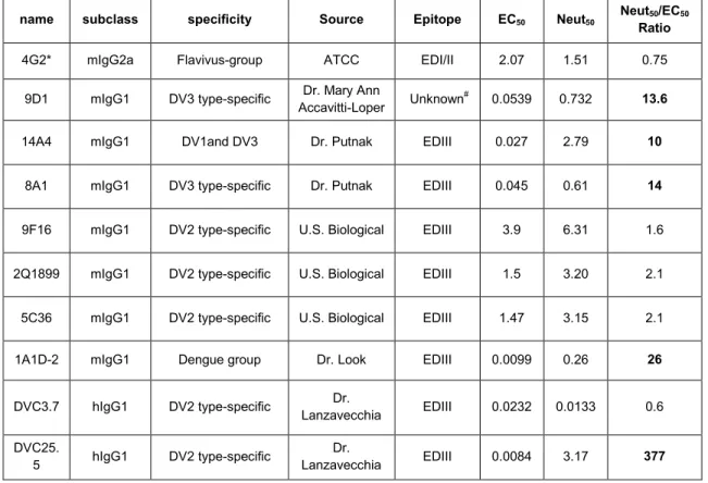
Antibodies Varies in EC50 of binding and FRNT50 of neutralization………58
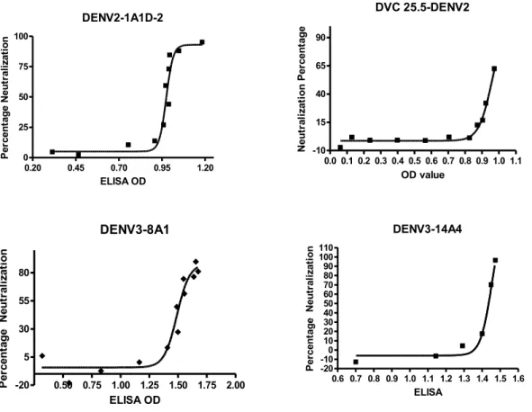
Two Stoichiometry Models of Binding/Neutralization………59
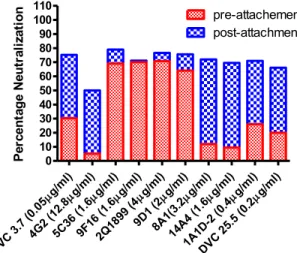
The neutralization mechanisms: pre-attachment or post-attachment………59
3.4 Discussion………..60
3.5 Reference………..71
CHAPTER 4: MODELING COMBINED NEUTRALIZATION AND ENHANCEMENT OF DENGUE VIRUS BY MULTIPLE MONOCLONAL ANTIBODIES ……….73
4.1 Introduction………73
4.2 Materials and Methods……….76
Cells………..76
Serum and MAbs………..76
Viruses suspension and Virus Antigens………....76
ELISA……….77
FACS Based Neutralization Assay………77
FACS Based ADE assay………77
4.3 Results………78
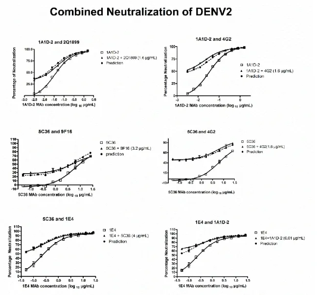
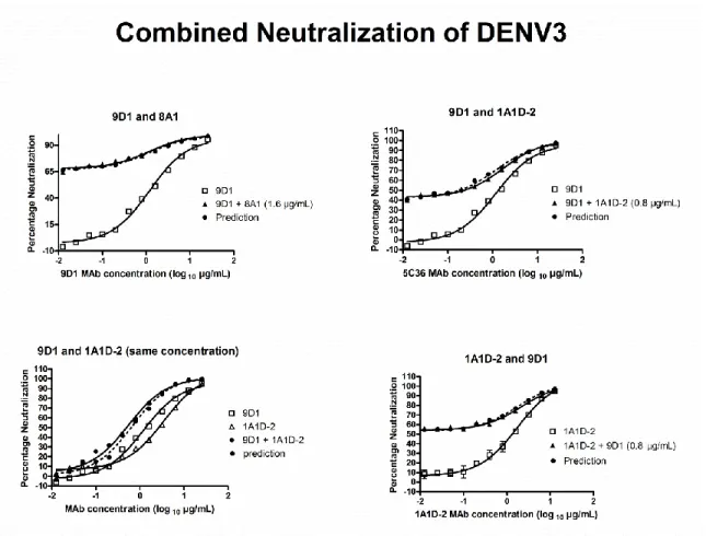
Independent neutralization of two MAbs in mixture………….…………..78
Additive ADE of two MAbs in Mixture……….………..79
Neutralizing MAb inhibits ADE mediated by Enhancing MAb…………80
Non-neutralizing Non-enhancing MAbs Modulate ADE Infection……....80
4.4 Discussion………..81
CHAPTER 5: THE MECHANISMS OF DIFFERENTIAL
NEUTRALIZATION OF DENGUE SEROTYPE 3
STRAINS OF MONOCLONAL ANTIBODY 8A1...………....94
5.1 Introduction………94
5.2 Methods and materials………....97
Cells……….97
DENV-3 Molecular Clone Strategy………..97
Recombinant Virus Recovery………..98
Virus Titration and Focus Reduction Neutralization Test (FRNT)……..99
Monoclonal Abs………....99
Software and statistics……….99
ELISA………..100
Mutagenesis and production of rEDIII………100
Surface Plasmon Resonance (SPR)………101
5.3 Results………101
Mouse Monoclonal antibody neutralization against four DENV3 isogenic clones containing E protein from different genotypes………..101
Amino Acid Variations in EDIII lateral ridge………..104
The Mutations Altered Binding Affinity………...105
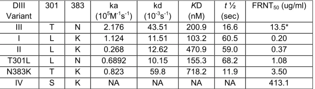
8A1 affinity is determined by off-rate….……….…106
EDIII Residues at Site 301 Alone Determines Dissociation Rate and Half-Life………..………...106
5.4 Discussion………107
5.5 References………...118
CHAPTER 6: DENGUE VIRUS MATURATION MODULATES SENSITIVITY TO ANTIBODY MEDIATED NEUTRALIZATION…....…….…...……..….122
6.1 Introduction………..122
Cells……….…124
Serum and MAbs………124
C6/36 cell derived virus………124
U937-DC-SIGN cell Derived Virus………..………125
Immature Virus………...125
Virus Titration and Focus Reduction Neutralization Test (FRNT)……125
Function Assay……….…126
Furin Inhibition Experiment……….…126
Western Blot………..…127
Capture ELISA………..……127
MAb Epitope Mapping………..128
6.3 Results……….…129
The human monocytic cell line U937-DC-SIGN produced mostly mature virus………..….129
Maturation state of DENV modulates Neutralization Sensitivity to some MAbs……….130
Epitopes recognized by maturation state sensitive MAbs……….130
Binding of MAbs to mature or immature virions……….131
Human immune sera from dengue patients displayed maturation dependent differences in neutralization potency…………132
Endosome furin cleavage is involved in modulating antibody mediated neutralization………...…………...…132
6.4 Discussion………134
6.5 References………...…151
CHAPTER 7: DISCUSSION……….154
7.1 The misled humoral immune response………...154
7.2 The gap between antibody binding and neutralization………..…159
LIST OF TABLES
Table
2.1 Subject demographics, serologies, and hybridoma yields from
dengue-immune travelers ...……..…….…....………42
2.2 Characteristics of envelope (E) protein binding human MAbs…….………...…43
2.3 Characteristics of premembrane (prM) protein binding human MAbs ……….44
3.1 MAbs and their EC50 of binding and Neutralization...……….…65
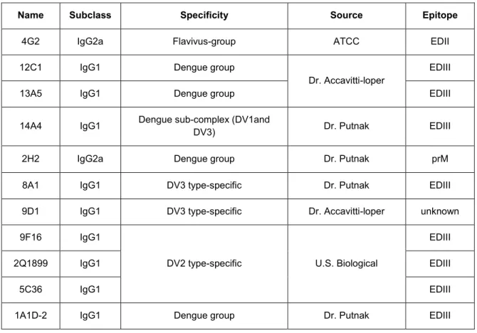
4.1 Panel of Mouse MAbs used in this project. ………...85
5.1 Kinetics of DENV-3 DIII-MPB variants with 8A1....……….111
6.1 Monoclonal Antibodies used in this work...………..141
6.2 Sera used in this work and the differential neutralization of two viruses…....142
LIST OF FIGURES
Figure
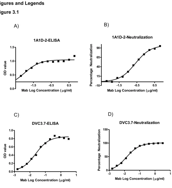
1.1 Genome structure of dengue virus and the 11 virus proteins...…...12 1.2 Top and side view of the Dengue virus envelope protein dimer structure…..13 1.3 The flavivirus life cycle..………...14 1.4 Structure of mature and immature dengue virion.………..15 2.1 Serotype-specific strongly neutralizing E protein binding human MAb……..45 2.2 Cross-reactive poorly neutralizing E protein binding human MAbs……..…46 2.3 Cross-reactive poorly neutralizing prM protein binding human MAbs…..…47 3.1 ELISA Binding Curve and Neutralization Curves of representative
MAb DVC3.7 and 1A1D-2. ………...………...………..66 3.2 Neutralization/Binding Plots of 5 MAb showing linear stoichiometry
4G2 and 5 MAbs showing threshold stoichiometry..………..………67 3.3 Function assay results of all 10 MAbs..………69 3.4 Hypothetical relationships between neutralization mechanisms
and stoichiometry models and the MAbs assumed to associate
with each scenario...70 4.1 Combined Neutralization of DENV by Two MAbs………..….86 4.2 Combined ADE of two MAbs at Non-neutralizing concentration.…….……88 4.3 Inhibition of ADE by Neutralizing MAbs………..….89 4.4 Cross-reactive and non-neutralizing MAb 12C1 and 13A5 can
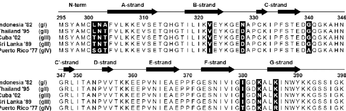
modulating the ADE mediated by MAb 4G2……..………..….….90 5.1 Cartoon of DENV3 EDIII and Alignment of EDIII amino acid sequence…...112 5.2 Calculated FRNT50 values for mouse MAbs against the
recombinant virus clones………..113 5.3 Calculated FRNT50 values for MAb 8A1 against the virus clones……...…115
5.4 Results of ELISA using mouse MAb 8A1 (0.2 μg/ml) against
captured whole viruses…………..………...………...116 5.5 Kinetic analysis of 8A1 interaction with DENV-3 EDIII-MBP
6.1 Maturation state of DENVs produced by different cell lines.………...143 6.2 Maturation-dependent neutralization of DENVs by MAbs……….144 6.3 DENV Envelope Residues that Affect 2A10 Binding………...…………...….145 6.4 Relative binding of MAbs to fully mature and immature DENVs.………...…146 6.5 Maturation-dependent neutralization of DENVs by human
dengue immune sera………..147 6.6 Furin cleavage inhibition abolishes majority of C6/36 virus
LIST OF ABBREVIATIONS
AA amino acid Ab antibody
ADE antibody dependent enhancement ATCC American type culture collection C capsid protein
CO2 Carbon dioxide
DC-SIGN dendritic cell-specific intercellular adhesion molecule-3-grabbing nonintegrin DENV dengue virus
DF dengue fever
DHF dengue hemorrhagic fever DSS dengue shock syndrome E envelope protein
EDI envelope protein domain I
EDI/II envelope protein domain I and domain II EDII envelope protein domain II
EDIII envelope protein domain III
ELISA enzyme-linked immunosorbent assay FRNT Focus Reduction Neutralization Assay HCV hepatitis C virus
HIV human immunodeficiency virus IFN interferon
IgG immunoglobulin G IgM immunoglobulin M
MOI multiplicity of Infection MAb monoclonal antibody MBP maltose binding protein nm nanometer
NS non-structural protein OD optical density
PCR polymerase chain reaction PFU plaque forming units prM pre-membrane protein
PRNT plaque reduction neutralization test rEDIII recombinant EDIII
CHAPTER 1
BACKGROUND AND SIGNIFICANCE
1.1 General Introduction
1.1.1 Emerging virus: Dengue virus (DENV) is a re-emerging arth2ropod-borne virus. In
the last 50 years, dengue incidences increased 30 fold and its geographical distribution expanded to most tropical and subtropical areas. As a result, dengue is becoming increasingly recognized as one of the world's most threatening infectious diseases. However, despite decades of research, we still do not have a vaccine to prevent its transmission or feasible post-exposure therapy to treat the disease.
1.1.2 Dengue epidemiology: Dengue is endemic in more than 110 countries in tropical
and subtropical areas such as Southeast Asian, Latin America, and Sub-Sahara Africa. These areas are all heavily populated and every year 50 - 100 million people are
infected with Dengue, leading to half a million hospitalization and 12,500 - 25,000 deaths (WHO 2009; Whitehorn and Farrar 2010). Most people can recover from dengue
infection without any adverse consequences. Overall, the mortality rate is very low, but in cases of severe disease, the mortality rate roars up to 26% without supportive therapy (WHO 2009; Ranjit and Kissoon 2011).
for about 4 days and mosquitoes taking blood meal from this patient will get infected and transmit the virus to other humans living in the neighborhood, leading to localized
outbreak. In the last 50 years, due to a combination of urbanization, population growth and increasing international travel, dengue became a global epidemic, putting 2.5 billion people worldwide in risk (WHO 2009).
1.1.3 Dengue virology: Dengue is a single-stranded, positive sense RNA virus of the
family Flaviviridae, genus flavivirus. It is closely related to several other important human pathogens such as Japanese encephalitis virus (JEV), West Nile virus (WNV), Yellow fever virus (YFV) and Tick-borne encephalitis virus (TBEV). The DENV genome encodes 10 genes and is translated into a single polyprotein and then cleaved into structural proteins (C-prM-E) and non-structural proteins (NS1-NS2A-NS2B-NS3-NS4A-NS4B-NS5) (Figure 1.1). The C (capsid) protein forms a virion scaffold that encapsidates the RNA genome. The prM (membrane) protein is important in the assembly of virion and subsequent maturation. The E protein (envelope) is the major surface exposed antigen and the principal target of host antibody response. The 7 NS proteins have diverse roles such as protease, RNA-dependent RNA polymerase or immune regulation proteins participating in evading immune response.
genotype causes severe disease epidemics whereas the American genotype has only
been linked to mild or subclinical epidemics (Rico-Hesse 2003).
The E glycoprotein has been crystallized and the structure has been solved (Modis, Ogata et al. 2004; Nayak, Dessau et al. 2009). E protein forms a homodimer on the mature DENV virion. The virus envelope consists of 90 E glycoprotein homodimers. Three dimers are arranged in a herringbone pattern to form a raft-like structure on the virion surface (Figure 1.2). The ectodomain of E protein has three domains: domain I, II and III (EDI-EDIII). Specifically, EDII contains fusion loop, which mediates membrane fusion and EDIII belongs to IgG superfamily and is believed to participate in cell receptor binding (Crill and Roehrig 2001).
et al. 2007). Then the NS proteins initiate replication of viral genomes and subsequently the newly synthesized RNA is packaged by C protein to form a nucleocapsid (Figure 1.3). Through unknown mechanisms, the nucleocapsid is encapsulated by ER membrane rafts covered with prM/E trimers and buds into the ER to form immature virion. The immature virion contains 180 prM/E dimers that project vertically outward from viral surface as 60 trimeric spikes (Zhang, Chipman et al. 2003; Zhang, Corver et al. 2003) (Figure 1.4). The immature virions are transported through trans-Golgi network, in which the low pH environment triggers structural reorganization of glycoproteins (Figure 1.3). The prM/E heterodimers dissociate and form 90 E dimers lying flat on the surface of the particle, with prM capping the fusion peptide of the E protein. This exposes the cleavage site for cellular endoprotease furin to cleave "pr" peptide that covers the fusion peptide (Stadler, Allison et al. 1997; Yu, Zhang et al. 2008; Zybert, van der Ende-Metselaar et al. 2008). This process is called maturation. It has been shown that the maturation process is not efficient in certain cell lines, resulting in the production of partially mature or totally immature virions (Lok, Kostyuchenko et al. 2008; Dejnirattisai, Jumnainsong et al. 2010; Junjhon, Edwards et al. 2010).
1.1.4 Pathogenesis: Dengue infection with any serotype results in long term protection
DHF/DSS are much higher than infants born to mothers who are naïve to dengue infection (Halstead, Nimmannitya et al. 1970). These evidences strongly suggested that pre-existing heterotypic immunity is critical for development of DHF/DSS. Halstead proposed Antibody Dependent Enhancement (ADE) as an underlying mechanism of DHF/DSS (Halstead 1970). The ADE hypothesis stated that during secondary infection, the pre-existing antibodies from a primary infection fails to neutralize heterotypic virus but forms infectious immune complex with the virus, and the immune complexes bind to and infect monocytic cells expressing Fc-ɣ receptors via the Fc portion of the antibody. This leads to increased total infected cell mass and enhanced virus replication and is hypothesized to be responsible for the severe disease outcome. Other competing hypothesis such as dengue strain-dependent virulence or host factors had been proposed but none of them have been extensively studied like the ADE hypothesis (Halstead, Rojanasuphot et al. 1983; Mongkolsapaya, Dejnirattisai et al. 2003).
Numerous studies have confirmed the ADE phenomenon in vitro or in vivo with animal models but the detailed mechanisms of how ADE leads to DHF/DSS are still unknown.
1.1.5 Vaccine development: Despite numerous approaches that have been tried in the
last sixty years, an effective dengue vaccine remains elusive. The ideal dengue vaccine
promising potential compared to other vaccine candidates. The major concern with any dengue vaccine is to avoid eliciting cross-reactive and non-neutralizing antibodies which may enhance subsequent virus infection instead of preventing it. To address this
problem, the goal of vaccines in development is to elicit robust and simultaneous protective immunity to all 4 serotypes. In addition, we need to know exactly which virus epitopes induce type-specific/neutralizing or cross-reactive/non-neutralizing antibodies. My work will help to shed some light on this question.
1.2 Antibody Neutralization of Dengue Virus
1.2.1 Neutralizing antibody: Most of my work in this dissertation is about antibody
neutralization of dengue virus. The study of dengue neutralization is critical for
understanding dengue virology and pathogenesis and advancing vaccine development. Type-specific antibodies are believed to be the major neutralizing component in dengue polyclonal serum and depletion study by de Alwis et al. demonstrated this hypothesis (de Alwis, Smith et al. 2012). So the questions are where do these type-specific antibodies bind and where do most human dengue-reactive antibodies bind? Studies using mouse MAbs indicated that most neutralizing antibody epitopes are on EDIII (Cockburn,
particular group of antibodies binding to conformational epitopes on virion surface which appear to be the major neutralizing epitopes targeted by Abs in polyclonal sera (de Alwis, Smith et al. 2012). These Abs bind to whole virus but do not bind to soluble E
glycoprotein which suggests that the epitope exists only on virus surface but not E protein monomer. Whether these Abs bind to tertiary structure of E trimers on the immature virion surface or E dimmers of the mature virion surface is still unknown.
1.2.2 Mechanisms of neutralization: Many DENV neutralizing MAbs had been
characterized up to today but the mechanisms and stoichiometry of neutralization are still largely elusive. Pierson and colleagues suggested that antibody neutralization of flavivirus is a multiple hit phenomenon in which the number of antibodies binding to the virion must exceed certain threshold for neutralization to happen (Pierson, Xu et al. 2007). This model has been demonstrated in West Nile virus and the threshold is estimated to be around 30 antibodies. This stoichiometry model explains the MAb’s neutralization potency as occupancy of accessible epitopes: for strongly neutralizing MAb, its accessible epitopes are much more than 30, so it takes small occupancy of binding (percentage of the total available epitopes that are bound by antibodies) to exceed the threshold; otherwise for weakly neutralizing MAb, its total available epitopes
are less than 30, then even maximum occupancy (at highest concentration) are still below threshold. However, this model has several flaws as it ignores the possibility that location and function of epitope itself may affect the threshold requirement as binding to different epitopes may exert different effect on virus entry, fusion, replication steps and thus the stoichiometry of neutralization may vary. Secondly, it does not take into
work, I tried different experimental design and proposed additional stoichiometry model
to address these flaws.
1.3 Methods to study dengue Virus
1.3.1 Generation of Infectious Viruses: Most previous DENV neutralization and ADE
studies have used virus derived from mosquito cell line C6/36. The C6/36 cell monolayer is infected at low MOI and kept in 28 ºC incubator for 6 days before the supernatants are harvested as infectious virus stocks. Occasionally, people may use Vero cell derived viruses for their work, but seldom did anyone uses human monocytic cells derived viruses as infectious virus. As mentioned above, viruses derived from different cells lines may vary in glycosylation and maturation. It has been shown that mosquito cells and mammalian cells have variation in processing prM Cleavage (Dejnirattisai, Jumnainsong et al. 2010). Our experience suggested even using the same method of virus production such as C6/36 methods mentioned above, the virus stocks produced at different batches still vary in neutralization assays, not only the titer varies in logs magnitude but also the sensitivity to neutralization. In this thesis, the mechanisms of this variation will be illustrated. But it is important for the field to recognize the existence of this pitfall and
take serious effort to standardize infectious virus stock between different labs.
relevant in pathogenesis than the mosquito cell derived viruses. However, so far there were not any reports using human monocytic cell derived viruses for dengue
neutralization or ADE study. Considering the lack of knowledge about these viruses, it is imperative and indispensable for us to study human monocytic cell line derived viruses and its neutralization.
1.3.2 Neutralization Assays: Focus Reduction Neutralization Assays (FRNT) is
currently the most widely used assay for dengue neutralization study. It is derived from the focus assay, which is also the standard method for titration of virus (Okuno, Igarashi et al. 1978). Compared to traditional Plaque Reduction Neutralization Assay (PRNT), FRNT uses Vero cell monolayer and antibody staining to detect the virus infection focus instead of plaque formation and this improvement provides better accuracy, higher out-put and the ability to use virus strains that do not plaque well. Based on this assay, a more sophisticated high-throughput assay called micro-neutralization test (MNT) has been developed. This assay utilizes 96 well plates and automated counting of focus using ELISpot reader (Taketa-Graham, Powell Pereira et al. 2010).
Both FRNT and FACS based neutralization assay were employed in this
research. For most of the neutralization assay, FACS based assay using U937-DC-SIGN is preferred for its efficiency and relevance, especially in experiment when only C6/36 derived viruses are used. But when U937-DC-SIGN derived viruses are used as infection agents, the traditional FRNT is preferred because monocytic cell line derived viruses show a very low efficiency in infecting other monocytic cell lines such as U937-DC-SIGN (data not shown), a phenomenon already reported by Dejnirattisai
(Dejnirattisai, Webb et al. 2011).
1.3.3 Factors Modulating in vitro Neutralization: DENV neutralization by antibodies in
vivo is critical for host to clear virus infection. While using animal models for dengue neutralization study is expensive and time-consuming, researchers are forced to rely on in vitro neutralization assays that can mimic in vivo neutralization. Dengue infection of cell in vitro is a complicate process involving viruses, cells, temperatures, pH, incubation time, antibodies and other serum components such as complement. Any variation in these factors can alter virus infection and affect antibody mediated virus neutralization. However, most of these factors are understudied. But they must be taken into
consideration when interpreting neutralization data.
Many cell lines have been found permissive to dengue viruses and several of them (Vero, U937-DC-SIGN, Raji-DC-SIGN and BHK) have been used in neutralization assay. These cell lines are known to use different surface molecule as attachment molecule for dengue virus (Kroschewski, Allison et al. 2003; Navarro-Sanchez, Altmeyer et al. 2003). The different usage of attachment molecule is a factor that must be
from cellular factors, viral factors such genetic mutation and non-genetic variation (such as maturation or glycosylation) affect neutralization even more. A lot has been studied about virus genetics and indicated how mutations increase or decrease neutralization sensitivity. This part is well elucidated and explained in this dissertation. There are also some non-genetic factors may affect neutralization, including maturation and
glycosylation.
1.4 Objectives of Study
One important question in DENV studies is that the humoral immunity fails to protect heterologous secondary infection. In order to elucidate this question, we aim to investigate the long term antibody response in vivo and characterize the neutralizing and enhancing monoclonal antibodies. Strongly neutralizing MAbs have the potential of treating DENV infection as therapeutic antibodies. To promote development of DENV antibody therapy, we aim to study mechanisms and stoichiometry of dengue-antibody interaction and to illustrate the mechanisms of virus escaping neutralization through natural genetic mutations. The monocytes are major targets of DENV infection in vivo but monocytic cell derived virus was seriously understudied in previous neutralization and ADE studies. To fill in this gap, we aim to determine the maturation of viruses derived from monocytes, and study how this may affect virus sensitivity to antibody mediated neutralization. In general, the objective of this study is to get better
Figures and Legends
Figure 1.1
Figures 1.1: Genome structure of dengue virus and the 11 virus proteins (adapted from
Figure 1.2
Figure 1.2: Top and side view of the Dengue virus envelope protein dimer structure with
Figure 1.3
Figure 1.3: The flavivirus life cycle. A - G represents each step of flavivirus life cycle
Figure 1.4
Figure 1.4: Structure of mature virion and immature virion. A. A surface shaded view of
1.5 References
Beltramello, M., K. L. Williams, et al. (2010). "The human immune response to Dengue virus is dominated by highly cross-reactive antibodies endowed with neutralizing and enhancing activity." Cell Host Microbe 8(3): 271-283.
Bryant, J. E., A. E. Calvert, et al. (2007). "Glycosylation of the dengue 2 virus E protein at N67 is critical for virus growth in vitro but not for growth in intrathoracically inoculated Aedes aegypti mosquitoes." Virology 366(2): 415-423.
Cockburn, J. J., M. E. Navarro Sanchez, et al. (2012). "Mechanism of dengue virus broad cross-neutralization by a monoclonal antibody." Structure 20(2): 303-314. Costin, J. M., E. Zaitseva, et al. (2013). "Mechanistic study of broadly neutralizing human
monoclonal antibodies against dengue virus that target the fusion loop." J Virol
87(1): 52-66.
Crill, W. and J. Roehrig (2001). "Monoclonal antibodies that bind to domain III of dengue virus E glycoprotein are the most efficient blockers of virus adsorption to Vero cells." 75: 7769-7773.
de Alwis, R., M. Beltramello, et al. (2011). "In-depth analysis of the antibody response of individuals exposed to primary dengue virus infection." PLoS Negl Trop Dis 5(6): e1188.
de Alwis, R., S. A. Smith, et al. (2012). "Identification of human neutralizing antibodies that bind to complex epitopes on dengue virions." Proc Natl Acad Sci U S A
109(19): 7439-7444.
Dejnirattisai, W., A. Jumnainsong, et al. (2010). "Cross-reacting antibodies enhance dengue virus infection in humans." Science 328(5979): 745-748.
Dejnirattisai, W., A. I. Webb, et al. (2011). "Lectin switching during dengue virus infection." J Infect Dis 203(12): 1775-1783.
Halstead, S. B. (1970). "Observations related to pathogensis of dengue hemorrhagic fever. VI. Hypotheses and discussion." Yale J Biol Med 42(5): 350-362.
Halstead, S. B., S. Nimmannitya, et al. (1970). "Observations related to pathogenesis of dengue hemorrhagic fever. IV. Relation of disease severity to antibody response and virus recovered." Yale J Biol Med 42(5): 311-328.
Halstead, S. B., S. Rojanasuphot, et al. (1983). "Original antigenic sin in dengue." Am J Trop Med Hyg 32(1): 154-156.
Junjhon, J., T. J. Edwards, et al. (2010). "Influence of pr-M cleavage on the
heterogeneity of extracellular dengue virus particles." J Virol 84(16): 8353-8358. Kroschewski, H., S. L. Allison, et al. (2003). "Role of heparan sulfate for attachment and
entry of tick-borne encephalitis virus." Virology 308(1): 92-100.
Lai, C. Y., W. Y. Tsai, et al. (2008). "Antibodies to envelope glycoprotein of dengue virus during the natural course of infection are predominantly cross-reactive and recognize epitopes containing highly conserved residues at the fusion loop of domain II." J Virol 82(13): 6631-6643.
Lambeth, C. R., L. J. White, et al. (2005). "Flow cytometry-based assay for titrating dengue virus." J Clin Microbiol 43(7): 3267-3272.
Lok, S. M., V. Kostyuchenko, et al. (2008). "Binding of a neutralizing antibody to dengue virus alters the arrangement of surface glycoproteins." Nat Struct Mol Biol 15(3): 312-317.
Modis, Y., S. Ogata, et al. (2003). "A ligand-binding pocket in the dengue virus envelope glycoprotein." Proc Natl Acad Sci U S A 100(12): 6986-6991.
Modis, Y., S. Ogata, et al. (2004). "Structure of the dengue virus envelope protein after membrane fusion." Nature 427(6972): 313-319.
Mongkolsapaya, J., W. Dejnirattisai, et al. (2003). "Original antigenic sin and apoptosis in the pathogenesis of dengue hemorrhagic fever." Nat Med 9(7): 921-927.
Navarro-Sanchez, E., R. Altmeyer, et al. (2003). "Dendritic-cell-specific ICAM3-grabbing non-integrin is essential for the productive infection of human dendritic cells by mosquito-cell-derived dengue viruses." EMBO Rep 4(7): 723-728.
Nayak, V., M. Dessau, et al. (2009). "Crystal structure of dengue virus type 1 envelope protein in the postfusion conformation and its implications for membrane fusion." J Virol 83(9): 4338-4344.
Okuno, Y., A. Igarashi, et al. (1978). "Neutralization tests for dengue and Japanese encephalitis viruses by the focus reduction method using peroxidase-anti-peroxidase staining." Biken J 21(4): 137-147.
Pierson, T. C., Q. Xu, et al. (2007). "The stoichiometry of antibody-mediated
neutralization and enhancement of West Nile virus infection." Cell Host Microbe
1(2): 135-145.
Putnak, R., J. Fuller, et al. (2003). "Vaccination of rhesus macaques against dengue-2 virus with a plasmid DNA vaccine encoding the viral pre-membrane and envelope genes." Am J Trop Med Hyg 68(4): 469-476.
Rico-Hesse, R. (2003). "Microevolution and virulence of dengue viruses." Adv Virus Res
59: 315-341.
Smith, S. A., Y. Zhou, et al. (2012). "Persistence of circulating memory B cell clones with potential for dengue virus disease enhancement for decades following infection." J Virol 86(5): 2665-2675.
Stadler, K., S. L. Allison, et al. (1997). "Proteolytic activation of tick-borne encephalitis virus by furin." J Virol 71(11): 8475-8481.
Stiasny, K., S. L. Allison, et al. (2002). "Membrane interactions of the tick-borne encephalitis virus fusion protein E at low pH." J Virol 76(8): 3784-3790. Taketa-Graham, M., J. L. Powell Pereira, et al. (2010). "High throughput quantitative
colorimetric microneutralization assay for the confirmation and differentiation of West Nile Virus and St. Louis encephalitis virus." Am J Trop Med Hyg 82(3): 501-504.
Villabona-Arenas, C. J., D. R. Miranda-Esquivel, et al. (2009). "Phylogeny of dengue virus type 3 circulating in Colombia between 2001 and 2007." Trop Med Int Health 14(10): 1241-1250.
Wahala, W. M., A. A. Kraus, et al. (2009). "Dengue virus neutralization by human immune sera: role of envelope protein domain III-reactive antibody." Virology
392(1): 103-113.
White, L. J., C. A. Sariol, et al. (2013). "An alphavirus vector-based tetravalent dengue vaccine induces a rapid and protective immune response in macaques that differs qualitatively from immunity induced by live virus infection." J Virol 87(6): 3409-3424.
Whitehorn, J. and J. Farrar (2010). "Dengue." Br Med Bull 95: 161-173.
WHO (2009). "Dengue and dengue haemorrhagic fever." WHO Fact Sheets 117. Xie, H., A. R. Cass, et al. (1998). "Yellow fever 17D vaccine virus isolated from healthy
vaccinees accumulates very few mutations." Virus Res 55(1): 93-99.
Yu, I. M., W. Zhang, et al. (2008). "Structure of the immature dengue virus at low pH primes proteolytic maturation." Science 319(5871): 1834-1837.
Zhang, W., P. R. Chipman, et al. (2003). "Visualization of membrane protein domains by cryo-electron microscopy of dengue virus." Nat Struct Biol 10(11): 907-912. Zhang, Y., J. Corver, et al. (2003). "Structures of immature flavivirus particles." EMBO J
22(11): 2604-2613.
CHAPTER 2
CHARACTERIZATION OF MONOCLONAL ANTIBODIES ISOLATED FROM
PBMC OF DENGUE IMMUNE PATIENTS
2.1 Introduction
Dengue viruses (DENV) are expanding globally with an estimated 50-100 million cases of dengue infection worldwide annually, and more than 20,000 deaths. It is now estimated that approximately one fifth of the world population is at risk of infection by DENV (Guzman, Halstead et al. ; Gibbons and Vaughn 2002; Gubler 2002). Dengue is also threatening the continental US. More than 25 cases of locally acquired infection were reported in Key West, Florida in 2009 and 2010 . Symptomatic dengue disease ranges in severity from an influenza-like illness to life threatening hemorrhagic fever or shock. Understanding the pathogenesis of dengue hemorrhagic fever/dengue shock syndrome (DHF/DSS) is of central importance in the rationale development of antiviral for treatment and vaccines to prevent dengue disease (Green and Rothman 2006; Whitehead, Blaney et al. 2007).
The DENV complex consists of 4 serotypes. Infection with a single serotype leads to antibody responses that cross-react with all serotypes (Halstead 2002). Despite the cross-reactivity, individuals only develop protective immunity against the serotype responsible for infection (Halstead 2002; Rothman 2004). Human studies have
antibodies bind to virions and enhance entry and replication in Fc-receptor-bearing cells, which leads to a higher viremia and release of cytokines and vasoactive mediators that increase vascular permeability (Halstead 2003). The molecular process of antibody-dependent enhancement (ADE) of infection has been demonstrated to occur with DENV and antibodies using cells in culture and animal models (Halstead and O'Rourke 1977). A better understanding of the molecular, genetic, and structural basis for recognition of dengue viruses by human antibodies is greatly needed, and could lead to the rational design of vaccines that enhance the induction of neutralizing antibodies while lowering the risk of DHF/DSS.
Dengue viruses are enveloped viruses of the Flaviviridae family that display pseudo-icosahedral symmetry, with 180 copies of the envelope (E) glycoprotein and 180 copies of the membrane (M) protein in the lipid bilayer membrane. Previous structural studies have shown that the dengue virus E protein is arranged into 30 rafts of three parallel dimers in mature virion particles (Kuhn, Zhang et al. 2002). Envelope
glycoproteins form dimers, and crystal structures of the dimeric E protein have been determined for three DENV serotypes (DENV1, DENV2 and DENV3) (Modis, Ogata et al. 2003; Zhang, Zhang et al. 2004; Modis, Ogata et al. 2005; Nayak, Dessau et al. 2009). The E glycoprotein monomer possesses three principal domains, designated domains I, II, and III (DI, DII, DIII). DIII is likely the recognition domain for the principal cell receptor (Rey, Heinz et al. 1995; Bhardwaj, Holbrook et al. 2001; Crill and Roehrig 2001; Hung, Hsieh et al. 2004). DII possesses the fusion loop (Modis, Ogata et al. 2004) and an N-linked glycan, which can contribute to cell binding by interaction with DC-SIGN, a
inoculated multiple times with dengue viruses (Brien, Austin et al. ; Shrestha, Brien et al. ; Roehrig, Bolin et al. 1998; Chen, Huang et al. 2007). Mapping of epitopes recognized by strongly neutralizing mouse MAbs has identified several major antigenic sites on the E protein (Pierson, Fremont et al. 2008). Studies have shown that sites in the region of the fusion peptide, located at the tip of E protein DII (Rey, Heinz et al. 1995; Oliphant, Nybakken et al. 2006) and near the E protein hinge and linker regions between DI and DII, are recognized by cross-reactive, moderately neutralizing mouse MAbs. The most potent neutralizing antibodies are serotype-specific, however, and bind epitopes on the lateral surface of DIII of the E protein (Modis, Ogata et al. 2005; Oliphant, Engle et al. 2005). Investigators also have started to uncover specific mechanisms of antibody neutralization of flaviviruses. Some flavivirus antibodies that neutralize probably do so by sterically blocking attachment of the virus to the cell receptor. Other neutralizing antibodies block a step after cell attachment and such antibodies possibly neutralize by preventing protein conformational changes required for viral fusion in endosomes
(Nybakken, Oliphant et al. 2005; Kaufmann, Nybakken et al. 2006). Some antibodies (for example the DENV MAb1A1D-2) appear to bind to hidden epitopes that are transiently exposed on the particle and promote a cascade of E protein rearrangements on the particle (Lok, Kostyuchenko et al. 2008). These studies have been instrumental in understanding mechanisms for flavivirus neutralization, however, since the antibodies were generated artificially in mice, the ability to translate this information to humans is limited.
(Dejnirattisai, Jumnainsong et al. ; Kou, Lim et al. ; Huang, Yang et al. 2006). This phenomenon results in as much as a 1,000-fold increase in infectivity and is
Fc-mediated (Balsitis, Williams et al. ; Littaua, Kurane et al. 1990). Recent work by Pierson et al., using mouse MAbs against WNV, has shed light on the stoichiometry of this process (Pierson ; Pierson, Xu et al. 2007). For each antibody studied, a threshold number of MAbs docked to the virion dictated whether neutralization or enhancement took place. The type of functional activity was determined not only by the location of the epitope, but also by the antibody affinity and epitope accessibility. An additional related ADE mechanism has been demonstrated to occur with anti-prM antibodies. Using either mouse or human MAbs, investigators have shown that the presence of anti-prM
antibodies causes immature viral particles that have prM on the surface, which are normally noninfectious, to acquire the ability to infect cells efficiently through Fc-mediated pathways (Dejnirattisai, Jumnainsong et al. ; Rodenhuis-Zybert, van der Schaar et al. ; Rodenhuis-Zybert, Wilschut et al.). The role that ADE and anti-prM antibody-mediated enhanced infectivity of immature virus particles play in the
pathogenesis of dengue infection is of significant interest. Characterization of antibodies obtained from humans following natural infection could prove to be an important tool for further investigation.
these investigators showed a significant correlation between the presence of such antibodies and dengue virus neutralizing activity (Crill, Hughes et al. 2009). Taken together, these studies suggest that very rare serotype-specific potent neutralizing antibodies may be the primary determinants of protection against severe disease in humans.
Recently, several groups have generated panels of human dengue-specific MAbs using B cells from people exposed to natural DENV infection, to investigate the humoral response to dengue infection. Schieffelin et al. generated three E protein reactive human MAbs and showed that all three where at least partially cross-reactive and two lacked neutralizing activity (Schieffelin, Costin et al.). A panel of six human anti-preM
monoclonal antibodies was developed and shown to be entirely cross-reactive and devoid of significant neutralization activity (Dejnirattisai, Jumnainsong et al.). Other investigators have isolated larger panels of human MAbs to dengue (Beltramello, Williams et al. ; de Alwis, Beltramello et al.). These studies indicate that most antibodies are serotype cross reactive and weakly to non-neutralizing. Fewer than 5% of the antibodies displayed moderate to strong neutralization of one or more serotypes.
In the study presented here, we use a high efficiency optimized method to generate human hybridomas to make a large panel of MAbs to dengue viruses derived from peripheral blood cells of travelers following natural primary or secondary infections.
2.2 Materials and Methods
Viruses and recombinant proteins used in study. DENV1 WestPac-74, DENV2
previously described (Wahala, Kraus et al. 2009). A pure virus antigen mixture containing equal quantities of each of the 4 serotypes were used to screen and
characterize human hybridomas. Recombinant proteins containing fragments of E or prM were used to determine antigens and domains recognized by human antibodies.
Recombinant envelope (rE) proteins (80% of E protein) from the 4 DENV serotypes were purchased from Hawaii Biotech, Inc (Modis, Ogata et al. 2003). This antigen binds to conformational MAbs and X-ray crystallography studies have demonstrated that these proteins retained a native-like structure (Modis, Ogata et al. 2003; Modis, Ogata et al. 2005). DIII of envelope from each of the 4 serotypes was expressed as fusion protein with maltose binding protein (MBP) in E. coli and purified as previously described (Modis, Ogata et al. 2003). These proteins also bind to conformational MAbs and retain native-like structure (Modis, Ogata et al. 2003). Purified DENV2 proteins containing DI and II of envelope and pr peptide of prM were kindly provded by Margaret Kielian and A. Zheng from Albert Einstein College of Medicine, New York (Liao, Sanchez-San Martin et al. ; Zheng, Umashankar et al.).
Human subjects and peripheral blood cell isolation. We identified a panel of 12
dengue immune subjects in North Carolina by screening volunteers who suspected exposure during past travel to dengue endemic regions. In most cases we were able to pinpoint the year and country of infection by taking a detailed travel and clinical history. From dengue immune subjects approximately 100 mL of blood was collected by
veinpuncture and immediately processed to isolate peripheral blood mononuclear cells (PBMCs) by density gradient separation on Ficoll. The cells were immediately
Generation of human hybridomas. Previously cryopreserved samples were thawed rapidly in a 37 ºC water bath and washed once in 10 mL pre-fusion medium (Stemcell Technologies, ClonaCell®-HY 03801). Cells were counted and viability assessed with trypan blue staining (Gibco 15250-061). For every 4 million viable cells, the following
then was added: 13 mL warmed pre-fusion medium, 20 L CpG stock (2.5 mg/mL)(ODN
2006; Invivogen #tlrl-hodnb-5), 20 L cyclosporin A stock (1 mg/mL in ethanol; Sigma
C1832), 20 L Chk2i stock (10 mM; Sigma #C3742), 4.5 mL clarified supernatant from
cultures of B95.8 cells (ATCC VR-1492) containing Epstein-Barr virus. The mixture then
was plated into 384-well plates (NUNC 164688) at 50 L/well, and plates were incubated
at 37 ºC with 5% CO2 for 10 days, prior to screening for antigen specific cell lines with
ELISA. Cells from wells with supernatants reacting in dengue virus-specific ELISA then were expanded by collecting all cells in the well and transferring them to a 96 well flat
bottom plate (Falcon 353072) in 200 L pre-fusion medium containing irradiated human
PBMCs, as follows:20 mL pre-fusion media, 20 L CpG stock,20 L Chk2i stock,and 8
million heterologous healthy donor PBMCs that had been gamma-irradiated with 3,000
rad.Plates were incubated at 37 ºC for 4 days before exchanging 100 L of medium with
fresh pre-fusion medium. Plates then were incubated for an additional 3-4 days prior to fusion with HMMA2.5 non-secreting myeloma cells (kindly provided by Dr. Marshall Posner).
Screening ELISA. Gradient-purified DENV prepared in carbonate binding buffer was
used to coat ELISA plates (Nunc 242757), then UV-inactivated using a calibrated UV light source (Stratalinker; Stratagene) for 10 min, prior to incubation at 4 °C overnight.
Plates then were blocked with 50 L/well blocking solution and incubated at room
Plates were washed x 4 with PBS, and 5 L of supernatant was transferred from one
well of a 384-well plate containing EBV-transformed B cell lines, using a pin-tool device
(V&P Scientific), into 25 l/well of block. Plates were incubated at room temperature for
1 hr prior to washing x 4 with PBS. Secondary antibody (goat anti-human Fc; Meridian Life Science, W99008A) was applied at a 1:5,000 dilution in blocking solution using 25
l/well, and plates again were incubated at room temperature for 1 hr. Following repeat
PBS washing x 4, phosphatase substrate solution (1 mg/mL phosphatase substrate in 1
M Tris aminomethane) (Sigma, S0942) was added at 25 L/well and plates were
incubated at room temperature for 3 hr before reading the optical density at 405 nm on a Molecular Devices plate reader.
Electrofusion of EBV-transformed B cells with myeloma fusion partner. HMMA2.5
cells were counted and suspended as 10 million cells/mL in a microcentrifuge tube in 1 mL warmed cytofusion media prepared as follows: 300 mM sorbitol (Fisher, BP439-500), 0.1 mM calcium acetate (Fisher, AC21105-2500), 0.5 mM magnesium acetate (Fisher, AC42387-0050), 1.0 mg/mL BSA (Sigma, A2153). Following 7 days of expansion in 96-well plates, cells from EBV-transformed B cell 96-wells were pipetted gently into
microcentrifuge tubes containing 1 mL of warmed cytofusion medium. Transformed B cells and HMMA2.5 cells were centrifuged at 900 x g for 4 min, supernatants were decanted and the pellets resuspended in 1 mL of cytofusion medium. This process was repeated three times to ensure equilibration to cytofusion medium. Cytofusion medium
then was decanted gently from each sample tube such that approximately 100 L
cuvette then was placed in a modified cytofusion device, using a BTX cuvette holder (BTX Safety stand, Model 630B) and Cyto-Pulse Sciences generator (Cyto-Pulse Sciences, PA-4000/PA-101) and the electrical discharge program run with following settings: pre-fusion AC current of 70 volts for 40 sec, followed by a DC current pulse of 300 volts for 0.04 msec, then post-fusion AC current of 20 volts for 30 sec. Following fusion, cuvettes were incubated at 37 °C for 30 min. The content of each cuvette then was added to 20 mL of HAT medium containing ouabain, composed of the following: 500 mL post-fusion medium (Stemcell Technologies, 03805), one vial 50X HAT (Sigma,
H0262), and 150 L of a 1 mg/mL stock of ouabain (Sigma, 013K0750). Fusion products
then were plated at 50 L/well into 384-well plates and incubated at 37 °C for 21 days
before screening hybridomas for antibody production by ELISA.
MAb production and purification. Wells containing hybridomas producing dengue
virus-specific antibodies were cloned biologically by two rounds of limited dilution plating. Once clonality was achieved, each hybridoma was expanded in post-fusion medium (Stemcell Technologies, 03805) until 50% confluent in 75 cm2 flasks (Corning, 430641).
For antibody expression, the cells in 75 cm2 flasks were collected with a cell scraper; the
hybridomas were washed in serum free media (GIBCO Hybridoma-SFM from Invitrogen, 12045084), and split equally amongst four 225 cm2 flasks (Corning, 431082) containing
250 mL serum free media. Flasks were incubated for 21 days before medium was
clarified by centrifugation and 0.2 m sterile filtered. Antibodies were purified from
clarified medium by protein G chromatography (GE Life Sciences, Protein G HP Columns, 17-0404-03).
Neutralization assay. The neutralizing potency of antibodies was measured using a
stably transfected with DC-SIGN, as previously described (Kraus, Messer et al. 2007).
ADE assays. The ability of antibodies to enhance DENV was measured using U937
cells that had not been engineered to express DC-SIGN in the absence of the virus attachment factor; these Fc receptor bearing cells are only susceptible to infection in the presence of dengue-specific antibodies. Test antibodies were diluted to different
concentrations and mixed with viruses containing 104 FFU in 96-well round bottom plates,
and incubated at 37°C for 45 minutes. Then 2 x 10⁴ U937 cells were added to the
mixture and incubated for 2 hrs at 37°C. The cells were washed to remove unbound virus/ antibody mixtures and incubated with fresh medium at 37°C. The cells were harvested 24 hrs later, fixed and processed for flow cytometric analysis to determine the percent of infected cells. ADE activity was expressed as the percent increase of infected cells in the dengue-specific antibody treated sample compared to the sample treated with a control antibody.
2.3 Results
Electrofusion technique for generation of human hybridomas. For generation of
CpG, and CHK2 inhibitor greatly increased transformed B cell numbers, resulting in the greater likelihood of generating the hybridoma of interest by electrofusion. This added step improved the overall efficiency of isolation of hybridomas by at least two-fold (data not shown). Additional improvements in throughput and efficiency were achieved by adapting a different electroporation cuvette system (BTX cat no. 45-0207, Model 630B) from that previously described for use with a PA-4000/PA-101 electrofusion device (Cyto-Pulse Sciences). This cuvette system allows for the use of standard disposable
electroporation cuvettes, permitting cytofusion of two samples at one time. In addition to the convenience of the disposable cuvette, a moderate improvement in the efficiency of hybridoma generation was observed when compared to the Cytopulse cuvette system (data not shown). Finally, improvement in consistency was seen with the use of a commercial hybridoma cell culture medium, typically used for generation and growth of mouse hybridomas (ClonaCell-HY Medium, STEMCELL Technologies). Together these modifications resulted overall in a greater than ten-fold increase in the number of human hybridomas that could be generated using PBMCs from immune individuals, compared to historical data in our laboratory.
Demographics and screening of traveler subjects. To better our understanding of the
contained antibodies that broadly neutralized two or more serotypes were considered to have had two or more previous infections (“secondary”) (Table 2.1). For secondary cases, it was not possible to use the neutralization titers to identify the specific serotypes causing infection or the sequences of the infecting viruses, since these sera typically recognized all four serotypes. All DENV immune subjects filled out a detailed
questionnaire about previous travel and clinical history, and this information was used to determine the year and location of likely infection, as well as the interval between
infection and sample collection for the current study (Table 2.1).
To produce dengue specific human hybridomas, cryopreserved PBMC samples from each subject were thawed and inoculated with EBV in the presence of CpG, CHK2 inhibitor, and cyclosporine A, as described in the Materials and Methods section. As shown in Table 2.1, between 744 and 2,976 EBV transformed B cell cultures were generated from samples from each of the twelve subjects, who had suffered primary DENV 2 (2 subjects) or DENV 3 (4 subjects) infection, or secondary infection (6 subjects). Screening for the presence of antibodies to dengue virus in the supernatant of
transformed B cell cultures showed that between 0.4 and 5% of wells in the 384-well transformations were positive. Based on the number of positive wells and the number of lymphocytes tested, the frequency of DENV-specific B cells in circulation was estimated for each subject (Table 2.1). Frequency was based on the total number of B cells plated (estimated as 5% of total PBMCs) with an estimated 10% EBV transformation efficiency (based on average colony counts in transformed wells). A total of thirty-seven human anti-dengue virus hybridomas was generated from twelve different subjects, with a yield ranging from 1 to 10 hybridomas per subject. Twenty-eight hybridomas were derived from individuals who had primary infection and nine from secondary infection.
with dengue 20 years or more prior to PBMC collection. Antibodies secreted by each of the thirty-seven human hybridoma cell clones were purified and characterized to
determine their dengue virus protein specificity. Twenty-nine MAbs bound the E protein, while eight bound the M protein.
Characteristics of E protein binding human MAbs. Results of the characterization of
29 purified human anti-E MAbs from anti-dengue hybridomas are shown in Table 2.2 Hybridomas derived from subjects 3, 19, 103 and 105 comprise the majority (23 of the 29 anti-E MAbs generated). Cells were obtained from these subjects following a primary infection. The remaining six anti-E antibodies were derived from six individuals following either primary or secondary infection. Three anti-E MAbs (designated 4G9, 2C2, and 3H4) were IgG2 molecules, while the rest were of the IgG1 isotype. Of the anti-E MAbs
generated, 12 used kappa and 17 used lambda light chains.
protein – 1C17, 1M23, 2J20, 1B23 and 1M19. Antibodies that did not bind recombinant domain III of E protein were tested in ELISA for binding to domain I/II of E. All of the anti-E protein antibodies that did not bind domain III (except for 2D22; i.e., 23 out of 29 anti-envelope antibodies) bound DI/II.
The ability of these 29 human anti-E protein MAbs to neutralize representative viruses of the four dengue serotypes is shown in Table 2.2. Interestingly, six antibodies exhibited little or no neutralizing activity (neut50 > 10μg/ml) against any of the four dengue
serotypes. Most MAbs, 13 out of 29, were found to neutralize viruses weakly from three or four serotypes, with equivalent potency against those viruses. Seven antibodies were found to neutralize only one or two serotypes weakly. Of the 29 antibodies determined to bind E protein, only three MAbs, 2D22, 5J7, and 2J20, were determined to have
moderate to strong (neut50 < 1μg/ml) neutralizing activity against at least one serotype.
2 at a concentration of 0.08 μg/ml (panel D). Interestingly, as can be seen in Figure 2.1, the maximum percentages of virus neutralization for MAbs 5J7 and 2D22 were very different. The maximum percentage of neutralization for MAb 5J7 was nearly 100%, whereas MAb 2D22 could neutralize only about 60% of virus in the assay at the highest concentration of antibody tested.
The ability of the 29 human anti-E MAbs to enhance infection in vitro is shown in Table 2. ADE assays were performed at a concentration of 1 μg/ml against a virus from each dengue serotype. Four antibodies did not exhibit enhancing activity (i.e., caused < 5 fold enhancement) to three or even four dengue serotypes. Most MAbs, 15 out of 29, were found to enhance infection moderately (5–25 fold) for viruses from two to four dengue serotypes. Of the 29 antibodies tested, 17 had strong (> 25 fold) enhancing activity against virus from at least one serotype. Interestingly, MAb 2J20 strongly neutralized DENV 3 virus but strongly enhanced DENV 2 infection.
In Figure 2.2, neutralization or enhancement activity against a heterologous DENV 4 virus is shown for several representative human anti-E protein MAbs. Flow cytometric neutralization assays were performed using U937-DCSIGN cells and
human MAbs we isolated from donors with history of previous primary or secondary dengue infection.
Characteristics of membrane (M) protein binding human MAbs. A significant number
of the MAbs we isolated were specific for M protein. Results of the characterization of purified M protein-specific human MAbs from anti-dengue hybridomas are shown in Table 2.3. Hybridomas derived from subjects 27 and 105 comprise 4 of the 8 anti-M MAbs generated. The remaining 4 antibodies were derived from cells from subjects 3, 5, 15, and 110. In contrast to the findings with E protein MAbs, an equal number of anti-M protein anti-MAbs was generated from subjects following primary or secondary infection. All of the anti-M protein MAbs were of the γ1 isotype. Of the anti-M protein MAbs generated, 4 had kappa light chains and 4 had lambda light chains.
As can be seen in Table 2.3, eight of the 37 human anti-dengue MAbs did not bind E protein in initial analysis, but instead bound to M protein in western blot. Of the eight anti-M protein antibodies characterized, all were fully cross-reactive, binding all four dengue serotypes in ELISA at the lowest concentration tested, 1μg/ml. To further
characterize these human anti-M protein antibodies, we tested their ability to bind the recombinant “pr” portion of the membrane protein of DENV2 in ELISA. All eight MAbs bound recombinant “pr” in ELISA (Table 2.3). We tested these 8 human anti-M protein MAbs for ability to neutralize viruses from each of the four dengue serotypes (Table 2.3). Interestingly, each exhibited some weak neutralizing activity against virus from at least one of the four dengue serotypes. Surprisingly, MAbs 2M2 and 2H12 showed moderate to strong neutralization activity against virus of a single serotype.
representative viruses from each dengue serotype. All but one of the eight MAbs had strong (> 25-fold) enhancing activity for virus from at least one serotype. Interestingly, anti-M protein MAb 5L20 was nearly devoid of enhancing activity for viruses of any of the dengue serotypes in this assay.
Concentration-dependent neutralization and ADE assays were performed for each of the human anti-M protein MAbs against viruses from each of the four dengue serotypes. Results for several representative MAbs are shown in Figure 2.3. The ability of each anti-M protein antibody to neutralize and/or enhance infection with DENV 1 (panel A), DENV 2 (panel B), DENV 3 (panel C), or DENV 4 (panel D) is shown. MAbs 4F8, 1G6, and 4E9, each exhibited similar concentration-dependent neutralization curves, with neut50 concentrations of > 10μg/ml against viruses from each dengue serotype. The
concentration-dependent neutralization of MAb 5L20, however, differed, exhibiting almost no neutralizing activity. ADE occurred with MAbs 4F8, 1G6, and 4E9 (as well as 5E6, 2M2, 2H12, and 5G22 - data not shown). Overall, all but one anti-M protein MAb
exhibited enhancing activity. The degree of enhancement varied between 2- and 126-fold, depending on the MAb and the serotype of the virus tested. Interestingly, anti-M protein MAb 5L20 did not enhance infection in this assay at any concentration tested.
2.4
Discussion
These studies reveal that the human B cell response to DENV infection is dominated by cross-reactive antibodies with low or no neutralizing potency and
also with partially or fully immature particles displaying prM protein that otherwise would be poorly infectious or non-infectious. Remarkably, B cells encoding these types of antibodies persisted in the circulation for decades after infection.
Interestingly however, a few rare, potently neutralizing antibodies that are nearly devoid of enhancing activity were produced naturally by humans in response to infection. Mapping of the epitopes recognized by naturally-occurring human dengue virus specific antibodies is essential in determining whether these enhancing and neutralizing activities can be separated. Understanding the epitopes and activity of these neutralizing
antibodies will be critical for vaccine development, as vaccines that induce high potency neutralizing antibodies that lack enhancing activity are desirable. Ideally, the reactivity of epitopes bound by enhancing antibodies should be reduced or eliminated in candidate antigens during the rational development of a dengue vaccine, so as to discourage such dominant recognition of these antigenic features by the humoral immune response. The goal is that such molecular information could be used in the rational design of dengue vaccines that enhance the induction of protective neutralizing antibodies and reduce the risk of development of severe disease.
and secondary dengue infection, when evaluated by western blotting studies using soluble antibodies in polyclonal sera from immune subjects (Lai, Tsai et al. 2008). Our studies show that the pattern of specificity of circulating B cells also reflects this predominance. Characterization of the E protein-specific antibodies showed that most were of the IgG isotype. This distribution is typical of the profile of most virus-specific responses in humans and also reflects the predominance of the IgG1 isotype in the circulating human memory B cell population. Light chain usage was split almost evenly at 12 kappa light chains and 17 lambda light chains. Both isotype and light chain usage suggest that isolation of clones using the hybridoma method is unbiased, as expected. Thus, the panel of human antibodies presented here likely reflects the frequency and specificity of the anti-dengue B memory cell pool present in circulation following recovery from primary or secondary infection.
neutralizing activity. Characterization of the 26 fully cross-reactive anti-E protein MAbs showed that most, but not all, were directed toward the DI/II region.
E protein specific human B cells specifying strongly neutralizing antibodies are rare. Of the 29 human anti-envelope antibodies generated, only three (MAbs 5J7, 2J20 and 2D22) met our definition of moderate to strongly neutralizing (neut50 < 1.0μg/ml) –
ADE activity, not neutralization, was the dominant functional activity noted for the E-protein specific antibodies in this study. All but two anti-E protein antibodies enhanced infection of at least one serotype at the concentration tested (1μg/ml). Strongly
neutralizing serotype-specific MAb 2D22 and fully cross-reactive weakly neutralizing MAb 3H4 were the only MAbs that had no detectable ability to enhance dengue infection at this initial concentration. Interestingly, further dilution of 2D22, well below the
concentration where enhancement occurred with all of the other MAbs, demonstrated enhancement of infection (data not shown). Maximum ADE activity (25-fold) was seen at a concentration of 0.025μg/ml, as compared to between 0.1 and 1.0μg/ml, seen for other MAbs in our panel. Sixteen antibodies met our definition for mediating strong (> 25-fold) enhancement of at least one serotype, including the DIII-binding strongly neutralizing antibody 2J20. Taken together, these data show that potency of neutralization does not always inversely correlate with the strength of enhancement, as strongly neutralizing antibodies also were strongly enhancing at lower concentrations, while MAb 3H4 that was weakly neutralizing did not display enhancing activity. It is important to note that the human antibody response to dengue infection seems to rarely include antibodies that are able to separate neutralization from enhancement, as exemplified by MAb 2D22. This MAb possesses potent serotype-specific neutralization activity without the ability to enhance infection at the typical concentration range seen with weakly neutralizing cross-reactive MAbs. Understanding the specificity of these rare but very important antibodies is critical for the rational design of a dengue vaccine.
generated from subjects following secondary infection (17 % anti-E, 50 % anti-M). This finding suggests that the human antibody response to M protein may be increased
relative to the response to E protein following a secondary infection. This observation has been suggested previously by others using western blotting methods and dengue
immune sera (Lai, Tsai et al. 2008). The most prominent characteristic noted in the panel of human anti-M MAbs here was the degree of cross-reactivity. All eight MAbs were fully cross-reactive, binding all four dengue serotypes in ELISA at the lowest concentration tested (1μg/ml) using whole virus as antigen. All but one M protein-specific MAb met our definition for strong (> 25-fold) enhancement of infection. Further characterization of these eight MAbs showed that all were directed toward the “pr” portion of the M protein, as they bound to recombinant “pr” fragment protein in ELISA. Interestingly, however, two anti-M protein MAbs met our definition for moderate to strongly neutralizing activity (neut50 < 1.0μg/ml) – MAbs 2H12 and 2M2. These two pr-specific MAbs demonstrated
serotype-specific neutralizing activity despite being fully cross-reactive. The physiologic significance of potent neutralization of dengue by pr-specific antibodies that should recognize only immature particles is uncertain, but bears further exploration.
could point the way toward better rational design of dengue vaccine antigens.
Unexpectedly, some pr-specific antibodies exhibit relatively potent neutralizing activity.
2.5 Acknowledgments.
This work was supported by NIH grant U54 AI057157, theFigures and Legends
Figure 2.1.
Figure 2.1: Serotype-specific strongly neutralizing E protein binding human MAbs A. The ability of purified human MAb 5J7 to neutralize the four serotypes of dengue virus is shown over a concentration range. B. The ability of purified human MAb 2D22 to neutralize the four serotypes of dengue virus is shown over a concentration range. C.
Figure 2.2
Figure 2.2: Cross-reactive poorly neutralizing E protein binding human MAbs A. The ability of purified human MAb 2A15, 2A10, or 1L5 to neutralize or enhance DENV 4 is shown over a concentration range. These MAbs represent a class of fully cross-reactive, weakly neutralizing and moderately enhancing antibodies. B. The ability of purified human MAb 3F13, 1A15, or 3H9 to neutralize or enhance DENV 4 is shown over a concentration range. These MAbs represent a class of fully cross-reactive,
Figure 2.3
Figure 2.3: Cross-reactive poorly neutralizing PrM protein binding human MAbs