RESEARCH
Long non-coding RNA CASC15 promotes
melanoma progression by epigenetically
regulating PDCD4
Yakun Yin
1†, Bin Zhao
2†, Dongqin Li
1and Guangwen Yin
1*Abstract
Background: Long non-coding RNAs (LncRNAs) have been identified as critical regulators in a variety of cancer types. Cancer susceptibility candidate 15 (CASC15), a lncRNA located at chromosome 6p22.3, has been discovered to participate in melanoma progression and phenotype switching. Nevertheless, the roles and molecular mechanisms of CASC15 in melanoma are far from being understood.
Results: We found that CASC15 expression was up-regulated in melanoma tissues and associated with advanced pathological stages. Function experiments displayed that CASC15 knockdown hindered proliferation, facilitated apop-tosis and suppressed invasion, while CASC15 overexpression facilitated proliferation and invasion in melanoma cells. Further mechanistic analysis showed that CASC15 epigenetically silenced the expression of programmed cell death 4 (PDCD4) by recruiting EZH2 and increasing H3K27me3 level at the promoter region of PDCD4. Additionally, PDCD4 overexpression inhibited proliferation, enhanced apoptosis and decreased invasion of melanoma cells. Moreover, CASC15-knockdown-induced anti-cancer effects were abated by PDCD4 down-regulation. Furthermore, depletion of CASC15 blocked tumor growth of melanoma by up-regulating PDCD4 in vivo.
Conclusions: CASC15 acts as an oncogene by negatively regulating PDCD4 expression via recruiting EZH2 and subsequently increasing H3K27me3 level. Together, our study indicates that CASC15/EZH2/PDCD4 may serve as a promising therapeutic target for melanoma intervention.
Keywords: Melanoma, LncRNA, CASC15, PDCD4, EZH2
© The Author(s) 2018. This article is distributed under the terms of the Creative Commons Attribution 4.0 International License (http://creat iveco mmons .org/licen ses/by/4.0/), which permits unrestricted use, distribution, and reproduction in any medium, provided you give appropriate credit to the original author(s) and the source, provide a link to the Creative Commons license, and indicate if changes were made. The Creative Commons Public Domain Dedication waiver (http://creat iveco mmons .org/ publi cdoma in/zero/1.0/) applies to the data made available in this article, unless otherwise stated.
Background
Melanoma, one of the most aggressive cutaneum carci-noma, arises from melanocytes that produce pigment melanin [1]. In 2017, it is estimated that there exist approximately 87,110 new cases of melanoma and 9730 deaths from melanoma in the United States [2]. Though the treatment options for melanoma have progressed tremendously over the past decade [3], patients in dis-tant stage still display an unfavourable prognosis due to high metastatic potential, with a 5-year survival rate of only 17% [4]. Thus, exploring the possible molecular
mechanisms of melanoma initiation and progression is imperative for developing innovative therapeutic strate-gies of melanoma.
Long non-coding RNAs (lncRNAs) are defined as a type of transcripts with more than 200 nucleotides in length and little protein-coding potential. LncRNAs have been shown to control gene expression via multiple mechanisms, including genetic imprinting, chromatin remodeling, transcriptional regulation, post-transcrip-tional regulation, and translapost-transcrip-tional regulation [5]. A vari-ety of lncRNAs have been demonstrated to affect cancer phenotype through modulating proliferation, cell cycle, apoptosis, migration, invasion and angiogenesis in dif-ferent malignant tumors [6]. Enhancer of zeste homolog 2 (EZH2), a key catalytic component of the polycomb repressive complex 2 (PRC2), acts as a histone methyl-transferase that alter histone H3 lysine 27 trimethylation
Open Access
*Correspondence: [email protected] †Yakun Yin and Bin Zhao contributed equally to this work
(H3K27me3) and modulate gene expression [7]. LncR-NAs have been reported to be able to interact with EZH2, thus affecting the genes associated with a serial of cellular processes in kinds of cancers [8]. For instance, lncRNA PVT1 recruited EZH2 to the large tumor suppressor kinase 2 (LATS2) promoter and inhibited LATS2 tran-scription, thereby promoting proliferation and decreased apoptosis in non-small cell lung cancer cells [9]. LncRNA CRNDE contributed to colorectal cancer progression by epigenetically repressing DUSP5 and CDKN1A
tran-scription via binding to EZH2 [10]. LncRNA PCAT-1
promoted proliferation and migration of osteosarcoma cells possibly through binding with EZH2 to transcrip-tionally suppress p21 expression [11].
Cancer susceptibility candidate 15 (CASC15), also annotated as LINC00340, is located on chromosome 6p22.3 and initially identified in silico as a highly active lncRNA [12]. Russell et al. [13] reported that CASC15 depletion facilitated proliferation and invasive capabili-ties in neuroblastoma, elucidating its antitumor effect in neuroblastoma. Interestingly, CASC15 expression was up-regulated and acted as a carcinogene in hepatocellu-lar carcinoma [14] and gastric cancer [15]. Moreover, a recent document revealed that CASC15 was associated with melanoma development and disease recurrence, and knockdown of CASC15 induced a cell phenotype switching between proliferative and invasive states [16]. However, the precise functional roles and potential molecular basis of CASC15 in melanoma are far from being elucidated.
In this study, study, we found that CASC15 expression was up-regulated in melanoma tissues compared with adjacent normal tissues. Furthermore, CASC15 knock-down repressed proliferation and invasion, and enhanced apoptosis of melanoma cells partly by epigenetically reg-ulating PDCD4 expression via binding to EZH2.
Methods
Clinical specimen
A cohort of 42 tumor tissues and adjacent normal tissues were collected from melanoma patients at the First Affili-ated Hospital of Zhengzhou University between March 2014 and May 2016. No patients had received radio-therapy or chemoradio-therapy prior to surgery resection. All tissues were maintained at − 80 °C until RNA extraction. The histological diagnosis of melanoma was confirmed in accordance with the World Health Organization (WHO) criteria. This study was performed with approval of the Ethic Committee of the First Affiliated Hospital of Zhengzhou University following the Declaration of Hel-sinki Principles. All patients signed the written informed consents before enrolling in this study.
Cell culture
Melanoma cell lines (A375, SK-2, M21, MEL-RM, B16, and SK-MEL-1) were purchased from the Cell Bank of the Chinese Academy of Sciences (Shang-hai, China). All melanoma cell lines were cultured in DMEM (Invitrogen, Carlsbad, CA, USA) containing 10% FBS (Invitrogen) and 100 U/ml of penicillin/strep-tomycin (Invitrogen). Normal human epidermal mel-anocytes (HEMa-LP) were obtained from Invitrogen and maintained in Medium 254 (Cascade Biologics, Portland, OR, USA) supplemented with human mel-anocyte growth supplement (Cascade Biologics). All culture was performed in a humidified incubator with 5% CO2 atmosphere at 37 °C.
Oligonucleotides, plasmid constructs and transfection
Small interference RNAs (siRNAs) specifically targeting CASC15 (si-CASC15#1, si-CASC15#2), siRNA specifi-cally against EZH2 (si-EZH2), siRNA specifispecifi-cally target-ing PDCD4 (si-PDCD4), and scrambled oligonucleotides used as negative control (si-con) were chemically synthe-sized by GenePharma (Shanghai, China). To overexpress CASC15 or programmed cell death 4 (PDCD4), the full length cDNA sequences of CASC15 or PDCD4 were amplified and inserted into pcDNA3.1 vector (Invitro-gen), named as pcDNA-CASC15 and pcDNA-PDCD4. Lipofectamine 2000 (Invitrogen) was used to transfect oligonucleotides and constructs into melanoma cells according to the manufacturer’s instruction.
RNA extraction and qRT‑PCR
Total RNA was isolated from melanoma tissues and cells using TRIzol reagent (Invitrogen) according to the man-ufacture’s specification. The first-strand cDNA was syn-thesized from 1 μg of total RNA using PrimeScript™ II
1st Strand cDNA Synthesis Kit (Takara, Dalian, China). qRT-PCR reaction was conducted with ABI power SYBR Green PCR Master Mix (Applied Biosystems, Foster City, CA, USA) on an ABI Prism 7900 Sequence Detection System (Applied Biosystems). The relative expressions of
CASC15 and PDCD4 mRNA were measured using the 2−
ΔΔCt method with GAPDH as an internal control.
Cell proliferation analysis
determined by a microplate reader (Molecular Devices, Sunnyvale, CA, UAS) at a wavelength of 450 nm.
Cell cycle analysis
After 48 h of incubation, A375 and M21 cells were col-lected, washed with PBS, fixed in 70% ethanol, and then stained with 50 μg/ml propidium iodide (PI) contain-ing 40 μg/ml RNase. Followcontain-ing incubation at 37 °C for 30 min in the dark, a FACSCalibur flow cytometer (BD Biosciences, San Jose, CA, USA) was used to analyze the percentages of cells in G0/G1, S, and G2/M phases.
Cell apoptosis analysis
After 48 h of incubation, A375 and M21 cells were har-vested, washed with PBS, and stained with fluorescein isothiocyanate (FITC)-Annexin V and propidium iodide in the dark at room temperature using FITC Annexin V Apoptosis Detection Kit (BD Biosciences). Then, the apoptotic rate was measured by a FACSCalibur flow cytometer (BD Biosciences).
Cell invasion analysis
Cell invasion assays were carried out using a transwell chamber (Corning Incorporated, Corning, NY, USA) pre-coated with Matrigel (BD Biosciences, San Diego, CA, USA). Totally, A375 and M21 cells (1 × 10⁵) suspended
in 200 μl serum-free medium with 1 μg/ml Mitomycin C were added into the upper chamber, while 600 μl com-plete medium with 10% FBS was added into the lower chamber. After incubation for 24 h, cells on the upper surface of the membrane were removed with a cot-ton swab, and cells on the lower surface were fixed with methanol, stained with crystal violet, and counted in five random fields under an inverted microscope.
Western blot analysis
The total protein was extracted from melanoma tissues and cells using RIPA buffer (KeyGEN Biotech, Nanjing, China). A Pierce BCA protein assay kit (Thermo Fisher Scientific, Rockford, IL, USA) was applied to determine the protein concentrations. Equal amount of protein (30 μg) was separated by 10% SDS-PAGE gel and trans-ferred to PVDF membranes (Millipore, Billerica, MA, USA). Then, the membranes were blocked with 5% skimmed milk and incubated overnight with primary antibodies against PDCD4 (Cell Signaling Technology, Beverly, MA, USA), EZH2 (Cell Signaling Technology) and β-actin (Cell Signaling Technology), followed by probed with horseradish peroxidase-labeled secondary antibody (Santa Cruz Biotechnology, Santa Cruz, CA, UAS). β-actin was used as an endogenous control. ECL chromogenic substrate (Thermo Fisher Scientific) was used to detect the protein bands.
Subcellular fractionation location
The separation of nuclear and cytoplasmic RNA was per-formed using Cytoplasmic & Nuclear RNA Purification Kit (Norgen, Belmont, CA, USA) according to manufac-turer’s guidance.
Dual‑luciferase reporter assay
The PDCD4 promoter reporter vector was purchased from Genechem (Shanghai, China). Melanoma cells were co-transfected with the reporter promoter and si-CASC15 or si-con. After 48 h, the luciferase activity was measured using the Dual-Lucierase Reporter Assay Sys-tem (Promega, Madison, WI, USA).
RNA immunoprecipitation (RIP) assays
Magna RIP™ RNA-Binding Protein Immunoprecipitation
Kit (Millipore) was applied to perform RIP experiments on the basis of the manufacture’s manual. Antibody for EZH2 RIP assay was purchased from Cell Signaling Tech-nology. IgG antibody (Cell Signaling Technology) was used as a negative control. The co-precipitated RNAs were purified and analyzed by qRT-PCR analysis.
RNA–protein pull down assays
CASC15 and antisense-CASC15 was in vitro transcribed with TranscriptAid T7 High Yield Transcription Kit (Thermo Fisher Scientific), and then labeled with biotin using Pierce RNA 3′ Desthiobiotinylation Kit (Thermo Fisher Scientific). A Pierce Magnetic RNA–Protein Pull down Kit (Thermo Fisher Scientific) was applied to per-form RNA–protein pull-down experiments. Briefly, biotinylated transcripts were mixed with melanoma cell lysates, followed by incubation with streptavidin agarose beads (Invitrogen) for 1 h at 25 °C. Finally, the retrieved protein was detected by western blot.
Chromatin immunoprecipitation (ChIP) assays
ChIP assays were conducted using the EZ ChIP™ kit
(EMD Millipore) according to the manufacturer’s instruc-tion. Briefly, melanoma cells were fixed with 1% formal-dehyde to obtain protein-DNA crosslinks. Then cells were washed, lysed and sonicated to acquire chromatin fragments of 200–1000 bp. Subsequently, the chromatin was immunoprecipitated with antibodies against EZH2 (Abcam), H3K27me3 (Millipore) or IgG (Millipore), and then further incubated with magnetic beads. Precipitated chromatin DNA was recovered and analyzed by qRT-PCR. The primer sequences for PDCD4 promoter region amplification are listed as follows: PDCD4, 5′-GGA TAG
GTT GAT TAT TTG GAGT-3′ (sense) and 5′-TTA TTT
Animal experiments
Male BALB/c athymic nude mice (5 weeks old) were pur-chased from the Shanghai Experimental Animal Center of Chinese Academy of Sciences (Shanghai, China) and maintained under specific pathogen-free conditions. A375 cells (1 × 10⁷) stably transfected with con or
sh-PVT1 were subcutaneously injected into the tail veins of mice. Tumor sizes were measured every 4 days. At 28 days after injection, the mice were killed and tumor weights were detected. qRT-qPCR was used to evaluate CASC15 expression, and western blot was employed to determine the levels of Ki-67 and PDCD4 in resected tumor masses.
Statistical analysis
All data were shown as mean ± standard deviation (mean ± SD). The difference between groups was ana-lyzed using one-way ANOVA or Student’s t test by SPSS
17.0 software (IBM, Chicago, IL, USA). The correlation between CASC15 and PDCD4 mRNA expression was evaluated by Pearson correlation analyses. P < 0.05 was considered as statistically significant.
Results
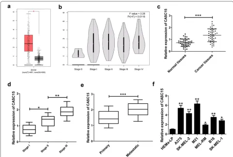
CASC15 expression was up‑regulated in melanoma tissues and associated with melanoma progression
Firstly, we analyzed CASC15 expression in 461 mela-noma tissue samples and 558 normal tissue samples
using the bioinformatics tool GEPIA (http://gepia
.cance r-pku.cn/detai l.php?gene=&click tag=boxpl
ot), and the data showed that CASC15 expression
was increased in melanoma tissue (Fig. 1a). Moreover, CASC15 expression was enhanced with the develop-ment of melanoma stage (Fig. 1b). Then, we evaluated the expression of CASC15 in 42 pairs of melanoma tissue and adjacent non-cancerous tissues. The results
revealed that CASC15 expression was obviously higher in melanoma tissues than that in corresponding
nor-mal tissues (Fig. 1c). Also, CASC15 expression was
dramatically up-regulated in higher stage patients compared with lower stage patients (Fig. 1d). Addition-ally, CASC15 expression was increased in metastatic melanoma tissues in comparison with that in primary melanoma tissues (Fig. 1e). The correlation between CASC15 expression and clinicopathological features
in melanoma patients was displayed in Table 1. The
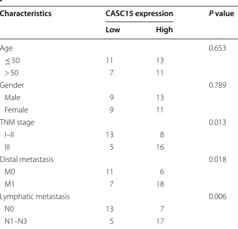
data demonstrated that CASC15 expression was asso-ciated with TNM stage (P = 0.013), distal metastasis (P = 0.018), and lymphatic metastasis (P = 0.006), but was not related to other clinical characteristics, such as age and gender in melanoma. Furthermore, we explored the level of CASC15 in several melanoma cells (A375, SK-MEL-2, M21, MEL-RM, B16 and SK-MEL-1) and normal human epidermal melanocytes (HEMa-LP). The results presented that CASC15 was much more abundant in melanoma cells than that in HEMa-LP cells (Fig. 1f). These results indicated that up-regulated CASC15 might participate in melanoma progression.
Knockdown of CASC15 repressed proliferation, induced apoptosis and decreased invasion in melanoma cells
To investigate the biological effects of CASC15 on mela-noma development, we lowered endogenous CASC15 expression in A375 and M21 cells by two specific siR-NAs targeting CASC15 (si-CASC15 #1 and si-CASC15 #2) (Fig. 2a, b). CCK-8 assays disclosed that silenced
CASC15 significantly suppressed the proliferation abil-ity of A375 and M21 cells (Fig. 2c, d). Cell cycle analy-sis uncovered that knockdown of CASC15 resulted in an evident cell cycle arrest at G0–G1 phase in A375 and M21 cells (Fig. 2e, f). Also, flow cytometry assay discov-ered that the apoptotic rate was remarkably enhanced in A375 and M21 cells following the down-regulation of CASC15 (Fig. 2g, h). Additionally, transwell invasion analysis manifested that depletion of CASC15 greatly alleviated the invasive capability of A375 and M21 cells (Fig. 2i, j). Together, knockdown of PVT1 inhibited pro-liferation, promoted apoptosis and impaired invasion in melanoma cells.
Overexpression of CASC15 induced proliferation and invasion in melanoma cells
Simultaneously, gain-of-function experiment was per-formed in melanoma cells by transfection with pcDNA-CASC15. As we might expect, CASC15 expression was successfully increased in both A375 and M21 cells
fol-lowing introduction with pcDNA-CASC15 (Fig. 3a).
Further function assays disclosed that enforced expres-sion of CASC15 greatly promoted proliferation (Fig. 3b), induced cell cycle progression (Fig. 3c) and enhanced invasion (Fig. 3e) in A375 and M21 cells. Moreover, no significant change was observed in apoptotic rate of mel-anoma cells following CASC15 overexpression (Fig. 3d). Collectively, enforced expression of CASC15 promoted melanoma progression.
CASC15 suppressed PDCD4 expression by interacting with EZH2
It has been reported that approximately 20% of human lncRNAs recruit PRC2 to specific genome sites to silence gene expression [17]. CASC15 was previously revealed to be involved in the tumorigenesis of gastric cancer via interacting with EZH2 and WDR5 in nucleus [15]. PDCD4, a tumor suppressor, was previously proved to be down-regulated by PRC2 complex via increasing the level of H3K27me3 at its promoter in glioma [18]. Thus, we wondered whether CASC15 could regulate PDCD expression through EZH2 in melanoma. To prove this hypothesis, we firstly analyzed the effects of CASC15 knockdown on PDCD4 expression in melanoma cells, and the results stated that PDCD4 expression was evi-dently enhanced in si-CASC15 #2-transfected A375 and M21 cells (Fig. 4a). Then, subcellular fractionation loca-tion assays displayed that CASC15 was distributed both in nucleus and cytoplasm of A375 and M21 cells (Fig. 4b, c), implying that CASC15 may exert regulatory function at transcriptional and post-transcriptional levels. Subse-quently, we probed the interaction possibility of CASC15
Table 1 Correlation between CASC15 expression
and clinicopathological characteristics in melanoma patients
* P < 0.05 was considered statistically significant
Characteristics CASC15 expression P value
Low High
Age 0.653
≤ 50 11 13
> 50 7 11
Gender 0.789
Male 9 13
Female 9 11
TNM stage 0.013
I–II 13 8
III 5 16
Distal metastasis 0.018
M0 11 6
M1 7 18
Lymphatic metastasis 0.006
N0 13 7
and EZH2 using RNA–protein interaction prediction website (http://pridb .gdcb.iasta te.edu/RPISe q/). The data showed that CASC15–EZH2 interaction score was 0.65 using RF classifier, and 0.98 using SVM classifier (Fig. 4d). Moreover, down-regulation of EZH2 led to an obvious increase of PDCD4 expression when compared to nega-tive control group in A375 and M21 cells (Fig. 4e), sug-gesting that EZH2 could modulate PDCD4 expression. Also, luciferase reporter assays revealed that depletion
of CASC15 increased the activity of PDCD4 promoter in both A375 and M21 cells, indicating that CASC15 could regulate PDCD4 expression at the transcriptional level (Fig. 4f).
Next, to verify whether CASC15 could directly bind to EZH2, RIP and RNA–protein pull down assays were conducted in A375 and M21 cells. RIP assays showed that CASC15 level was greatly enriched by EZH2 anti-body (Fig. 4g, h). RNA–protein pull down experiments Fig. 3 CASC15 overexpression facilitates proliferation and invasion in melanoma cells. A375 and M21 cells were transfected with pcDNA empty vector or pcDNA-CASC15, followed by the evaluation of CASC15 expression (a), cell proliferation (b), cell cycle distribution (c), apoptosis (d) and invasion (e)
Fig. 4 CASC15 epigenetically silences PDCD4 expression by binding to EZH2. a Western blot assay was carried out to evaluate the protein level of PDCD4 in A375 and M21 cells transfected with si-con or si-CASC15 #2. b, c qRT-PCR assay was used to measure CASC15 expression in cytoplasm and nucleus of A375 and M21 cells. GAPDH was used as a cytoplasm marker and U6 was used as a nucleus marker. d The probability of interaction of CASC15 and EZH2 was predicted by RNA–protein interaction prediction website (http://pridb .gdcb.iasta te.edu/RPISe q/). Predictions with probabilities > 0.5 were considered positive. RPISeq predictions are based on Random Forest (RF) or Support Vector Machine (SVM). e Western blot assay of PDCD4 protein expression in A375 and M21 cells after transfection with si-con or si-EZH2. f Luciferase reporter assay was performed in both A375 and M21 cells after co-transfection with PDCD4 promoter reporter and si-con or si-CASC15. g, h RIP experiments were conducted in A375 and M21 cells, followed by qRT-PCR analysis of CASC15 in the coprecipitated RNA. The fold enrichment of CASC15 in EZH2 RIP is relative to its matched IgG control. i, j In vitro transcribed biotin-labeled CASC15 and anti-CASC15 were incubated with A375 and M21 cell lysates, and the extracted protein was analyzed by western blot using an antibody against EZH2. A nonspecific protein (β-actin) is shown as the control. k, l ChIP of EZH2 occupancy and H3K27me3 binding in the PDCD4 promoter in A375 and M21 cells after transfection with si-con or si-CASC15 #2. Enrichment was quantified relative to input control. Antibody against IgG was used as a negative control. m PDCD4 mRNA expression in melanoma tissues (n = 461) and normal tissues (n = 558) from GEPIA database (http://gepia .cance r-pku.cn/detai l.php?gene=&click tag=boxpl ot). n Relative expression levels of PDCD4 mRNA in melanoma tissues (n = 42) and adjacent normal tissues (n = 42). o Spearman correlation analysis of CASC9 and PDCD4 expression in 42 melanoma tissues. Results are represented as mean ± SD of three independent experiments. **P < 0.01
also demonstrated that labeled CASC15 could pull down EZH2 rather than antisense CASC15 from the nuclear extract fraction of A375 and M21 cells (Fig. 4i, j). Together, these results indicated that CASC15 could interact with EZH2 in melanoma cells. To further address whether CASC15 engage in transcriptional repression through the enrichment of EZH2 to PDCD4 promoter, we performed ChIP analysis in A375 and M21 cells. The
results declared that EZH2 could bind to PDCD4 pro-moter region and induce H3K27 trimethylation, while CASC15 knockdown decreased EZH2 binding and H3K27ME3 occupancy of PDCD4 promoter (Fig. 4k, l).
samples (n = 461) compared with normal tissue samples (n = 558). As expected, we found that PDCD4 expres-sion was lower in 42 cases of melanoma tissue samples than that in normal counterparts (Fig. 4n). Moreover, a negative correlation was observed between CASC15 and PDCD4 expression in melanoma patients (Fig. 4o). All these data supported the conclusion that CASC15 epigenetically inhibited PDCD4 expression by recruit-ing EZH2 to its promoter region and increasrecruit-ing the H2K27me3 level of its promoter in melanoma.
PDCD4 overexpression hindered proliferation, enhanced apoptosis and suppressed invasion in melanoma cells
To investigate the functions of PDCD4 in melanoma pro-gression, we constructed PDCD4-overexpression plas-mid (pcDNA-FOXF1) and transfected it into A375 and M21 cells. qRT-PCR analysis confirmed that PDCD4 expression was prominently enhanced at mRNA and protein level in A375 and M21 cells after transfection
with pcDNA-PDCD4 (Fig. 5a, b). CCK-8 experiments
illustrated that enforced expression of PDCD4 resulted in an apparent decrease of A375 and M21 cell prolifera-tion (Fig. 5c). Flow cytometry analysis showed a G0–G1 cell cycle arrest and an increased apoptotic rate in A375 and M21 cells following PDCD4 up-regulation (Fig. 5d, e). Transwell invasion assay found that the invasive abil-ity of A375 and M21 cells was substantially abated after introduction with pcDNA-PDCD4 (Fig. 4f). In summary, overexpression of CASC15 blocked proliferation, pro-moted apoptosis and attenuated invasion in melanoma cells.
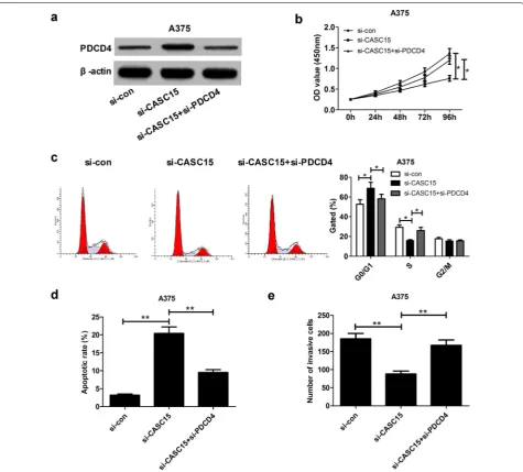
The tumor‑suppressive effect mediated by CASC15 knockdown was weakened following depletion of PDCD4
To further explore whether the effect of CASC15 on melanoma progression was mediated by PDCD4, we performed the restoration experiment in A375 cells by co-transfection with si-CASC15 and si-PDCD4. As pre-sented in Fig. 6a, si-CASC15-induced increase of PDCD4 expression was attenuated by down-regulating PDCD4. Moreover, the anti-proliferation (Fig. 6b, c), pro-apop-tosis (Fig. 6d) and anti-invasion (Fig. 6e) effects elicited by CASC15 knockdown were greatly reversed following PDCD4 depletion. In all, CASC15 promoted melanoma progression through regulating PDCD4.
Depletion of CASC15 suppressed melanoma tumorigenesis in vivo
To further determine the roles of CASC15 in melanoma tumorigenesis in vivo, sh-CASC15- or sh-con-trans-fected A375 cells were subcutaneously inoculated into
the nude mice. The data displayed that CASC15 knock-down significantly decreased the tumor volumes (Fig. 7a) and lowered the tumor weights (Fig. 7b, c). Moreover, the expression of Ki-67 was strikingly reduced in tumors developed from sh-CASC15-transfected A375 cells com-pared with sh-con group (Fig. 7d). Additionally, CASC15 expression was declined (Fig. 7e) and PDCD4 protein level was increased (Fig. 7f) in tumor masses of sh-CASC15 group when compared to that of control group. All these data demonstrated that knockdown of CASC15 inhibited melanoma cell proliferation in vivo possibly by regulating PDCD4.
Discussion
A growing body of evidence highlights that lncRNAs regulate cell proliferation, differentiation, migration and invasion in the occurrence and development of diverse malignant tumors, including melanoma [19, 20]. CASC15 was previously discovered to affect cell pheno-type switching between proliferative and invasive states in melanoma [16]. Nevertheless, further investigations on the biological significance and underlying mechanism of CASC15 in melanoma are required.
Firstly, we found that CASC15 expression was increased in melanoma tissues and cells. Moreover, higher CASC15 level was associated with advanced TNM stage and metastasis. Subsequent loss- and gain-of-function experiments showed that CASC15 knock-down inhibited proliferation, facilitated apoptosis and decreased invasion of melanoma cells, while CASC15 overexpression displayed the opposite effects. Consist-ent with our findings, Yao et al. delineated that CASC15 was highly expressed in gastric cancer tissues and cells, and down-regulation of CASC15 impaired the prolif-eration capability of gastric cancer cells [21]. He et al. reported that CASC15 was overexpressed in hepatocellu-lar carcinoma tissues and cells, and depletion of CASC15 repressed proliferation, promoted apoptosis, and lowered invasion of hepatocellular carcinoma cells [14]. Con-versely, CASC15 was validated as a tumor suppressor in neuroblastoma [13] and acute leukemia [22].
blot analysis showed that knockdown of CASC15 or EZH2 both induced the expression of PDCD4 in mela-noma cells. Moreover, CASC15 was verified to be dis-tributed in nucleus and cytoplasm, implying its potential transcriptional regulation function. Luciferase reporter analysis verified that CASC15 could regulate PDCD4 expression at the transcriptional level. Subsequent RIP and RNA–protein pull down assays further certified
the binding between CASC15 and EZH2. Further ChIP assays disclosed that EZH2 could directly bind to the promoter of PDCD4 in melanoma cells, and depletion of CASC15 lowered the binding ability of EZH2 to the promoter region of PDCD4 and reduced H3K27 tri-methylation. Taken together, CASC15 inhibited PDCD4 expression partially by recruiting EZH2 to its promoter region in melanoma. Concordant with our results, Fig. 5 PDCD4 overexpression inhibits proliferation, induced apoptosis and attenuate invasion in melanoma cells. A375 and M21 cells were
CASC15 was found to affect the tumorigenesis of gastric cancer by regulating CDKN1A via interaction with EZH2 and WDR5 in nucleus, meanwhile, CASC15 partici-pated in gastric cancer progression by modulating ZEB1 expression through sponging miR-33a-5p in cytoplasm
[15]. However, in melanoma, the regulatory
mecha-nisms of CASC15 in cytoplasm are required to be further discussed.
PDCD4 has been identified as a tumor suppressor gene and potential anticancer target for several years [27, 28]. Accumulating evidence has elucidated that PDCD4 could exert antitumor activity in a variety of
cancer types, such as bladder cancer [29], colon carci-noma [30] and oropharyngeal cancer [31], via different regulatory pathways. In this study, PDCD4 expression was found to be down-regulated and negatively cor-related with CASC15 expression in melanoma tumor tissues. Gain-of-function assay confirmed that over-expression of PDCD4 blocked proliferation, induced apoptosis and repressed invasion of melanoma cells. In agreement with our data, PDCD4 was demonstrated to be decreased in melanoma tumor tissues, and inhib-ited melanoma cell proliferation, migration and inva-sion [32, 33]. Furthermore, restoration experiment Fig. 6 Silencing PDCD4 is partly involved in the oncogenic function of CASC15 in melanoma cells. A375 cells were transfected with si-con,
uncovered that PDCD4 silencing greatly reversed the anti-cancer function induced by CASC15 knockdown. Finally, transplantation tumor experiments revealed that knockdown of CASC15 inhibited tumor growth in vivo possibly via up-regulating PDCD4 expression.
Conclusions
In conclusion, our study demonstrated that CASC15 contributed to melanoma progression partially by EZH2-medicated silence of PDCD4 expression, illu-minating the promising prospect of CASC15 as a potential target and therapeutic application for mela-noma patients. However, the other possible regulatory mechanisms of CASCA15 in melanoma cell functions remain require further investigation.
Abbreviations
lncRNAs: long non-coding RNAs; EZH2: enhancer of zeste homolog 2; PRC2: polycomb repressive complex 2; H3K27me3: histone H3 lysine 27 trimethylation; LATS2: large tumor suppressor kinase 2; CASC15: cancer susceptibility candidate 15; HEMa-LP: human epidermal melanocytes; siRNAs: small interference RNAs; RIP: RNA immunoprecipitation; ChIP: chromatin immunoprecipitation.
Authors’ contributions
YKY and GWY designed the study. YKY and BZ conducted the experiments, analyzed the data, and prepared the manuscript. DQL and GWY edited and the manuscript. All authors read and approved the final manuscript.
Author details
1 Department of Dermatology, The First Affiliated Hospital of Zhengzhou University, No. 1 Jian She East Road, Zhengzhou 450052, China. 2 Department of Dermatology, The Third People’s Hospital of Henan Province, No 198 Fu Niu Road, Zhengzhou 450006, China.
Acknowledgements
We want to thank all participants involved in this study.
Competing interests
The authors declare that they have no competing interests.
Availability of data and materials
The datasets used and/or analyzed during the current study are available from the corresponding author on reasonable request.
Consent for publication
Not applicable.
Ethics approval and consent to participate
This study was approved by the Ethic Committee of the First Affiliated Hospital of Zhengzhou University following the Declaration of Helsinki Principles. Written informed consents were obtained from each participant involved in this study.
Funding
None.
•fast, convenient online submission •
thorough peer review by experienced researchers in your field • rapid publication on acceptance
• support for research data, including large and complex data types •
gold Open Access which fosters wider collaboration and increased citations maximum visibility for your research: over 100M website views per year •
At BMC, research is always in progress.
Learn more biomedcentral.com/submissions
Ready to submit your research? Choose BMC and benefit from: Publisher’s Note
Springer Nature remains neutral with regard to jurisdictional claims in pub-lished maps and institutional affiliations.
Received: 4 May 2018 Accepted: 7 July 2018
References
1. Gray-Schopfer V, Wellbrock C, Marais R. Melanoma biology and new targeted therapy. Nature. 2007;445:851–7.
2. Siegel RL, Miller KD, Jemal A. Cancer statistics, 2017. CA Cancer J Clin. 2017;67:7–30.
3. Rozeman EA, Dekker TJA, Haanen JBAG, Blank CU. Advanced melanoma: current treatment options, biomarkers, and future perspectives. Am J Clin Dermatol. 2018;19:303–17.
4. Siegel RL, Miller KD, Jemal A. Cancer statistics, 2016. CA Cancer J Clin. 2015;66:7–30.
5. Sun W, Yang Y, Xu C, Guo J. Regulatory mechanisms of long non-coding RNAs on gene expression in cancers. Cancer Genet. 2017;216–217:105–10.
6. Camacho CV, Choudhari R, Gadad SS. Long noncoding RNAs and cancer, an overview. Steroids. 2018;133:93–5.
7. Cao R, Wang L, Wang H, Xia L, Erdjumentbromage H, Tempst P, et al. Role of histone H3 lysine 27 methylation in polycomb-group silencing. Sci-ence. 2002;298:1039–43.
8. Benetatos L, Voulgaris E, Vartholomatos G, Hatzimichael E. Non-coding RNAs and EZH2 interactions in cancer: long and short tales from the transcriptome. Int J Cancer. 2013;133:267–74.
9. Wan L, Sun M, Liu GJ, Wei CC, Zhang EB, Kong R, et al. Long non-coding RNA PVT1 promotes non-small cell lung cancer cell proliferation through epigenetically regulating LATS2 expression. Mol Cancer Ther. 2016;15:1082–94.
10. Ding J, Li J, Wang H, Tian Y, Xie M, He X, et al. Long noncoding RNA CRNDE promotes colorectal cancer cell proliferation via epigenetically silencing DUSP5/CDKN1A expression. Cell Death Dis. 2017;8:e2997. 11. Huang J, Deng G, Liu T, Chen W, Zhou Y. Long noncoding RNA PCAT-1
acts as an oncogene in osteosarcoma by reducing p21 levels. Biochem Biophys Res Commun. 2017;495:2622–9.
12. Glusman G, Qin S, El-Gewely MR, Siegel AF, Roach JC, Hood L, et al. A third approach to gene prediction suggests thousands of additional human transcribed regions. PLoS Comput Biol. 2006;2:e18.
13. Russell MR, Penikis A, Oldridge DA, Alvarezdominguez JR, Mcdaniel L, Diamond M, et al. CASC15-S is a tumor suppressor lncRNA at the 6p22 neuroblastoma susceptibility locus. Cancer Res. 2015;75:3155–66. 14. He T, Zhang L, Kong Y, Huang Y, Zhang Y, Zhou D, et al. Long non-coding
RNA CASC15 is upregulated in hepatocellular carcinoma and facilitates hepatocarcinogenesis. Int J Oncol. 2017;51:1722–30.
15. Wu Q, Xiang S, Ma J, Hui P, Wang T, Meng W, et al. Long non-coding RNA CASC15 regulates gastric cancer cell proliferation, migration and epithe-lial mesenchymal transition by targeting CDKN1A and ZEB1. Mol Oncol. 2018;12:799–813.
16. Lessard L, Liu M, Marzese DM, Wang H, Chong K, Kawas N, et al. The CASC15 long intergenic noncoding RNA locus is involved in melanoma progression and phenotype switching. J Invest Dermatol. 2015;135:2464–74.
17. Khalil AM, Guttman M, Huarte M, Garber M, Raj A, Morales DR, et al. Many human large intergenic noncoding RNAs associate with
chromatin-modifying complexes and affect gene expression. Proc Natl Acad Sci USA. 2009;106:11667–72.
18. Chen Y, Bian Y, Zhao S, Kong F, Li X. Suppression of PDCD4 mediated by the long non-coding RNA HOTAIR inhibits the proliferation and invasion of glioma cells. Oncol Lett. 2016;12:5170–6.
19. Tang Y, Cheung BB, Atmadibrata B, Marshall GM, Dinger ME, Liu PY, et al. The regulatory role of long noncoding RNAs in cancer. Cancer Lett. 2017;391:12–9.
20. Yu X, Zheng H, Tse G, Chan MT, Wu WK. Long non-coding RNAs in mela-noma. Cell Prolif. 2018. https ://doi.org/10.1111/cpr.12457 .
21. Yao XM, Tang JH, Zhu H, Jing Y. High expression of LncRNA CASC15 is a risk factor for gastric cancer prognosis and promote the proliferation of gastric cancer. Eur Rev Med Pharmacol Sci. 2017;21:5661–7.
22. Fernando TR, Contreras JR, Zampini M, Rodriguez-Malave NI, Alberti MO, Anguiano J, et al. The lncRNA CASC15 regulates SOX4 expression in RUNX1-rearranged acute leukemia. Mol Cancer. 2017;16:126.
23. Sarkar D, Leung EY, Baguley BC, Finlay GJ, Askarianamiri ME. Epige-netic regulation in human melanoma: past and future. EpigeEpige-netics. 2015;10:103–21.
24. Yan KS, Lin CY, Liao TW, Peng CM, Lee SC, Liu YJ, et al. EZH2 in cancer progression and potential application in cancer therapy: a friend or foe? Int J Mol Sci. 2017;18:E1172.
25. Ni N, Song H, Wang X, Xu X, Jiang Y, Sun J. Up-regulation of long noncod-ing RNA FALEC predicts poor prognosis and promotes melanoma cell proliferation through epigenetically silencing p21. Biomed Pharmacother. 2017;96:1371–9.
26. Wu Y, Hu L, Liang Y, Li J, Wang K, Chen X, et al. Up-regulation of lncRNA CASC9 promotes esophageal squamous cell carcinoma growth by negatively regulating PDCD4 expression through EZH2. Mol Cancer. 2017;16:150.
27. Lankat-Buttgereit B, Göke R. The tumour suppressor Pdcd4: recent advances in the elucidation of function and regulation. Biol Cell. 2009;101:309–17.
28. Li JZH, Wei G, Ho WK, Wen BL, Wei WI, Chan YW, et al. The clinical associa-tion of programmed cell death protein 4 (PDCD4) with solid tumors and its prognostic significance: a meta-analysis. Chin J Cancer. 2016;35:95. 29. Liu J, Zhai R, Zhao J, Kong F, Wang J, Jiang W, et al. Programmed cell
death 4 overexpression enhances sensitivity to cisplatin via the JNK/c-Jun signaling pathway in bladder cancer. Int J Oncol. 2018. https ://doi. org/10.3892/ijo.2018.4303.
30. Wang Q, Zhu J, Wang YW, Dai Y, Wang YL, Wang C, et al. Tumor suppressor Pdcd4 attenuates Sin1 translation to inhibit invasion in colon carcinoma. Oncogene. 2017;36:6225–34.
31. Zhang X, Gee H, Rose B, Lee CS, Clark J, Elliott M, et al. Regulation of the tumour suppressor PDCD4 by miR-499 and miR-21 in oropharyngeal cancers. BMC Cancer. 2016;16:86.
32. Wan J, Yang J, Huang Y, Deng L. MicroRNA-150 inhibitors enhance cell apoptosis of melanoma by targeting PDCD4. Oncol Lett. 2018;15:1475–82.