the scapula as a reference landmark: a
retrospective analysis of 50 radiographs
Michael T. Haneline,
DC, MPH*Robert Cooperstein,
MA, DCMorgan D. Young,
BSJustin Ross,
RN, DCObjective: The purpose of this study was to determine which spinal segment most closely corresponds to the level of the inferior angle of the scapula (IAS) using measurements taken on A-P full-spine radiographs.
Methods: Fifty sequentially selected radiographs were analyzed independently by two examiners. A straight edge was used to ascertain which spinal levels
corresponded with the right and left IASs. For analysis, each spinal level was subdivided into three regions: upper vertebral body, lower vertebral body, and intervertebral space.
Results: The mean spinal level corresponding to the left IAS was midway between the T8–9 interspace and the upper T9 body (range, lower T7 to upper T10). The mean spinal level corresponding to the right IAS was slightly lower, but still within the upper T9 body (range, lower T7 to lower T10). These levels correspond to the T8 spinous process.
Conclusions: There is a considerable amount of variability in where the IASs are located, but most commonly, they correspond to the level of the upper body of T9.
(JCCA 2008; 52(1):24–29)
k e y wo r d s : scapula, inferior angle, spinal segment,
radiographs.
Objectif : L’objectif de la présente étude consistait à déterminer quel segment médullaire correspond le plus au niveau de l’angle inférieur de l’omoplate (IAS) en se servant des mesures relevées des radiogrammes A-P. du rachis intégral.
Méthodes : Cinquante radiogrammes sélectionnés d’une manière séquentielle ont été analysés
indépendamment par deux examinateurs. Une règle à tracer a été utilisée pour établir quels niveaux rachidiens correspondaient aux IAS de la gauche et ceux de la droite. Aux fins d’analyse, chaque niveau rachidien a été subdivisé en trois régions : corps lombaire supérieur, corps lombaire inférieur et espace intervertébrale.
Résultats : Le niveau rachidien moyen correspondant à l’IAS de la gauche était mi-chemin entre l’inter-espace T8–9 et le corps T9 supérieur (plage, du T7 inférieur au T10 supérieur). Le niveau rachidien moyen
correspondant à l’IAS de la droite était légèrement inférieur, cependant, toujours à l’intérieur du corps T9 supérieur (plage, du T7 inférieur au T10 inférieur). Ces niveaux correspondent à l’apophyse épineuse T8.
Conclusion : Il existe une variabilité significative là où se situent les IAS, mais plus couramment, ils
correspondent au niveau du corps supérieur de T9. (JACC 2008; 52(1):24–29)
m o t s c l é s : omoplate, angle inférieur, segment
médullaire, radiogrammes.
* Corresponding Author: Michael T. Haneline, DC, MPH, Professor, Palmer College of Chiropractic West, 90 East Tasman Drive, San Jose, CA 95134, Phone: 408-944-6190, Fax: 408-944-6017, Email: [email protected]
Introduction
Musculoskeletal practitioners commonly use contiguous bony landmarks to locate spinal levels.1 One such land-mark is the inferior angle of the scapula (IAS), which is said to correlate with the T7, T8, or T9 spinous process or spinal level, depending on the source.2–4 Gray’s Anatomy indicates that the IAS corresponds to the level of the ninth rib, which attaches to the spine at the T8–9 inter-space.5 In another study on the location of the IAS, we provide a comprehensive list of citations, including many that suggest it lies at the level of the T7 spinous process.6 Some of this discrepancy is due to fact that some authors refer to the level of the spinous process, while others re-fer to the body of the vertebra. In spite of this inconsist-ency, no studies have attempted to determine as their primary goal which vertebral segment truly corresponds to the IAS.
Lewis et al.4 reported that manual palpation of surface landmarks is a valid method for determining the actual location of the scapula. Based on these findings, the au-thors opined that the location of thoracic landmarks could be accurately determined using surface palpation of scapular position as a reference point. On the other hand, several related studies have investigated the reliability of manual and physical therapists in locating spinal levels by palpation of spinous processes.7–10 Most found their procedures of low reliability. Thus, Lewis’ optimism not-withstanding, we are not confident that using the scapula position as a landmark would help in identifying segmen-tal spinal levels by number.
Locating the precise spinal level by palpation is espe-cially important to practitioners who intend to apply a specific therapy to a specific segment. However, anes-thetists who attempted to identify a given lumbar inter-space by palpation of spinous processes and iliac crests identified a level cephalad to their target in 33% to 68% of cases.11 Another study reported that the actual level, as determined by markers placed on patients’ backs prior to MRI, was from one space below to four spaces above the level identified by the anesthetist.12 Similar findings have been reported when physical therapists8 and spine surgeons13 were involved. Chiropractors, as well, are of-ten interested in locating spinal levels by palpation and would benefit if a valid and reliable analytic method were available.
The purpose of the current study was to determine
which spinal segment corresponds to the level of the IAS by means of measurements taken on retrospectively ana-lyzed A-P full-spine radiographs that were taken with pa-tients in an upright neutral posture.
Methods
Fifty-three radiographs were sequentially selected from the educational radiographic archives of a chiropractic college, consisting of several hundred films. Once a suffi-cient number of female radiographs were located, we se-lected only males, passing over approximately 10 radiographs of females, in order to include enough males so that the gender distribution would be approximately equal. All radiographs were A-P full-spine views that were taken with the patient standing erect, feet together, arms at the side, and the back touching a radiographic bucky. No identifying information was collected from the radiographs; only the above-mentioned measurements and subjects’ gender, age, and height.
Radiographs were retrospectively analyzed by two chi-ropractic students, each of whom had completed their training in radiographic interpretation. The radiographs were evaluated independently by each examiner in a blinded fashion. Any discrepancies between examiners were resolved by one or both of the study chiropractors.
SPSS, Inc, Chicago, Ill.). Data were analyzed using de-scriptive statistics including frequencies, percentages, mean/median, and standard deviation. Also, correlations were examined between age, height, gender, and the pres-ence of scoliosis in relation to scapular position and ver-tebral level. Spinal levels were coded for easier statistical analysis with 1 representing the upper T6 body, 2 the lower T6 body, 3 the T6–7 interspace, and so on; continu-ing in sequence to 14 for the lower T10 body.
This project was approved by the Institutional Review Board of the Palmer College of Chiropractic, Davenport, Iowa. Informed consent was not required from the in-cluded subjects because of the study’s retrospective na-ture, as well as the shared use of the radiographic library within the institution. Subjects’ names were protected and only de-identified information was gathered from the x-rays (e.g., age, gender, height, and weight).
Results
A total of 53 radiographs were selected, although 3 were ultimately excluded because of poor imaging technique precluding analysis. There were 25 males and 24 fe-males, with gender information missing for 1 radiograph. The mean (SD) age was 47.5 (15.4) years and the mean
height was 168.5 (12.0) cm. There were no disagree-ments between examiners as to the spinal level reported in association with the IAS.
The mean spinal level corresponding to the left IAS was midway between the T8/9 interspace and the upper body of T9. This corresponded to the T8 spinous process. The range was from lower T7 to upper T10. The median value was upper T9, which was also the mode, the most common position of the IAS, accounting for 33% of the observations.
The mean spinal level corresponding to the right IAS was approximately at the level of the upper body of T9, and T8 spinous process. The range was from lower T7 to lower T10. The median and mode on the right both corre-sponded to lower T9, which comprised 32% of the obser-vations for that side. Figure 2 shows the distribution of instances of vertebral levels corresponding to the left and right IASs.
Slight differences were apparent between males and females that are presented in Table 1. Most notably, the IAS was positioned slightly lower bilaterally on males by approximately one-quarter of a vertebral body.
Only 1 radiograph depicted thoracic scoliosis greater than 10 degrees, determined to be 15° by the Cobb meth-od.14 The scapulae were positioned at the level of upper T9 on the left and lower T8 on the right in this case. The left scapulae were not observable on 4 of the radiographs because they were obscured by the superimposed heart shadow; as a result, the related data did not figure in the analysis.
Discussion
This study demonstrates that there is a considerable amount of variability in where the IASs are located. They are most commonly found at the level of the upper body of T9 on the left and the lower body of T9 on the right; corresponding most closely to the T8 spinous process. This finding, which was determined using a retrospective study design, is consistent with our earlier prospective study that utilized an entirely different methodology.6
The range for IAS position is rather wide, spanning T7 to T10, suggesting that patient variations in scapular posi-tion would refute any simplistic associaposi-tion of the scapular tip with a specific vertebral level. The right scapula was in a lower position more frequently, possibly due to handed-ness.15 According to Kendall et al,15 patients typically Figure 1 Two thoracic vertebrae depicting the
maintain a posture wherein the lower shoulder is on the side of the dominant hand.
The mid-thoracic spinous processes are nearly vertical, directed obliquely downward5 and extending to the inter-space of the segment below. A cadaveric study of 12
tho-racic spines from T5 through T8 reported that the length of spinous processes was variable, ranging from 2.6 to 4.5 cm, with a mean value of 3.5 cm.16 Thus, the spinous process completely spans an entire vertebral body; e.g., lower body of T8 corresponds to the T7 spinous process. Figure 2 Frequencies of spinal levels associated with the left and right inferior angle of the scapula (IAS).
Table 1 Spinal levels corresponding to the inferior angle of the scapula for the intact group, males, and females.
n Mean Range
Intact group
Left 44 Between T8/9 and Upper T9 Lower T7-Upper T10
Right 48 Lower T9 Lower T7-Lower T10
Males
Left 22 Upper T9 Lower T7-Upper T10
Right 24 Lower T9 Lower T7-Upper T10
Females
Left 22 T8/9 Lower T7-T9/10
(see Figure 1) Therefore, when the IAS is found parallel to the body of a given vertebra it is also parallel to the tip of the spinous process of the vertebra above.
The interexaminer reliability of identifying spinous processes has been reported to be poor, with errors rang-ing up to 177.1 mm in the thoracic spine. Therefore, in addition to the problem posed by the IAS lying in a somewhat wide range, it is likely that examiner error fur-ther compounds the difficulty a musculoskeletal practi-tioner would have in accurately locating a particular spinal segment using the IAS as a landmark
Our findings should be of interest to clinicians who use the scapula as a landmark for locating other anatomical structures, such as vertebral levels. Such clinicians include chiropractors, osteopaths, physical therapists, orthoped-ists, neurologorthoped-ists, anesthetorthoped-ists, surgeons, nurses, discogra-phers, and others.
Although this study did not investigate the validity of attempting to identify subluxations or other aspects of spinal dysfunction radiographically, it is common prac-tice among chiropractors to take radiographs to identify potential segments or areas deemed worthy of adjustive care. The present study suggests it may be very challeng-ing to situate areas of radiographic interest on the actual patient, at least in the thoracic spine, if the scapula is to be used as a landmark for identifying spinal levels.
The following factors may have an influence on the ac-curacy of locating a spinal level seen on a radiograph on an actual patient:
• The scapular tip varies in location from patient to pa-tient, exhibiting a rather wide range.
• The scapular tip tends to be lower on the side of the dominant upper extremity.
• The scapular tip may line up with different levels in the standing patient as compared with the prone patient, so that spinal levels identified in relation to the scapula in an upright radiograph may change relative to the scap-ula if the patient is placed prone or supine for the pur-poses of an adjustive procedure.
• There may be examiner error.
Since the rule of thumb most used in chiropractic and elsewhere has been that the inferior scapular tip lies at the spinous process of T7, whereas it is actually closer to T8, then any spinal landmark located starting from this
ap-parently erroneous landmark is likely to be phase-shifted on average by about one vertebral level, winding up one level below what was intended. Moreover, patient ana-tomical variation is such that even “correcting” the rule of thumb would at best direct the chiropractor to a 3 level spinal region that probably contains the level seen on the radiograph, not the point location that is generally de-sired.
To the extent chiropractors have been using the stand-ing T7 spinous process = scapular tip benchmark, and the prone T6 spinous process = scapular tip benchmark rules for finding thoracic locations, they have not been intro-ducing forces where they thought they were. Consistently being one vertebra below the target suggested by radiog-raphy results in systematic bias, while not taking into ac-count that the scapular tip lies within an anatomical range rather than a single place introduces random error. Accordingly, clinicians who believe segmental specifici-ty remains clinically important and thus take radiographs to identify adjustable segments will have to explain their claimed good results in some other way, since they have been treating the putatively wrong level much of the time.
The sources of error we highlight are not entirely insu-perable. We believe a clinician could improve his or her accuracy by simply revising the traditional spinal land-mark rule of thumb, and look for the scapular tip to be on average one level lower than previously thought. Even more importantly, provided the scapula tip may be visual-ized on the radiograph (collimation may prevent this), the clinician need not presume what spinal level corresponds to the scapular tip. That information may be directly vis-ualized. Even at that, the chiropractor would still have to determine how that landmark changes going from the standing to the prone position or seated position.
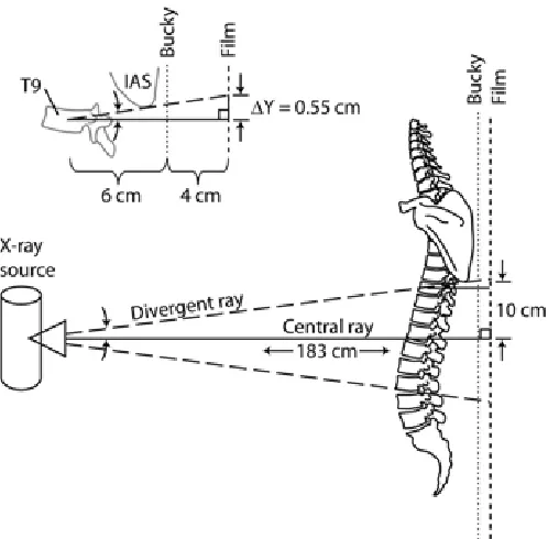
Since the radiographic beam in a full spine radiograph penetrates the IAS in an inferior to superior direction, we calculated the extent to which radiographic distortion may have influenced our findings, which is illustrated in Figure 3. Using these figures, we calculated that the IAS lies approximately one-half cm lower than reported here-in, which corresponds to approximately one-quarter of a typical T9 vertebral body.17
Funding sources and conflicts of interest
Figure 3 Because of radiographic image distortion, the actual vertebral level corresponding to the IAS is approximately one-half cm lower than reported herein. The illustration is not to scale.
References
1 Najm WI, Seffinger MA, Mishra SI, et al. Content validity of manual spinal palpatory exams – A systematic review. BMC Complement Altern Med 2003; 3:1.
2 Ellis H. Clinical anatomy: a revision and applied anatomy for clinical students. 10th ed. Oxford, UK; Malden, MA: Blackwell Science, 2002.
3 Oatis CA. Kinesiology: the mechanics and pathomechanics of human movement. 1st ed. Philadelphia: Lippincott Williams & Wilkins, 2004.
4 Lewis J, Green A, Reichard Z, Wright C. Scapular position: the validity of skin surface palpation. Man Ther 2002; 7:26–30.
5 Gray H. Anatomy of the Human Body. Philadelphia: Lea & Febiger, 1918; Bartleby.com, 2000.
6 Cooperstein R, Haneline M. Identification of thoracic spinal levels by palpation versus an x-ray gold standard. In press. Journal of Chiropractic Medicine.
7 Binkley J, Stratford PW, Gill C. Interrater reliability of lumbar accessory motion mobility testing. Phys Ther 1995; 75:786–792; discussion 793–795.
8 Downey BJ, Taylor NF, Niere KR. Manipulative physiotherapists can reliably palpate nominated lumbar spinal levels. Man Ther 1999; 4:151–156.
9 McKenzie AM, Taylor NF. Can physiotherapists locate lumbar spinal levels. Physiotherapy 1997; 83:235–239. 10 Billis EV, Foster NE, Wright CC. Reproducibility and
repeatability: errors of three groups of physiotherapists in locating spinal levels by palpation. Man Ther 2003; 8:223–232.
11 Lirk P, Messner H, Deibl M, et al. Accuracy in estimating the correct intervertebral space level during lumbar, thoracic and cervical epidural anaesthesia. Acta Anaesthesiologica Scandinavica 2004; 48:347–349. 12 Broadbent CR, Maxwell WB, Ferrie R, Wilson DJ,
Gawne-Cain M, Russell R. Ability of anaesthetists to identify a marked lumbar interspace. Anaesthesia 2000; 55:1122–1126.
13 Ebraheim NA, Inzerillo C, Xu R. Are anatomic landmarks reliable in determination of fusion level in posterolateral lumbar fusion? Spine 1999; 24:973–974.
14 Goldberg MS, Poitras B, Mayo NE, Labelle H, Bourassa R, Cloutier R. Observer variation in assessing spinal curvature and skeletal development in adolescent idiopathic scoliosis. Spine 1988; 13:1371–1377.
15 Sobush DC, Simoneau GG, Dietz KE, Levene JA, Grossman RE, Smith WB. The lennie test for measuring scapular position in healthy young adult females: a reliability and validity study. J Orthop Sports Phys Ther 1996; 23:39–50.
16 Sran MM, Khan KM, Zhu Q, McKay HA, Oxland TR. Failure characteristics of the thoracic spine with a posteroanterior load: investigating the safety of spinal mobilization. Spine 2004; 29:2382–2388.