Resumen: El objetivo de este estudio fue establecer una guía para odontólogos que permita identificar el momento ideal para extraer los primeros molares permanentes con mal pronóstico, con el objectivo de obtener un cierre espontáneo del espacio, teniendo en cuenta que el primer molar permanente es más susceptible a desarrollar caries en niños debido a su erupción temprana en la cavidad oral y es el más afectado por la hipomineralización. Material y Métodos: Se procedió a buscar y analizar artículos científicos de las bases de datos PUBMED y SciELO, publicados entre 1998 y 2018 en base a criterios de inclusión y exclusión. Conclusión: Nuestra revisión sugiere que se debe realizar una evaluación completa del primer molar permanente comprometido antes de planificar una extracción. Se puede concluir que la etapa ideal para extraer un primer molar permanente con mal pronóstico son los estadios E o D de Demirjian, evaluados con radiografías, que se genera a una edad cronológica de 8 a 10,5 años, idealmente en presencia de terceros molares.
Palabras Clave: Extracción dental; diente molar; odontólogos; radiografía dental; pronóstico.
Abstract: The aim of this study was to establish a guide for general dentists that allow identifying the ideal moment to extract the first permanent molars with poor prognosis, in the hope of obtaining a spontaneous closure of space, regarding that the first permanent molar is more susceptible to develop dental caries in children due to its early eruption in the oral cavity and molar incisor hypomineralization affects them the most. Methods: we proceed to search for the articles in databases PUBMED and SciELO, selected articles published from 1998 a 2018 were analyzed based on inclusion and exclusion criteria. Conclusion: Our review suggests that a full evaluation of the compromised first permanent molar should be performed before planning an extraction. It can be concluded that the ideal stage to extract a first permanent molar with bad prognosis is in the Demirjian stage E or D evaluated with radiographs, which is generated at a chronological age of 8 to 10.5 years, ideally in the presence of third molars.
Keywords: Tooth extraction; molar; dentists; radiography, dental; prognosis. Javiera Scheu.1
Cecilia Cerda.2
Víctor Rojas.3
Extracción oportuna de los primeros molares permanentes
severamente afectados en dentición mixta.
Timely extraction of the first permanent molars
severely affected in mixed dentition.
Affiliations:1Universidad Mayor, Santiago,
Chile. 2Universidad Nacional Andrés
Bello, Santiago, Chile.3Universidad de los
Andes, Santiago, Chile.
Cite as:
Scheu J, Cerda C & Rojas V. Timely extraction of the first permanent molars severely affected in mixed dentition. J Oral Res 2019; 8(3):263-268. DOI: 10.17126/joralres.2019.039
Corresponding author: Cecilia Cerda. Universidad Nacional Andrés Bello, República #239, Santiago, Chile. E-mail: [email protected]
Receipt: 10/30/2018 Revised: 11/05/2018
INTRODUCTION.
Between 60 and 90% of school students around
the world have caries.1 The first permanent molar
(FPM) erupts early in the oral cavity, thus becoming prematurely exposed to noxae, making it more prone to
caries before 15 years of age.2 In addition, it is the tooth
most susceptible to developmental defects such as
molar-incisor hypomineralization (MIH).3
The prevalence of MIH in children 6 to 12 years old in the Metropolitan Region of Santiago de Chile was
12.7%, mostly at 8 and 9 years of age.4 The molars
affected by MIH commonly show enamel disintegration in the occlusal area, which favors the development of caries and has a poor restorative prognosis, without even considering that they may need extensive restorations
shortly after eruption.5
Treatment of the molars affected by MIH can be painful due to the difficulty of achieving adequate anesthesia, and the alterations in enamel makes the success of the restoration unpredictable, with repeated restorations often needed.
As a result of these repeated treatment, the behavior of children worsens, as they are more afraid and anxious
about dental treatment.5 In cases where molars with
severe MIH cannot be maintained in good condition, extraction should be considered as an alternative treatment.
A successful eruption of the second permanent molar (SPM) can be achieved when performing the extraction of the FPM under appropriate circumstances, which may take the place of the PMP and appropriatly replace it.
When the third molar erupts there is the possibility of completing the molar area, preventing the development of a possible malocclusion. Therefore, when extracting the severely affected PMP, a cycle of restaurations
is avoided.6-8 The management of children with
compromised FPMs presents several challenges including
high rates of restaurantion failures, which entail high dental costs. If one considers that this restoration should last 50 years, that in many instances the enamel is of poor quality, having to perform restaurations repeatedly or even endodontic treatments, it is often better option
to replace this FPM with the SPM.9
In patients in which a FPM presents a poor prognosis, the management of pain, infection, or both is essential, limiting the number of general anesthesia and determining the best cost/benefit balance for the patient.8
The objective of this study is to establish a guide for general dentists to identify the ideal time to extract the first permanent molars with a poor prognosis, in the hope of obtaining a spontaneous closure of the resulting space.
MATERIALS AND METHODS.
Articles were searched in the PUBMED and SciELO databases and subsequently selected, according to a series of previously established inclusion criteria.
Boolean combinations (and, or) of the following terms were used in the search strategy: first permanent molar, extraction, ortodonthics, panoramic radiography, spontaneous closure of space.
The inclusion criteria were the following: extraction of at least one permanent first molar, radiographic record prior to extraction; bibliographic reviews, systematic reviews or clinical trials.
The exclusion criteria were: extraction of teeth other than permanent first molars, extractions for orthodontic treatment, prior orthodontic treatment.
The search limits used were: period of publication of the articles between 1998 and 2018; children aged 5 - 16 years; and Spanish and English language.
The selection of articles was made at first through the reading the abstracts, discarding redundant ones (Figure 1).
Figure 1. Flow diagram of the articles reviewed..
PUBMED SCIELO
Title and Abstract Articles
184
Full text, Inclusion and exclusion criteria
Articles 35
Selected final Articles
Study design Study Year Conclusions
Systematic review Eichenberger et al. 7 2015 The overall success rate of the clinical outcome for maxillary
ex-traction of FPMs was higher than mandibular exex-traction. Extra-ctions of the mandibular FPMs should be performed between 8 and 11.5 years of age to achieve a good clinical outcome.
Systematic review Alkhadra et al. 15 2017 Treatment with active devices is important after extraction of
FPM with poor prognosis. If such therapy is not necessary, extrac-tion at the ideal age of development should be considered to achieve spontaneous closure of the space.
Systematic review Afnan et al. 2 2018 The ideal time for FPM extraction is at Demirjian stage E.
Bibliographic review Sandler et al. 14 2000 Careful case evaluation must be considered before treatment to
ensure that the benefits will outweigh any disadvantages.
Bibliographic review Gill et al. 13 2001 In order to plan the extraction of FPM it is necessary to evaluate
the need for subsequent orthodontic treatment, if this is not necessary, the extraction must be considered at the ideal time.
Bibliographic review Ong et al. 20 2010 Depending on each individual case, the ideal time for the extraction
of lower FPMs may be early-on, between 8 and 9 years of age, but generally it is about 10 years.
Bibliographic review Cobourne et al. 6 2014 Treatment planning for forced FPM extraction can present a
plex problem, particularly in the presence of underlying malocclusion.
Bibliographic review Wu et al. 8 2017 There is no good quality scientific evidence to support the
"opti-mal time" for extraction of FPMs with a poor prognosis.
Case Control Yavuz et al. 16 2006 The early loss of the first permanent molars could have an
acce-lerating effect on the development of the third molar on the side of the extraction com pared to the contralateral teeth.
Case Control Rahhal et al. 11 2014 Clinically it is recommended to extract highly destroyed upper
FPM at the age of 10.5 years to ensure complete closure of the extraction space by the pas sive mesial drift of the upper SPM.
Longitudinal study Jälevik et al. 5 2007 The extraction of FPMs severely affected by MIH is a good treatment
alternative.
Retrospective Study Teo et al. 12 2013 Although only more than half of the patients had FPMs removed at
the "ideal time," this did not seem to influence the successful posi-tioning of the upper or lower SPMs.
Retrospective Study Teo et al. 17 2016 In addition to FPM extraction time, the presence of the permanent
third molar and the SPM angulation should also be considered in order to ensure a reliable degree of spontaneous closure of the lower SPM space.
Retrospective Study Patel et al. (18) 2017 It is suggested that the presence of the third molar and mesially
angled SPM are favorable for the closure of the space.
CFPM: First permanent molar. SPM: Second permanent molar. MIH: molar-incisor hypomineralisation.
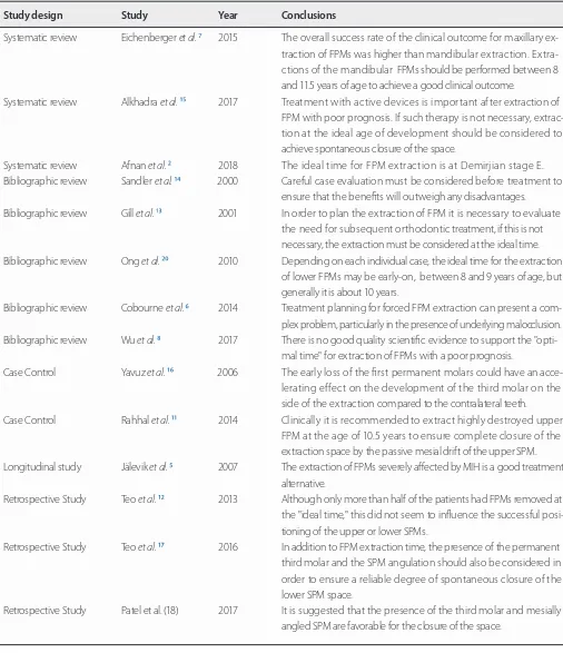
Table 1 . Summary of information gathered from each of the selected studies.
RESULTS.
The full text of the articles was read and based on inclusion and exclusion criteria, the final articles were selected, as summarized in Table 1.
DISCUSSION.
of early calcification in the dentin of the SPM root
bifurcation,6 which normally occurs at the chronological
age between 8 and 10 years,10 and is equivalent to the
Demirjian stage of development E.
Another parameter used as a reference for FPM extraction is when the SPM is at the cement-enamel junction of the FPM’s disto-buccal root, which generally occurs at the age of 10.5 years and it accelerates the
eruption of the SPM.11
Extractions of the upper FPMs have a more predictable
closure of the space compared to the lower extractions,12
therefore, the determination of the timing of when to extract the FPM is more critical in the mandible than in the maxilla.6,12,13
The extraction of an FPM when fulfilling the conditions described above should allow the SPM to advance to the PMP area, achieving good proximal contact with the second premolars. Before proceeding to FPM extraction, it is necessary to evaluate clinical and radiographic conditions in order to increase the probability of spontaneously closing the space.
Malocclusion, arc length discrepancy (crowding or excess space), molar ratio, permanent tooth loss, dental development stage, teeth size, state of eruption of the second premolar at the time of FPM extraction, presence of third molars reasonably well formed and in good position, divergent skeletal malocclusions with dolichofacial pattern, anterior open bite, and state of the other teeth (dental anomalies).5,8,14,15
The extraction of the mandibular FPM has been noted to facilitate the eruption of the third molar as it
increases the available space.16
According to Teo et al.,17 the development of the
second premolar within the bifurcation of the second temporal molar should be identified in order to achieve a favorable eruptive guidance and prevent distal displacement, rotation and inclination of the second premolar towards the extraction site; mesial angulation of the SPM in relation to the FPM increases the closure of the space. Regarding the presence of the third molar, the extraction of the FPM favors the eruption of the third molar, increasing the space for its eruption and decreasing possible impaction. According to these authors, the presence of one or more radiographic
factors is significantly associated with greater closure of the space.
Furthermore Patel et al.,18 took the FPM as reference
and defined that the mesial or straight angulation of the developing SPM and the presence of the third molars are significantly related to the closure of the space when inferior FPMs are extracted. Chronological age is used as a determinant of dental maturation in relation to the patient's age; most studies take one into account more than the other.
In the study by Jalevik et al.,5 FPMs were extracted
between the ages of 5.6 and 12.7 years, on average at 8.2 years. They defined as an ideal result the spontaneous and complete closure of the space, obtaining a point of contact between the second premolar and SPM without angulation or rotation of either of the two teeth, without distal displacement of the second premolar and without individual dental malocclusions.
This follow-up study showed a favorable closure of space in 85.2% of cases after the extraction of upper and lower PMP only taking into account the chronological age of the patients.
On the other hand, among the methods used to evaluate dental maturation on radiographs are those of Nolla, Demirjian and Moorrees. Nolla's method divides dental development into 11 stages that range from 0 which denotes absence of crypt and 11, which denotes apical closure in unirradicular and multiracial teeth. The Moorrees method proposes the allocation of maturation stages for the crown and root, and these vary in number for single-rooted or multi-rooted teeth.
Regarding the Demirjian method we highlight the following states: D crown developed, E early or initial bifurcation, F late bifurcation, G almost complete root
development.12
In the study conducted by Martínez et al.,19 the
Demirjian method presented greater precision in the estimation of dental age of the three methods applied, because it provided more precision between the evaluation of dental age and chronological age.
In the study by Teo et al.,12 despite the majority
obtained in most cases where the SPM were in stage E and F compared to the molars that were in stage than D or G.
According to Patel et al.,18 the stage of dental
development is significant only for the maxillary arch, not for the mandibular arch where the angulation of the developing SPM and the presence of the third molars is more relevant in order to obtain spontaneous closure of the space when FPMs are extracted.
The fundamental belief of those who measure only chronological age is that early extraction before 8 years of age could produce distal inclination and rotation of non-erupted second premolars, and conversely a late extraction after 11 years of age, that is, during or after the eruption of the SPM, will result in an incomplete closure of the space, mesial rotation of the SPM, distal migration of the second premolar, atrophy of the alveolar
ridge and overeruption of the opposite FPM.6,12,13
However, this is not entirely true, since as mentioned above, the chronological age must be complemented with the stage of dental development of the SPM taking into account the clinical characteristics on a
case-by-case basis. Indication versus contraindication according
to type of malocclusion:
Class I: With minimal crowding, it is necessary to extract at the ideal moment, not to perform tooth extractions of balance neither in the mandible nor the maxilla. If the mandibular FPM will be removed, consider extractions of compensation in the maxilla. If the maxillary FPM will be extracted, do not consider mandible compensation extractions.
With moderate crowding in the anterior area, extraction at the ideal time can result in spontaneous resolution of crowding and good space closure. Consider extractions of FPM for contralateral balance in bilateral
crowding.20
Class II: With slight crowding, the mandibular extractions should be performed at the ideal time and
the maxillary extractions should be postponed until the eruption of the SPM. Balance and compensation extractions are not recommended.
Subdivision I: Maxillary FPMs are better retained until the eruption of the SPM and employ restriction of a mesial movement of the SPM with a palatine arch.
Subdivision II: In the case of overbite, lower
extractions should be avoided.20
Clase III: it is best to refer to an orthodontist.
The extraction of PMPs is contraindicated in the following cases: brachyfacial type, patients with overbite, in absence of permanent teeth, diastema or
excess interdental space and class III malocclusion.8
CONCLUSION.
It can be concluded that the ideal stage to extract a FPM with a doubtful prognosis is at Demirjian stage E (early or initial bifurcation) or stage D (developed crown), which is around chronological age of 8 to 10.5 years.
As the spontaneous closure in the lower arch is less predictable than in the upper arch, it is very important to determine the ideal extraction time of the mandibular FMP, in addition in the mandible the mesial angulation of the developing SPM and the presence of the third molars is more relevant for achieving an optimal result. Because FPM extraction is not a successful treatment by itself in all cases, all patients should be referred to follow-up by an orthodontist.13 This review is especially useful for general
dentists who are often faced with children with extensive caries in FPMs, particularly in instances when it is not easy to consult an orthodontist.
Conflict of interests: There is no conflict of interest.
Ethics approval:It is not necessary.
Funding: None.
Authors’ contributions: All the authors have contri-buted to the study.
Acknowledgements: None.
REFERENCES.
1. OMS. Salud bucodental. WHO; 2012.
2. Saber AM, Altoukhi DH, Horaib MF, El-Housseiny AA,
Alamoudi NM, Sabbagh HJ. Consequences of early extraction of compromised first permanent molar: a systematic review. BMC Oral Health. 2018;18(1):59.
3. Batchelor PA, Sheiham A. Grouping of tooth surfaces by
susceptibility to caries: a study in 5–16 year-old children. BMC Oral Health. 2004;4(1):2.
4. Fresno MC. Prevalencia de hipomineralizacion incisivo
[Tesis]. Universidad de Chile; 2015.
5. Jälevik B, Möller M. Evaluation of spontaneous space
closure and development of permanent dentition after extraction of hypomineralized permanent first molars. Int J Paediatr Dent. 2007;17(5):328–35.
6. Cobourne MT, Williams A, Harrison M. National clinical
guidelines for the extraction of first permanent molars in children. Br Dent J. 2014;217(11):643–8.
7. Eichenberger M, Erb J, Zwahlen M, Schätzle M. The
timing of extraction of non-restorable first permanent molars: a systematic review. Eur J Paediatr Dent. 2015;16(4):272–8.
8. Wu M, Chen L, Bawole E, Anthonappa RP, King NM. Is
there sufficient evidence to support an optimum time for the extraction of first permanent molars? Eur Arch Paediatr Dent. 2017;18(3):155–61.
9. Chen J-W, Leggitt VL. Pulp treatment for young
first permanent molars: To treat or to extract? Endod Top. 2010;23(1):34–40.
10. Albadri S, Zaitoun H, McDonnell ST, Davidson LE.
Extraction of first permanent molar teeth: results from three dental hospitals. Br Dent J. 2007;203(7):E14.
11. Rahhal AA. Extraction Timing of Heavily Destructed Upper First Permanent Molars. Open J Stomatol. 2014;04(03):161–8.
12. Teo TKY, Ashley PF, Parekh S, Noar J. The evaluation of spontaneous space closure after the extraction of first permanent molars. Eur Arch Paediatr Dent. 2013;14(4):207–12.
13. Gill DS, Lee RT, Tredwin CJ. Treatment Planning for the Loss of First Permanent Molars. Dent Update. 2001;28(6):304–8.
14. Sandler PJ, Atkinson R, Murray AM. For four sixes. Am J Orthod Dentofacial Orthop. 2000;117(4):418–34.
15. Alkhadra T. A Systematic Review of the Consequences
of Early Extraction of First Permanent First Molar in Different Mixed Dentition Stages. J Int Soc Prev Community Dent. 2017;7(5):223–6.
16. Yavuz İ, Baydaş B, İkbal A, Dağsuyu İM, Ceylan İ. Effects of early loss of permanent first molars on the development of third molars. Am J Orthod Dentofac Orthop. 2006;130(5):634–8.
17. Teo TK-Y, Ashley PF, Derrick D. Lower first permanent
molars: developing better predictors of spontaneous space closure. Eur J Orthod. 2016;38(1):90–5.
18. Patel S, Ashley P, Noar J. Radiographic prognostic
factors determining spontaneous space closure after loss of the permanent first molar. Am J Orthod Dentofac Orthop. 2017;151(4):718–26.
19. Martínez Gutiérrez VM, Ortega-Pertuz AI. Comparación
de los métodos de Nolla, Demirjian y Moorrees en la estimación de la edad dental con fines forenses. Rev odontológica Mex. 2017;21(3):155–64.
20. Ong DC-V, Bleakley JE. Compromised first