From blindsight to sight: Cognitive
rehabilitation of visual field defects
Sylvie Chokron
a,b,c,∗, Celine Perez
a,b,c, Micha¨el Obadia
b,c, Isabelle Gaudry
a,b, Laurent Laloum
dand
Olivier Gout
b,caLaboratoire de Psychologie et NeuroCognition, CNRS, UMR5105, UPMF, Grenoble, France bERT TREAT VISION, Fondation Opthalmologique Rothschild, Paris, France
cService de Neurologie, Fondation Opthalmologique Rothschild, Paris, France
dService d’Ophtalmologie, Centre National Hospitalier des Quinze-Vingt, Paris, France
Abstract. Purpose: Traditionally, post-chiasmatic lesions were believed to result in complete and permanent visual loss in the topographically related areas of the visual field. However, a number of studies with monkeys, and later with humans, have demonstrated spared implicit visual functioning, referred to as ‘blindsight’. The present study assessed whether training this phenomenon would induce an objective restoration of conscious vision in the blind field of hemianopic patients.
Methods: For a period of 22 weeks nine patients with unilateral occipital damage participated in several forced-choice visual tasks known to elicit blindsight: pointing to visual targets, letter recognition, visual comparison between the two hemifields, target localization, and letter identification. Before and after rehabilitation, patients were submitted to a behavioral pre- and post-test, including visual detection and letter identification as well as to automated perimetry visual field testing (Humphrey Automated 24-2 Full Threshold).
Results: An objective improvement was found in the behavioral tasks for all patients at the post-test stage as well as an objective enlargement of the contralesional visual field for all except one of the nine patients. An overall decrease is seen in the number of undetected points (out of 30) on automated perimetry visual field testing after rehabilitation (F (1, 16)=22.57;p <0.001) for both eyes (Right Eye, RE: T=0;z=2.52;p <0.05; Left Eye, LE: T=0,z=2.37;p <0.05) regardless of lesion side. Conclusions: The results suggest that explicit (conscious) visual detection can be restored in the blind visual field by using implicit (unconscious) visual capacities. Results are discussed regarding visual field defect rehabilitation, blindsight, attention, and brain plasticity hypotheses.
1. Introduction
Unilateral post-chiasmatic injuries can cause visual defects in both monocular hemifields contralateral to the site of injury. Following unilateral occipital damage of the primary visual cortex one of the more common visual field defects observed is Homonymous Hemi-anopia (HH), which refers to a meticulously symmetri-cal loss of vision in the two eyes (see Zihl, 2000 for re-view). It has been known for some time that vision can
∗Corresponding author: Dr. Sylvie Chokron, Equipe TREAT Vi-sion, Service de Neurologie, Fondation Ophtalmologique Rothschild, 25, rue Manin, 75019 Paris, France. E-mail: [email protected].
occasionally improve spontaneously in some patients with uni- and bilateral postchiasmatic injury. Howev-er, as pointed out by Pambakian and co-workers, there is a poor prognosis for spontaneous recovery of field defects of vascular origin and the degree of recovery depends on the underlying pathology (Pambakian et al., 2005; Pambakian & Kennard, 1997). According to these authors, maximal recovery of a partial defect occurs within the initial 48 hours. Any recovery of a complete hemianopia occurs within the first 10 days, after which complete recovery is unlikely. A review of the literature demonstrates that less than 10% of pa-tients recover their full field of vision (Zhang et al., 2006; Zihl, 2000). However, more recently, in the
largest follow-up study ever conducted on HH (n =
254), Zhang et al. (2006) showed that the probability of spontaneous improvement of HH is related to the timing of the patient’s first evaluation, suggesting that greatest HH recovery occurs within the first weeks after cerebral injury. According to Pambakian & Kennard (1997), the extent of visual recovery correlates nega-tively with age, a history of diabetes or hypertension, and the presence of cognitive, language, or memory impairment.
Due to its poor recovery rate, hemianopia has been considered as a definitive and irreversible loss of vi-sion, even though it has been demonstrated that adapt-ed training programs may induce some visual field re-covery (Pambakian et al., 2005; Zihl, 2000). Over the past 40 years, several research groups have set out to determine whether patients with hemianopia have the ability to compensate for their field defect using eye movements, head movements, and extrastriate vision. Consistent with animal studies (Cowey, 1967; Mohler & Wurtz, 1977), it has been demonstrated that in hu-mans the detection of light stimuli presented at the bor-der of the blind visual field (Zihl & von Cramon, 1979) or the direction of saccades towards the border zone of the anopic field region is able to induce a recovery in some but not all patients (Jack et al., 2006; Van der Wildt & Bergsma,1997; Zihl, 1981). Interestingly, as seen in monkeys, recovery was mainly observed in the portion of the visual field subjected to practice (Zihl, 2000). In the same vein, “Vision Restoration Therapy” (VRT) (Kasten & Sabel, 1995; Kasten et al., 2006; Kas-ten et al., 2001; KasKas-ten et al., 1999; KasKas-ten et al., 1998; Kasten et al., 1998; Sabel & Kasten, 2000; Mueller et al., 2007; Sabel et al., 2005 for review) is a computer-ized visual detection training program, which runs on personal computers and is carried out at the patients’ home. VRT projects stimuli in the border areas of the hemianopic field (transition zone or borderzone), such as partially defective areas located typically between the intact and the blind regions of the visual field. Pa-tients press a key on the keyboard whenever they detect the stimulus presented in or near the borderzone. The patients perform the training exercises twice daily for half an hour each during a six month period. Recent-ly, six chronic, right hemianopic patients underwent functional magnetic resonance imaging (fMRI) while responding to stimuli in the trained visual border zone versus the non-trained seeing field before and after one month of VRT (Marshall et al., 2008). There was a significant increase in BOLD activity for border zone detection relative to detection in the seeing field af-ter the first month of therapy, which correlated with a
relative improvement in response times in the border zone location on behavioral tasks performed without imaging.
The authors concluded that VRT appears to induce an alteration in brain activity associated with a shift of attention from the non-trained seeing field to the trained border zone. The effect appears to be mediated by the anterior cingulate and dorsolateral frontal cortex in conjunction with other higher order visual areas in the occipitotemporal and middle temporal regions.
Other training techniques aim at systematically re-inforcing compensatory oculomotor strategies, there-by fortifying and enlarging the field of search. Based on observations of heminanopic patients’ oculomotor scanning behavior (Zihl, 1995), Zihl and co-workers developed a two-stage treatment method (see Zihl, 2000 for review). In the first stage, the use of large saccadic eye movements was used to enlarge patients’ field of search and to help them gain information as to where stimuli are located within a given spatial framework. In the second stage, patients learned to improve their scanning strategy, especially regarding spatial organi-zation. Importantly, the authors note that the magni-tude of gain was independent of other variables, such as etiology, time since lesion, type of field defect, field sparing, and patient age.
Given the difficulty in predicting visual field recov-ery and the vrecov-ery low interest in experimentally reha-bilitating vision in hemianopic patients, there is lim-ited research in the rehabilitation of hemianopia com-pared to both unilateral spatial neglect rehabilitation or ‘blindsight’ studies (see Chokron et al., 2007 for re-view). ‘Blindsight’ refers to the preservation of some unconscious visual processing in these patients’ anopic field. Traditionally, geniculostriate lesions were con-sidered to result in complete and permanent visual loss in the topographically related area of the visual field (Holmes, 1918; Huber, 1992). However, a number of studies with monkeys, and later with humans, have demonstrated that despite destruction of the striate cor-tex, or even hemispherectomy, some patients retain cer-tain visual function (Cowey & Stoerig, 1995; 1997; 2004; Goodall, 1957; Humphrey & Weiskrantz, 1967; Perenin & Jeannerod, 1975; Stoerig, 2006; Ukei, 1966; Weiskrantz, 2004). Weiskrantz, Warrington, Sanders and Marshall (1974) initially documented this intrigu-ing phenomenon in their famous case, DB (see also Weiskrantz, 1986; 1996 for discussion). DB exhibited a relatively preserved ability to point to stimulus loca-tions, detect movement and discriminate the orienta-tion of lines, gratings and letters, despite his denial of
seeing the stimuli. This preservation of unconscious visual abilities in the blind hemifield is now usually referred to as blindsight. There are two types of blind-sight. Specifically, Type 1 blindsight refers to the per-sistance of visual capacities in the absence of acknowl-edged awareness by the patient; whereas, Type 2 blind-sight is used to characterize such capacities with im-paired awareness of the stimulus presence (Weiskrantz, 2004). In addition, regarding motion detection in the blind visual field, the Riddoch phenomenon refers to the preserved ability of hemianopic patients to detect the presence of motion within their scotomatous fields without the ability to characterize the other attributes of the stimulus (Riddoch, 1917).
According to several authors (Cowey & Stoerig, 1991; Morland et al., 2004; Stoerig & Cowey, 1991), the blindsight phenomenon can be explained by at least three different hypotheses: (1) extrageniculate mediation through subcortical pathways, (2) geniculo-extrastriate mediation, and (3) partial sparing of visu-al cortex with sufficient preservation of corticvisu-al pro-cessing for stimuli to reach objective but not subjective thresholds. According to the first hypothesis, visual information transmitted through the retinotectal path-way or some other subcortical pathpath-way (e.g. retino-pulvinar) is projected to extrastriate visual cortex and is sufficient to drive visually guided behavior without awareness. According to the second proposition, direct projections from the lateral geniculate to extrastriate cortex may be sufficient for some visual discrimination and even for some “sensations” that patients do not experience as actually “seeing”. Finally, based on the third hypothesis, “islands” of spared occipital cortex would explain implicit residual visual function such as blindsight. However, this last hypothesis has been chal-lenged in studies showing complete defects on perimet-ric testing or complete visual defects in the contrale-sional visual field as demonstrated by neuroimaging techniques (see for discussion Ro and Rafal, 2006).
Despite the extensive use of eye movement direction and light detection training to rehabilitate visual field defects with moderate results, only a few studies have focused on the use of blindsight (or implicit vision) to restore conscious vision in the blind hemifield (Sahraie et al., 2006; Stoerig, 2006). Nevertheless, as Ro and Rafal (2006) pointed out, blindsight might provide a rational approach to rehabilitation. Zihl (2000) trained patients to localize unconsciously seen targets in their blind hemifield. They demonstrated that target local-ization improved with practice and that patients who improved felt more confident and less disabled,
sug-gesting that blindsight training may be of therapeutic value as well as rehabilitative. Unfortunately, it is dif-ficult to ascertain whether the improvement was due to an objective visual field recovery (better conscious detection of visual targets) or due to an enlarged search field (better visual exploration). Using neuroimaging techniques (e.g., fMRI, ERP, ERMF) during visual de-tection and discrimination tasks, some recent studies pointed out how orienting attention in space may ac-tivate early visual cortex (Fu et al., 2005; Jack et al., 2006; Noesselt et al., 2002). Therefore, it is difficult to assess the role of attention on the improvement of visual capacities in the blind field. This point will be further addressed in the discussion section.
Ro and collaborators (Boyer et al., 2005; Ro et al., 2004; Ro & Rafal, 2006 for review), have used TMS to simulate blindsight in normal participants. According to these studies, spatial orientation and color processing might be spared even without primary visual cortex (in-hibited by TMS) supporting the existence of a geniculo-extrastriate pathway that bypasses V1 and awareness (Stoerig & Cowey, 1989; 1991). As proposed by Ro and Rafal (2006), since both orientation and color can-not be effectively processed by the superior collicu-lus the most plausible pathway supporting these visu-al discriminatory behaviors without V1 and awareness may be a direct geniculate pathway into area V4 of extrastriate cortex, which contains a high proportion of feature-selective and color-opponent cells (Zeki, 1980). This retino-tectal and/or geniculo-extrastriate pathway could be seen as the anatomical basis of direct visual processing without awareness (such as blindsight) and could be advantageously used in the restoration of vi-sual function after primary vivi-sual cortex damage (Ro & Rafal, 2006). Most patients with visual cortex dam-age and resulting cortical blindness will have an intact superior colliculus making it possible to train or en-courage patients to utilize their retino-tectal functions, and perhaps remnant extrastriate processes that may be intact, to enhance visual awareness (Ro & Rafal, 2006; Stoerig, 2006; Stoerig & Cowey, 1989).
Consistent with this idea, Sahraie, Trevethan, MacLeod et al. (2006) recently carried out a daily detec-tion “training” task involving discriminadetec-tion of simple grating visual stimuli over a 3-month period in a group of 12 cortically blind patients. Psychophysical mea-surements were carried out before and after the training, and included detection of a range of spatial frequencies (0.5–7 cycles per degree), contrast detection at 1 cycle per degree, clinical perimetry, and subjective estimates of visual field defect. The authors demonstrated that
Left eye Right eye Left eye Right eye Left eye Right eye Pre-Test Post-Test Post-Test Post-Test Pre-Test Pre-Test
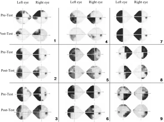
Fig. 1. For each patient, (a) Humphrey Automated Perimetry (24-2 Full Threshold program), from top to bottom: before training (VFT1), after training (VFT2) or (VFT3) for patients 2, 4, 5, 6, 8 and 9 who received an additional phase of training. (b) Lesion localization (CT-Scan and/or MRI).
repeated stimulation with appropriate visual stimuli in the blind visual field (and not only at the border zone as in VRT) can result in improvements in visual sensi-tivities in the very depths of the field defect thus con-firming that visual performance in the blind visual field can be improved by training residual capacities even in the absence of patients’ awareness.
Recently et al., (2007) used letters as stimuli pre-sented to the blind field to train two heminanopic
pa-tients. Flicker stimuli were presented at 30◦ or with
flickering letters at 10◦eccentricity in the blind visual field twice a week for a year. Training continued with more peripheral stimuli thereafter. Flicker sensitivity and neuromagnetic responses were registered at 1–2-month intervals, and Goldman perimetries were record-ed before, during and after training. Results showrecord-ed that flicker sensitivity in the blind hemifield improved
to the level of the intact hemifield within 30◦
eccen-tricity in one participant and 20◦ eccentricity in the
other. In addition, flickering letters were recognized equally at 10◦eccentricity in the blind and intact hemi-fields although no change was observed in the Gold-mann perimetry. Another study performed with the same team using the same experimental design
(Hen-riksson et al., 2007) demonstrated that this type of in-tensive training can reorganize visual cortices in such hemianopic patients. Indeed, magnetoencephalogra-phy documented changes in functioning during train-ing, while cortical organization illustrated through fM-RI demonstrated that following training visual infor-mation from both hemifields was processed mainly in the intact hemisphere. The fMRI mapping showed rep-resentations of both the blind and the normal hemifields in the same set of cortical areas within the intact hemi-sphere, specifically in the visual motion-sensitive area V5, in a region around the superior temporal sulcus and in retinotopic visual areas V1 (primary visual cortex), V2, V3 and V3a.
Within the same line of reasoning as Ro and Rafal (2006), Sahraie, Trevethan, MacLeod et al. (2006) and Stoerig (2006), the aim of the present study was to test if training visual capacities in the contralesional hemi-field could induce an objective restoration of conscious vision in the blind field of hemianopic patients. Specif-ically, we examined the use of the blindsight phenome-na in rehabilitating visual awareness in the blind hemi-field. Instead of using simple visual detection and con-trast discrimination in the blind visual field as Sahraie
Table 1
Demographic and clinical data
Age Sexa Onset of illness (months) Locus ofblesion Etiologyc VFDd
P#1 59 M 64 O Ischemic LSQ P#2 43 F 70 O Hemorrhagic LH P#3 80 F 22 TO Ischemic LH P#4 33 F 16 TO CVT LH P#5 75 F 10 TPO Ischemic LH P#6 24 F 14 TPO TBI LH P#7 66 M 121 TPO Meningioma LH P#8 54 M 12 TO Ischemic RH P#9 67 M 26 PO junction Hemorrhagic RH a: M, Male; F, Female.
b: F, Frontal ; T, Temporal ; P, Parietal ; O, Occipital.
c: TBI, traumatic brain injury; CVT, cerebral venous thrombosis.
d: VFD, Visual field defects; LH, left hemianopia; RH, right hemianopia; LSQ, left superior
quadranopia.
Fig. 2. Lesion localization (CT-Scan and/or MRI) as indicated by the white arrow for each patient (patient’s number in the right corner). et al. (2006) did, the current study aimed to submit
hemianopic patients to more ecological tasks (i.e., lo-calization and letter identification of static stimuli) and to measure the improvement with automated perimetry.
2. Materials and methods 2.1. Participants
Nine brain-damaged patients (4 men, 5 females;
av-erage age: 56; SD=19.1) with a persistent
homony-mous visual field defect (VFD) were included in the study. Patients 1–7 had a left VFD, and Patients #8 and #9 presented a right VFD (Fig. 1). All patients were right-handed except Patient 2 who was left-handed. All patients had a documented, single unilateral hemi-spheric lesion, and no past history of previous stroke. None of the patients suffered from impaired vigilance,
confusion, general mental deterioration or psychiatric disorders. Visual acuity of all patients was corrected-to-normal. Time from onset of brain injury to the ini-tial visit in our service (corresponding to the first vi-sual field testing presented here) always exceeded six months (see Table 1) to ensure that possible recovery after training could not be attributed to spontaneous re-covery. The location of brain lesion was determined
based on brain MRI and/or CT scans1(see Fig. 2).
The type of visual field defects, etiology, the lesion location determined by neuroimaging and the onset of brain injury are detailed in Table 1. Patients carrying the diagnosis of neglect (tested with the BEN: Batterie d’Evaluation de la N ´egligence; Azouvi et al., 2002) were not included in the study.
1Unfortunately, we did not obtain brain scans for patients #2 and
2.2. General procedure
Visual field testing (VFT) was performed prior to the beginning of the rehabilitation program (VFT1) and again between 22 and 30 weeks after the first visual training session (VFT2) in order to assess the effects of rehabilitation. Six patients received an additional 10 weeks of training followed by a third visual field examination (VFT3).
In addition to the VFTs, all patients were assessed with behavioral visual tests (BVT). Four sets of BVTs were conducted: one prior to the initial training session (BVT1) as a pre-test measure and three following the first training session to measure the effects of the neu-rovisual training. Therefore, as post-test one BVT was conducted following the first training phase (BVT2), one following the second training phase (BVT3), and a final set one week after the end of the rehabilitation program (BVT4) (see Table 2 for design details). 2.2.1. Visual field testing
Visual field defects can be directly measured by perimetry techniques and are usually classified accord-ing to the quality of the deficit, the portion of the field affected, and the quantitative extent of the deficit (see
Zihl, 2000 for review). The Humphrey Automated
Standard 24-2 Full Threshold program (Haley, 1986)
was used. With this program, 54 points, 6◦ apart of
the visual field are tested at a distance of 24 degrees to measure the size of patients’ visual fields and detect areas of non-seeing zones. Points in the visual field for each eye that were detected in less than 15 out of 30 stimulations were considered ‘undetected points’ (see Fig. 1). Perimetry testing in which loss of fixation oc-curred in more than 10% of the trials was discarded. Similarly, examinations where false positives or false negatives exceeded 5% were excluded.
VFTs were administered before and after the reha-bilitation sessions (along with BVT1 and BVT3) and carried the same testing parameters at each session. As shown in Fig. 1a, patients with homonymous hemi-anopia (HH) or homonymous quadranthemi-anopia (HQ) (except patient #3) did not present macular sparing at the initial stage.
2.2.2. Behavioral visual tests
Behavioral visual tests consisted of three comput-erized visual tasks (i.e., motor localization of targets, verbal localization of targets, and letter identification) performed using a PC-based system with a 17-inch, high-resolution monitor. Participants sat in a
comfort-able chair, directly in front of the middle of the comput-er touch screen and at a distance of 57 cm. Through-out the session, participants had to visually fixate on a dot at the center of the screen and were presented with three visual tasks in different, random order for each patient. The duration of each task was approximately seven minutes, with a three minute inter-task rest pe-riod. During this period, the monitor screen was gray and participants were free to close their eyes if they wished.
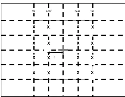
For the motor localization (ML) and verbal localiza-tion (VL) tasks patients had to localize the quadrant in which a visual target was presented. The target could appear at four different locations in each quadrant near (3◦) or far (6◦) from the fixation point (see Fig. 3) and each target was presented six times in each location (96 trials). The stimulus consisted of a black dot of
1.5◦ diameter presented alone on a white screen with
a yellow central fixation dot of 0.8◦diameter. Two
re-sponse modes were tested: a verbal rere-sponse (VL task), in which the patient had to report verbally where the stimulus appeared (upper left, upper right, lower left, or lower right), and a motor response (ML task) in which the patient had to point to the quadrant in which the stimulus appeared. Patients #1, #3, #5, #7, #9 began with the motor response condition and Patients #2, #4, #6, #8 began with the verbal response condition. Each display was flashed for 500 ms. When the participant responded, a 2000 ms inter-trial interval began. If the participant did not respond within the first 2000 ms, a 1000 ms delay was triggered.
For the letter identification (LI) task patients were presented three different letters (L, A or C) in a forced-choice recognition task. The letters were chosen with regard to their frequency and to their morphological properties (no physical or phonological similarities be-tween them). The letter could appear either near the central fixation dot (3◦in the left or right visual field) or at 6◦from the central fixation in the left or right visual field (Fig. 3) and each target was presented six times in each location (96 trials). Each display was flashed for 500 ms. When the participant responded, a 2000 ms inter-trial interval began. If the participant did not respond within the first 2000ms, a 1000 ms delay was triggered. During the behavioral tasks, visual fixation was monitored by the experimenter and trials in which a saccadic movement occurred were discarded. In order to be able to perform the tasks while maintaining fixa-tion, patients were trained several weeks before with a pure fixation task in which they had to fixate on a visual target centered to their eyes and their midsagittal body.
Table 2 Experimental design Pre-test VFT1 + BVT1 20 training sessions (10 weeks) Post-test I BVT2 20 training sessions (10 weeks) Post-test II BVT3 Post-test III VFT2 + BVT4 Additional 20 training sessions (10 weeks)* Post-test IV VFT3* VFT: Visual field testing (Humphrey 24-2 Full-Threshold Automated Perimetry).
BVT: Behavioral visual tests.
*Additional training sessions and VF with six patients (# 2, 4, 5, 6, 8, 9).
X X X X X X X X X X X X X X X X 3 near near far far
Fig. 3. Stimulus location in the localization and letter identification tasks. 2.2.3. Neurovisual training
Patients received 40 sessions of neurovisual training divided in two phases of 20 sessions each over a 22-week period. During the first 10 22-weeks, they performed 20 sessions of visual training followed by a two-week break. During weeks 12–22, a second set of 20 sessions were performed (see Table 2 for experimental design illustration). Each session comprised of four different tasks presented in a random order for each patient. The four computerized visual tasks were performed on a PC and consisted of (1) shape comparison, (2) detection, (3) judgment of orientation, and (4) letter identifica-tion. Participants sat in a comfortable chair, directly in front of and centered to the computer touch screen at a distance of 57 cm. During each task, participants had to visually fixate on a dot corresponding at the center of the screen.
The aim of shape comparison task was to train pa-tients to perform forced-choice comparisons between two stimuli appearing in each hemifield (blind and healthy). Two different shapes were used, a rectangle
(1◦ wide and 3◦ tall) and a triangle (base of 1◦, 3◦
tall). Sixteen different patterns of stimuli were present-ed eight times each (total 128 trials). The stimuli con-sisted of the two shapes, each appearing on each side of
a white screen with a yellow central fixation dot of 6.5◦
diameter. In half of the trials (n=64) patients were
presented the same shape (2 rectangles or 2 triangles) in each hemifield, whereas in the remaining half, the two shapes were different. The shapes were always pre-sented on the same horizontal meridian (top or bottom) but each shape appeared either near the fixation dot (3◦) or far from it (6◦). For the pre-test, two response modes were proposed. In the motor response condi-tion, in the trials where identical shapes were present-ed in each hemifield, patients had to touch the screen, whereas no manual response was required if there were different shapes on each side. In the verbal response condition, patients were required to say ‘same’ in the cases where identical shapes were presented in each hemifield. Each display was flashed for 500 ms. When the participant responded, a 2000 ms inter-trial interval began. If the participant did not respond within the first 2000 ms, a 1000 ms delay was triggered.
Table 3
Visual Field Testinga– Mean number of undetected targets (detected
less than 15/30 stimulations)
Patient VFT1b VFT1 VFT2c VFT2 VFT3d VFT3 LEe REf LE RE LE RE 1 21 19 6 8 * * 2 23 20 16 18 11 12 3 27 28 16 22 * * 4 17 16 9 12 2 2 5 28 33 11 18 17 11 6 22 28 20 26 18 24 7 26 28 26 27 * * 8 25 23 25 23 22 11 9 18 14 13 6 16 5 MEAN 23.11 16.78 12.58 SD 5.14 7.12 7.19
a: Humphrey Automated Perimetry, 24-2 Full Threshold b: VFT1, Visual Field Testing-prior to any training
c: VFT2, Visual Field Testing-22-30 weeks following first training
phase
d: VFT3, Visual Field Testing-following third training phase e: LE, Left Eyef: RE, Right Eye.
For the detection task patients had to detect the pres-ence or abspres-ence of a stimulus and, in the case of detec-tion, to localize the quadrant in which the visual target was presented. As in the localization task used in the BVTs, the target could appear at four different loca-tions in each quadrant and each target was presented six times at each location. In 96 trials there was a stimulus presented at one of the possible locations whereas in 64 trials the screen was empty. Participants were informed about the presence of these catch trials. The stimulus type and experimental procedure is the same as for the localization task used for the BVTs (see above).
In the judgment of orientation task patients were pre-sented black bars for 200 ms with three different orien-tations – vertical, 45◦to the right, or 45◦to the left – in a forced-choice recognition task. The stimulus could appear at nine different locations in each hemifield and was presented twice in each hemifield (108 trials). The patients were asked to mimic the orientation of the bar with their right hand.
Finally, in the letter identification task patients were presented three different letters (T, S or M) in a forced-choice recognition task (note that the letters used in pre and post-tests and in training sessions are different). One letter appeared either near the central fixation dot or further from it in the left or right visual field (see Fig. 3). Each letter was presented six times at each position resulting in 48 trials for each hemifield and 96 for the whole task. Each display was flashed for 500 ms. When the participant responded, a 2000 ms
inter-trial interval began. If the participant did not
respond, a 1000 ms delay ensued. As mentioned before,
visual fixation was monitored throughout the task by the experimenter and patients were trained several weeks before not to move their eyes during the task. During all the behavioral tasks (testing and training) feedback regarding the response accuracy was not provided to the patients.
3. Results
To ascertain the effect of the training tasks on visual function recovery, analyses were performed only on the VFT and BVT results, and not on the neurovisual training tasks. Left and right hemianopic patients were grouped together, thus the results refer to ipsi- and contralesional visual fields rather to left and right. 3.1. Visual field testing
A Wilcoxon paired samples non-parametric test was performed on the number of points detected at less than 15 out of 30 stimulations (considered as undetected points) on the VFTs of the nine patients to compare the size of the visual field at VFT1 and VFT2. Ta-ble 3 presents the mean number of undetected targets for each eye. Comparisons between VFT1 and VFT2 for both eyes show a significant decrease in the non-seeing/undetected zones of the visual field (Right Eye,
RE: T=0;z=2.52;p <0.05; Left Eye, LE: T=0,
z=2.37;p <0.05)
To investigate the effect of a third training phase, a Wilcoxon paired samples non-parametric test was performed on the VFT2 and VFT3 results of the six patients who received the additional training and visual field testing (see Table 3). A significant decrease in the non-seeing/undetected zones of the RE visual field was
seen between VFT2 and VFT3 (RE:T =0;z =2.2;
p <0.05). There was no significant difference for LE
(T =7.5,z=0.63;p >0.05).
An Analysis of Variance (ANOVA) was performed the nine patients’ VFT results with Eye (RE, LE) as the between subjects factor and Testing Time (VFT1, VFT2) as the within subjects factor. No effect of Eye was found. A main effect of Testing Time was seen with an overall decrease in the number of undetected
points between VFT1 (M =23.11, SD=5.14) and
VFT2 (M =16.78, SD=7.13),F (1, 16)=22.57;
p <0.001).
Finally, an ANOVA was performed on the VFTs of the six patients who received the additional training, with Eye (RE, LE) as the between subjects factor, and
Table 4
Behavioral Visual Tests - Average performance in percent accuracy (and standard deviations) at each phase
Ipsilesional BVT1a BVT2 BVT3 BVT4 Means by
visual field (pre-test) (10 weeks) (20 weeks) (22–30 weeks) task
Motor localization 95 (1.5) 97 (1.7) 98 (2.2) 99 (1.2) 97 (1.8)
Verbal localization 91 (3.1) 95 (1.7) 97 (2.1) 99 (1.1) 96 (2.9)
Letter identification 93 (2.9) 98 (1.9) 97 (2.1) 100 (0.6) 97 (2.6) Means by testing time 93 (2.0) 96 (1.7) 97 (0.6) 99 (0.6) 97 (1.5)
Contralesional BVT1 BVT2 BVT3 BVT4 Means by
visual field (pre-test) task
Motor localization 53 (6.4) 64 (6.3) 70 (5.2) 75 (6.1) 66 (6.2)
Verbal localization 42 (6.4) 55 (7.0) 64 (6.3) 70 (6.5) 58 (6.7)
Letter identification 44 (7.7) 55 (7.1) 62 (6.1) 72 (4.6) 59 (6.4) Means by testing time 46.3 (5.9) 58 (5.2) 65 (3.5) 72 (2.52) 61 (4.8)
a: BVT, Behavioral Visual Tests.
0 20 40 60 80 100 BVT1 (Pre Test) BVT2 BVT3 BVT4 BVT1 ( PreTest) BV T2 BVT3 BVT4 Time % accuracy ML VL LI
Ipsilesional visual field Contralesional visual field
Fig. 4. Behavioral visual tests 1, 2 and 3 (BVT1, BVT2, BVT3): Pre- and post-test accuracy levels in the ipsi- and contralesional visual fields for motor localization (ML), verbal localization (VL) and letter identification (LI).
Testing Time (VFT1, VFT2 and VFT3) as the within subjects factor. Once again, there was no effect of Eye
and a main effect of Testing Time was found,F(2, 20)
=19.23;p <0.000. Planned comparisons with LSD
post-hoc tests revealed fewer undetected targets during
VFT2 (M =16.42, SD=6.04) (p <0.01) compared
to VFT1 (M =22.25, SD= 5.63), and even fewer
undetected targets during VFT3 (M =12.58, SD =
7.19) (p <0.05) compared to VFT2 and VFT1.
Figure 1 provides the VFT1, VFT2 or VFT3 (Humphrey Automated 24-2 Full Threshold perimetry) results for each patient, showing an objective increase of visual detection performance in all except one patient in the pathological (contralesional) visual field after 22 weeks of the rehabilitation program focused on blind-sight training. The only patient in which no objective amelioration was found was Patient #7. Interestingly, this patient is the only one who had suffered from a voluminous meningioma leading to surgery. As can be
observed in Patient #7’s CT scan, the surgery led to the removal of a substantial part of the right temporo-parieto-occipital region. In addition, for this patient, there was a wider interval between the lesion and the rehabilitation program (121 months) as compared to the other patients.
3.2. Behavioral visual tests
As shown in Fig. 4, the pre- and post- BVT compar-isons demonstrated a dramatic increase of performance in the contralesional visual field with neurovisual train-ing for each of the visual tasks. The improvement was seen for the three tasks at BVT2, was increased at the BVT3, and maintained after the end of the rehabilita-tion program BVT4.
An ANOVA performed on the Time of Testing (BVT1, BVT2, BVT3, BVT4) x Visual Field (ipsile-sional, contralesional) x Tasks (ML, VL, LI) revealed
a significant effect of the testing time on global
perfor-mance,F(3, 21)=137.26;p <0.000), confirming the
significant positive effect of training (Table 4). As expected, the visual field stimulated had a
signif-icant effect on performance,F (1, 7)=120.56;p <
0.000) with better performance in the ipsilesional (M=
96.8, SD=1.56) than in the contralesional (M =61,
SD=7.81) visual field. In addition, a significant effect
of Task (F (2, 14)=10.61,p <0.002) was revealed,
with better performance in the ML task (M =81.5,
SD=4.3) than in the VL task (M =77, SD =5.1)
and the LI task (M =78, SD=5.2) (see Fig. 4 and
Table 4). In terms of interactions, a significant
inter-action between Testing Time and Task emerged (F(6,
42)=3.55,p <0.005) with greater improvement in
the ipsilesional visual field for the ML task than for the other tasks (see Fig. 4 and Table 4). A significant in-teraction between the Visual Field of presentation and
Task was also found,F(2, 14)=3.94,p <0.005. This
interaction is due to the fact that, as expected, there was a greater improvement in the contralesional than in the ipsilesional visual field.
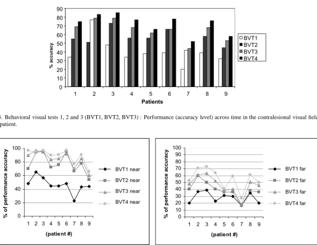
Figure 5 illustrates that improvement across time was present in all patients, even in Patient #7 who did not exhibit any objective improvement in the automated perimetry testing. However, our results confirm pre-vious studies in showing that performance may vary across patients and across time. Interestingly, as sev-eral studies with neglect patients have described (Bar-tolomeo et al., 2001; Chokron et al., 2002) performance was much more variable in the contralesional than in the ipsilesional field even at BVT4 when some recovery had occurred (see Standard Deviations, Table 4).
Figure 6 illustrates the significant effect of stimulus location on performance during the ML and VL tasks,
F(1, 8)=156.13;p <0.000, with better performance
for stimuli presented near the fixation point than far from it at each testing time (i.e., BVT1, BVT2, BVT3, BVT4). In addition, we found a significant interaction between the visual field of presentation and the
stim-ulus location (F (3, 24)=189.30; p < 0.001) with
a stronger effect of stimulus location in the contrale-sional versus ipsilecontrale-sional visual field. Finally, as illus-trated in Figure 6 performance improved for stimuli in the contralesional visual field presented both near the
fixation point (BVT1,M =54.5, SD=5.65; BVT4,
M =89.1, SD=5.03; ) and far from fixation (BVT1,
M =38.2, SD=4.10; BVT4,M =55.4, SD=7.3).
3.3. Functional vision recovery
In addition to the behavioral recovery, from a sub-jective point of view patients were aware of the
im-provement in daily life and observations of general vi-sual and vivi-sual field improvement were noted by all of the patients (even for patient #7 who did not show any objective recovery during the perimetry testing). Indeed, patients reported to be more confident in their vision and more efficient when reading, walking or sually scanning. When performing the automated vi-sual testing perimetry after the training sessions, they reported that they have ‘seen’ the stimuli for which they have answered. Several patients reported that af-ter only a few rehabilitation sessions they became able to use their vision in their contralesional visual field at different occasions, especially in ‘emergency’ con-ditions. For example, Patient #7 was able to catch his razor in the blind hemifield while it was falling down. After ten weeks of rehabilitation (BVT2), when de-tecting letters and forms in their ‘blind’ hemifield pa-tients were confident in their answer and reported that at this stage they were ‘seeing’ the stimulus they were answering. From a phenomenological point of view, at first, patients claimed that they did not see anything in their contralesional visual field and were ‘guessing’, then they reported that they ‘felt’ something in their blind field, they were then ‘pretty sure’ of their answer and finally they reported ‘seeing’ the stimulus in their ipsilesional visual field. Interestingly, this was also true for Patient #7 who experienced the same progres-sion of ‘subjective’ improvement, despite the lack of improvement in the automated perimetry examination (see Fig. 1).
4. Discussion
The present study confirms that training implicit vi-sual capacities can lead to an objective amelioration of conscious vision as demonstrated by the automated vi-sual perimetry and behavioral vivi-sual task performance. Using forced-choice visual tasks known to elicit ‘blind-sight’, we were able to rehabilitate conscious vision in the contralesional visual field in most patients (attest-ed by the visual perimetry). Automatic perimetry is considered to be an objective way to measure the pa-tients’ visual recovery in the contralesional visual field (Sahraie, 2007). The only patient in whom no objec-tive improvement at all was found using the perimetry examination was a patient who had undergone surgery of the right temporo-parieto-occipital region. Given the fact that these patients were ‘chronic’ patients (see Table 1) their recuperation cannot be seen as being the result of any spontaneous recovery. In addition, lack
0 10 20 30 40 50 60 70 80 90 1 2 3 4 5 6 7 8 9 Patients % a c c u ra c y BVT1 BVT2 BVT3 BVT4
Fig. 5. Behavioral visual tests 1, 2 and 3 (BVT1, BVT2, BVT3) : Performance (accuracy level) across time in the contralesional visual field for each patient. 0 20 40 60 80 100 1 2 3 4 5 6 7 8 9 (patie nt #) % o f p e rf o rm a n c e ac cu rac y PRETEST near PT1 near PT2 near PT3 near 0 10 20 30 40 50 60 70 80 90 100 1 2 3 4 5 6 7 8 9 (patient #) % o f p e rf o rm a n c e ac cu ra cy PRETEST far PT1 f ar PT2 f ar PT3 f ar BVT1 near BVT2 near BVT3 near BVT4 near BVT1 far BVT2 far BVT3 far BVT4 far
Fig. 6. Behavioral visual tests 1, 2 and 3 (BVT1, BVT2, BVT3): Pre- and post-test performance in the controlesional visual field for each patient near the fixation point (left) and far from the fixation point (right).
of a false positive tendency during the post-training VFTs rules out an artifactual result stemming from a tendency to over-respond (even when no stimulus is flashed in the blind hemifield). Finally, by using differ-ent tasks during the training and the testing phases, our results can be interpreted in terms of recovery rather than a learning effect biasing the performance during the post-tests.
In this way, the recovery we observed in the present hemianopic population might reflect a recuperation of ‘explicit’ vision in the contralesional visual field after training ‘implicit’ vision.
We will first discuss the objective improvement of visual detection as revealed by the automated perimetry and then the behavioral improvement measured by the ML and VL tasks as well as the LI task.
4.1. Visual field testing (automated perimetry) To our knowledge only a few studies have demon-strated that training hemianopic patients may lead to
an improvement of visual detection as recorded by au-tomated perimetry shown here (Kasten & Sabel, 1995; Kasten et al., 2006; Kasten et al., 2001; Kasten et al., 1999; Kasten et al., 1998; Kasten et al., 1998; Zihl, 2000). In the present study, there is a clear improve-ment for all patients except patient #7, as objectively measured with automated perimetry.
The fact that patient #7, who underwent surgery in the right temporo-parieto-occipital region, did not show any objective visual recovery confirms Zihl and von Cramon (1982; 1985) who hypothesized that visual field recovery can only be expected in cases with in-complete striate cortex injury. As in the present study, these authors were able to show that no recovery was observed in patients with complete destruction of the striate cortex on the affected side. However, it has to be noted that Patient #7 is the one with the most size-able delay between lesion acquisition and rehabilitation procedure, which might be a worsening factor.
Careful inspection of Figure 3 reveals that the im-provement always occurs for both eyes and is frequently
homonymous as is the deficit. This suggests a cortical plasticity within the visual areas with a preservation of retinotopic organization. This finding should be more thoroughly explored in future research in order to target cortical plasticity processes leading to visual field re-covery. In addition, the present study (showing visual recovery in patients with a lesion in the left or right oc-cipital lobe, including the primary visual cortex) con-trasts with previous studies (Zihl & Von Cramon, 1985 and see also Zihl, 2000 for discussion), which suggest that improvement in visual fields following stroke is generally limited to cases in which the lesion is located outside the primary visual cortex (including associa-tion cortex, subcortical nuclei or deep white matter). Whether or not visual field defect improvement is as-sociated with primary visual cortex damage or not is a controversial topic (Bosley et al., 1987). Furthermore, it is interesting to note that as reported in previous stud-ies (see Zihl, 2000 for review), the extent of visual field recovery neither depended crucially on the age of the patient nor on the interval between the lesion acquisi-tion and the start of the training program. For this rea-son, and as discussed by Zihl, a valid and reliable mea-sure that indicates whether a patient has a good or poor prognosis for visual recovery is still lacking. However, the positive effects of our treatment underline the need to elaborate such a measure in order to decide which patient should be enrolled in such a program.
4.2. Behavioral visual tests and functional recovery The first finding concerns the high performance of left and right hemianopic patients when localizing tar-gets in the contralesional visual field even at the pre-test phase. This result confirms previous ones report-ed both in humans and monkeys (Cowey & Stoerig, 1995; 1997; 2004). In addition, during all tests (pre and post) we observed a better performance in the ML than in the VL task, which confirm previous studies with blindsight patients (see Weiskrantz, 2004 for re-view). Furthermore, the results demonstrated greater improvement for the ML task than with the other tasks, suggesting that the more motor the task, the greater the improvement. This finding mirrors previous studies (i.e., Perenin & Jeannerod, 1975) in showing that mo-tor tasks could involve more automatic responses than tasks requiring verbal responses. In addition, accord-ing to Schendel and Robertson (2004) detection can be increased in hemianopic patients by placing their arm near the visual stimuli, within the blind hemifield. This should be further investigated in order to design a more efficient training program for such patients.
4.3. Training implicit residual capacities
In the present study, we chose to train hemianopic patients with complex visual tasks (letter identification and judgment of orientation) rather than using sim-ple visual detection tasks as in many previous stud-ies (see Zihl, 2000 for review; Sahraie et al., 2006). This choice was motivated by several studies showing a preservation of activation in extrastriate visual cortex despite the presence of a lesion in the primary visual cortex area as well as an improvement of performance in the blind field with detection and identification train-ing (Raninen et al., 2007). As discussed by Stoerig (2006), functional neuroimaging studies of human pa-tients confirm that extrastriate visual cortex in the le-sioned hemisphere continues to respond to stimulation of the blind hemifield. Blindsight also has been at-tributed to small islands of primary visual cortex that are supposed to survive the lesion. Although small is-lands of spared V1 tissue could be recruited to serve the implicit residual visual functions, the bulk of ropsychological, anatomical, physiological, and neu-roimaging data on both humans and monkeys indicate that blindsight is not just mediated subcortically, but that several extrastriate visual cortical areas retain or regain visual responsivity (Stoerig, 2006). In addition, the retino-recipient nuclei that have been physiologi-cally investigated after striate cortical ablation showed responsivity to stimuli presented in the cortically blind field (Payne et al., 1996; Stoerig, 2006). Furthermore, all of these nuclei project directly (like the dLGN and the pulvinar) or indirectly (like the superior colliculus) to extrastriate visual cortical areas. The various extras-triate visual cortical areas differ in the extent to which they continue to respond to information from the blind hemifield. Dorsal stream areas appear to retain more responsivity (see Salin & Bullier, 1995 for review).
There is also evidence showing that area V5/MT dis-plays the most robust responses after both cooling (Gi-rard et al., 1992) and ablation of V1 (Rodman et al., 1989). Whereas neither V2 nor V4 retained more than a very small number of neurons responding to the con-tralateral hemifield when V1 was ablated or cooled (see Bullier et al., 1996 for review), visual responses were still evoked from neurons in the polymodal cortex of the superior temporal cortex (Bruce et al., 1986; see Stoerig, 2006 for discussion). The preserved activity in extravisual cortical areas (dorsal and ventral path-ways) could account for the patients’ ability to learn in a few weeks to detect, localize and identify stimuli in their blind hemifield. Recently, Trevethan, Sahraie and
Weiskrantz (2007) were able to show that D.B., the first extensively studied blindsight case, could successfully identify outlined low contrast images of objects, make successful ’same/different’ discriminations for pairs of stimuli presented in his blind field and identify com-plex images (e.g., digital photographs) presented en-tirely within his cortically blind field. As we proposed here, Trevethan et al. (2007) underline the potential of using complex visual tasks for improvement in cases of blindsight.
Although the current study was able to demonstrate learning in hemianopic patients to localize and identify complex stimuli, it still remains difficult to understand how activating these extrastriate cortical areas can lead to a recovery of visual awareness in the blind hemifield as shown by the automated perimetry testing. Visual recovery in the blind field could result from a retroacti-vation from extrastriate cortical areas to spared islands of ipsilesional primary visual cortex, as suggested by recent studies (i.e., Silvanto et al., 2005), and/or could involve the participation of the intact hemisphere (Sil-vanto et al., 2007). We are currently running fMRI studies in the patients who recovered in their blind visu-al field to address this question more specificvisu-ally. An-other important issue raised by the present study is the contribution of attention to enhance visual recovery in the blind visual field.
4.4. Role of attention in visual recovery
As mentioned in the introduction, attention may play a crucial role in the recovery of the blind visual field by modulating the level of activation in striate, as well as extrastriate, cortex neurons (Bahrami et al., 2007; Fu et al., 2005; Jack et al., 2006; Noesselt et al., 2002) and may thus enhance visual detection and visual awareness in the contralesional visual field of hemianopic partic-ipants (Henriksson et al., 2007; Kentridge et al., 2004; Poggel et al., 2004; Sabel et al., 2004). Using complex visual tasks rather than simple visual detection to train hemianopic patients, as in the present study, may thus favor both the activity of extrastriate visual areas as well as the recruitment of exogenous and endogenous attention (Bartolomeo & Chokron, 2002). According to Ro and Rafal (2006), based on the strong relationship between attention and consciousness, it is conceivable that patients with visual field defects following prima-ry visual cortex damage might be able to use reflexive attentional orienting mechanisms of the superior col-liculus to eventually influence visual awareness. As we described here, although reports from patients suggest
that although there is no awareness of visual events, they sometimes have the sense or impression that some-thing was presented, which may be a function of reflex-ive orienting and may influence their ability to local-ize and discriminate at above chance levels (i.e., show blindsight) (Mohler & Wurtz, 1977). Unfortunately, in the present study, we cannot disentangle the specif-ic role of extrastriate area involvement and attentional processes in visual recovery.
Along the same lines, the present experiment re-vealed an effect of stimulus location on performance with better behavioral results for stimuli presented near rather than far from the fixation point regardless of test-ing time. This result could indicate that the observed improvement stems more from attentional rather than from a purely ‘visual’ process. However this result is challenged by the fact that during the post-test auto-mated perimetry (VFT2 and VFT3), an improvement deeper in the ipsilesional visual field was observed in
patients 1–5 and in patients 8 and 9 (up to 20◦, see
Fig. 1a). In this way, it is still difficult to assess if the observed recovery should be seen as the result of attentional process or is merely linked to a real plastic-ity of visual cortical areas. However, the recent study by Henriksson et al. (2007) suggests that some cortical plasticity, for example, ipsilesional visual cortex acti-vation, could be at work in such a recovery process after training visual discrimination in the blind visual field. Further fMRI studies with recovered hemianopic patients should help confirm this hypothesis.
5. Conclusions
The present study clearly demonstrates that objective visual field recovery is possible in chronic heminaopic patients by training them to detect, identify, or localize stimuli within their blind visual field and not only in the border zone. Similar to Raninen et al. (2007), we showed that using complex stimuli in forced-choice tasks known to elicit blindsight may lead to an objective recovery of explicit, conscious vision as measured with automatic perimetry.
In the largest study of homonymous hemianopia (HH) with detailed follow-up evaluation, Zhang et al. (2006) clearly showed that the probability of sponta-neous improvement of HH decreases with the time of the patient’s first evaluation, suggesting that greatest HH recovery within the first weeks after cerebral injury. As we mentioned in the introduction, none of the cases with stable underlying brain disease and stable
neuro-logic status continued to spontaneously improve after 6 months. If blindsight can open a path toward visual field restitution, as suggested by Stoerig (2006), Sahraie et al. (2006), Raninen et al. (2007) and the present study, visual field rehabilitation strategies should be initiated early after injury. Furthermore, given that true sponta-neous improvement after six months would be unusual in static neurologic diseases, documented improvement in the visual fields of these patients undergoing rehabil-itative therapy would be considered a sign of therapeu-tic efficacy. In order to demonstrate how visual train-ing based on traintrain-ing implicit visual capacities leads to conscious vision recovery in hemianopic patients, future research should include larger clinical trials and neuroimaging studies, as well as studies among patients with more peripheral deficits. We believe that there is an extraordinary clinical as well as theoretical benefit in the association between basic science, recovery, and rehabilitation studies in the field of vision, and hope that this area is likely to be one of active research in the future, as deficits in visual perception clearly lead to functional limitations and result in emotional distress.
Acknowledgements
This research was funded by the Edmond and Ben-jamin de Rothschild Foundations (New York and Gene-va). We thank Seta Kazandjian for her invaluable help.
References
This research was funded by the Edmond and Benjamin de Rothschild Foundations (New York and Geneva), (grant to the second author). We thank Seta Kazandjian for her invaluable help. Azouvi, P., Samuel, C., Dreyfus, L.A., Bernati, T., Bartolomeo, P.,
Beis, J.M., et al. (2002).
Sensitivity of clinical and behavioral tests of spatial neglect after right hemisphere stroke. Journal of Neurology, Neurosurgery and Psychiatry, 73, 160-6.
Bahrami, B., Lavie, N., & Rees, G. (2007). Attentional load mod-ulates responses of human primary visual cortex to invisible stimuli. Current Biology, 17, 509-513.
Bartolomeo, P., & Chokron, S. (2002). Orienting of attention in left unilateral neglect. Neuroscience and Biobehavioral Reviews, 26, 217-234.
Bartolomeo, P., Sieroff, E., Chokron, S. & Decaix, C. (2001). Vari-ability of response times as a marker of diverted attention. Neuropsychologia, 39, 358-363.
Bosley, T. M., Dann, R., Silver, F.L., Alavi, A., Kushner, M., Chawluk, J.B., et al. (1987).
Recovery of vision after ischemic lesions: Positron Emission To-mography. Annals of Neurology, 21, 444-50.
Boyer, J.L., Harrison, S., & Ro, T. (2005). Unconscious processing of orientation and color without primary visual cortex. Pro-ceedings of the National Academy of Sciences, 102, 16875-9. Bruce, C.J., Desimone, R., & Gross, C.G. (1986). Both striate cortex and superior colliculus contribute to visual properties of neurons in superior temporal polysensory area of macaque monkey. Journal of Neurophysiology, 55, 1057-75.
Bullier, J., Hupe, J.M., James, A., & Girard, P. (1996). Functional interactions between areas V1 and V2 in the monkey. Journal of Physiology – Paris, 90, 217-20.
Chokron, S., Colliot, P, Bartolomeo, P., Rhein, F., Eusop, E., Vassel, P., et al. (2002). Visual, proprioceptive and tactile performance in left neglect patients. Neuropsychologia, 40, 1965-1976. Chokron, S., Dupierrix, E., Tabert, M. H., & Bartolomeo, P. (2007).
Experimental remission of neglect. Neuropsychologia, 45, 3127-48.
Cowey, A. (1967). Perimetric study of field defects in monkeys after cortical and retinal ablations. Quarterly Journal of Experi-mental Psychology, 19, 232-45.
Cowey, A., & Stoerig, P. (1991). The neurobiology of blindsight. Trends in Neuroscience, 4, 140-5.
Cowey, A., & Stoerig, P. (1995) Blindsight in monkeys. Nature, 373, 247-9.
Cowey, A., & Stoerig, P. (1997). Visual detection in monkeys with blindsight, Neuropsychologia, 35, 929-39.
Cowey, A., & Stoerig, P. (2004). Stimulus cueing in blindsight. Progress in Brain Research, 144, 261-77.
Fu, S., Greenwood, P. M., & Parasuraman, R. (2005). Brain mech-anisms of involuntary visuospatial attention: An event-related potential study. Human Brain Mapping, 25, 378-90. Girard, P., Salin, P. A., & Bullier, J. (1992). Response selectivity of
neurons in area MT of the macaque monkey during reversible inactivation of area V1. Journal of Neurophysiology, 67, 1437-46.
Goodall, R.J. (1957). Cerebral hemispherectomy: Present status and clinical indications. Neurology, 7, 151-162.
Haley, M. J.(Ed.). (1986). The Field Analyzer Primer (2nd ed.). San Leandro, CA.: Allergan Humphrey.
Henriksson, L., Raninen, A., N¨as¨anen, R., Hyv¨arinen, L., & Vanni, S. (2007). Training-induced cortical representation of a hemi-anopic hemifield. Journal of Neurology, Neurosurgery and Psychiatry, 78, 74-81.
Holmes, G. M. (1918). Disturbances of vision by cerebral lesions. British Journal of Ophthalmology, 2, 353-384.
Huber, A. (1992). Homonymous hemianopia. Neuro-ophthalmology, 12, 351-366.
Humphrey, N.K., & Weiskrantz, L. (1967). Vision in monkeys after removal of the striate cortex. Nature, 215, 595-597. Jack, A. I., Shulman, G. L., Snyder, A. Z., McAvoy, M., & Corbetta,
M. (2006). Separate modulations of human V1 associated with spatial attention and task structure. Neuron, 51, 135-147. Kasten, E., & Sabel, B. A. (1995). Visual-field enlargement
af-ter compuaf-ter-training in brain-damaged patients with homony-mous deficits: An open pilot trial. Restorative Neurology and Neuroscience, 8, 113-127.
Kasten, E., Bunzenthal, U., & Sabel, B. A. (2006). Visual field recovery after vision restoration therapy (VRT) is independent of eye movements: An eye tracker study. Behavioral Brain Research, 175, 18-26.
Kasten, E., M ¨uller-Oehring, E., & Sabel, B. A. (2001). Stability of visual field enlargements following computer-based restitution training. Journal of Clinical and Experimental Neuropsychol-ogy, 23, 297-305.
Kasten, E., Poggel, D. A., M ¨uller-Oehring, E., Gothe, J., Schulte, T., & Sabel, B. A. (1999).
Restoration of vision II: Residual functions and training-induced vi-sual field enlargement in brain-damaged patients. Restorative Neurology and Neuroscience, 15, 273-87.
Kasten, E., W ¨ust, S., Behrens-Baumann, W., & Sabel, B. A. (1998). Computer-based training for the treatment of partial blindness. Nature Medicine, 4, 1083-7.
Kasten, E., W ¨ust, S., & Sabel, B. A. (1998). Residual vision in transition zones in patients with cerebral blindness. Journal of Clinical and Experimental Neuropsychology, 20, 581-98. Kentridge, R. W., Heywood, C. A., & Weiskrantz, L. (2004). Spatial
attention speeds discrimination without awareness in blind-sight. Neuropsychologia, 42, 831-5.
Kerkhoff, G., M¨unssinger, U., Haaf, E., Eberle-Strauss, G., & St¨ogerer, E. (1992).
Rehabilitation of homonymous scotomas in patients with post-geniculate damage of the visual system: Saccadic compen-sation training, Restorative Neurology and Neuroscience, 4, 245-54.
Marshall, R.S., Ferrera, J.J., Barnes, A., Xian Zhang, O’Brien, K.A., Chmayssani, M., Hirsch, J., & Lazar, R.M., (2008). Brain activity associated with stimulation therapy of the visual bor-derzone in hemianopic stroke patients. Neurorehabil Neural Repair, 22, 136-44.
Mohler, C. W., & Wurtz, R. H. (1977). Role of striate cortex and su-perior colliculus in visual guidance of saccadic eye movements in monkeys. Journal of Neurophysiology, 40, 74-94. Morland, A. B., Le, S., Carroll, E., Hoffmann M. B., & Pambakian, A.
(2004). The role of spared calcarine cortex and lateral occipital cortex in the responses of human hemianopes to visual motion. Journal of Cognitive Neuroscience, 16, 204-218.
Mueller, I., Mast, H. & Sabel, B.A., (2007). Recovery of visual field defects: A large clinical observational study using vision restoration therapy. Restor Neurol Neurosci, 25(5-6), 563-72. Noesselt, T., Hillyard, S. A., Woldorff, M. G., Schoenfeld, A., Hag-ner, T., Jancke, L., et al. (2002). Delayed striate cortical activation during spatial attention. Neuron, 35, 575-87. Pambakian, A.L., Currie, J., & Kennard, C. (2005). Rehabilitation
strategies for patients with homonymous visual field defects. Journal of Neuro-Ophthalmology, 25, 136-142.
Pambakian, A. L., & Kennard, C. (1997). Can visual function be restored in patients with homonymous hemianopia? British Journal of Ophthalmology, 81, 324-8.
Payne, B. R., Lomber, S. G., Macneil, M. A., & Cornwell, P. (1996). Evidence for greater sight in blindsight following damage of primary visual cortex early in life. Neuropsychologia. 34, 741-74.
Perenin, M. T., & Jeannerod, M. (1975). Residual vision in cortically blind hemifields. Neuropsychologia, 13, 1-7.
Poggel, D. A., Kasten, E., & Sabel, B. A. (2004). Attentional cueing improves vision restoration therapy in patients with visual field loss. Neurology, 63, 2069-2076.
Raninen, A., Vanni, S., Hyv¨arinen, L., & N¨as¨anen, R. (2007). Tem-poral sensitivity in a hemianopic visual field can be improved by long-term training user flicker stimulation. Journal of Neu-rology, Neurosurgery and Psychiatry, 78, 66-73.
Ro, T., Shelton, D., Lee, O. L., & Chang, E. (2004). Extragenicu-late mediation of unconscious vision in transcranial magnetic stimulation-induced blindsight. Proceedings of the National Academy of Sciences, 101, 9933–9935.
Ro, T., & Rafal, R. (2006). Visual restoration in cortical blindness: Insights from natural and TMS-induced blindsight. Neuropsy-chological Rehabilitation, 16, 377-96.
Riddoch, G. (1917). Dissociation of visual perceptions due to occipi-tal injuries with special reference to appreciation of movement. Brain, 40, 15-57.
Rodman, H. R., Gross, C. G., & Albright, T.D. (1989). Afferent basis of visual response properties in area MT of the macaque. I. Effects of striate cortex removal. Journal of Neuroscience, 6, 2033-50.
Sabel, B. A, & Kasten, E. (2000). Restoration of vision by training of residual functions. Current Opinion in Ophthalmology, 11, 430-6.
Sabel, B. A., Kenkel, S., & Kasten, E. (2005). Vision restoration therapy. British Journal of Ophthalmology, 89, 522-4. Sabel, B. A., Kenkel, S., & Kasten, E. (2004). Vision restoration
therapy (VRT) efficacy as assessed by comparative perimetric analysis and subjective questionnaires. Restorative Neurology and Neuroscience, 22, 399-420.
Sahraie, A. (2007). Induced visual sensitivity changes in chronic hemianopia. Current Opinion in Neurology, 6, 661-66. Sahraie, A., Trevethan, C. T., MacLeod, M. J., Murray, A. D., Olson,
J. A., & Weiskrantz, L. (2006). Increased sensitivity after repeated stimulation of residual spatial channels in blindsight. Proceedings of the National Academy of Sciences, 103, 14971-6.
Salin, P.A., & Bullier, J. (1995). Corticocortical connections in the visual system: structure and function. Physiological Reviews, 75, 107-54.
Schendel, K., & Robertson, L. C. (2004). Reaching out to see: Arm position can attenuate human visual loss. Journal of Cognitive Neuroscience, 16, 935-43.
Silvanto, J., Lavie, N., & Walsh, V. (2005). Double dissociation of V1 and V5/MT activity in visual awareness. Cerebral Cortex, 15, 1736-1741.
Silvanto, J., Cowey, A., Lavie, N., & Walsh, V. (2007). Making the blindsighted see. Neuropsychologia, 45, 3346-3350. Stoerig, P. (2006). Blindsight, conscious vision and the role of
primary visual cortex. Progress in Brain Research, 155, 217-34.
Stoerig, P., & Cowey, A. (1989). Wavelength sensitivity in blindsight. Nature, 342, 916–918.
Stoerig, P., & Cowey, A. (1991). Increment-threshold spectral sen-sitivity in blindsight: Evidence for colour opponency. Brain, 114, 1487–1512.
Stoerig, P., & Cowey, A. (1997). Blindsight in man and monkey. Brain, 120, 535-59.
Trevethan, C. T., Sahraie, A., & Weiskrantz, L. (2007). Form dis-crimination in a case of blindsight. Neuropsychologia, 45, 2092-103.
Ukei, K. (1966). Hemispherectomy in the human with special ref-erence to the preservation of function. Progress in Brain Re-search, 21, 285-338.
Van der Wildt, G. J., & Bergsma, D. P. (1997). Visual field enlarge-ment by neuropsychological training of a hemianopsia patient. Documenta Ophthalmologica, 93, 277-92.
Weiskrantz, L., Warrington, E. K., Sanders, M. D., Marshall, J. (1974). Visual capacity in the hemianopic field following a restricted occipital ablation. Brain, 97, 709-28.
Weiskrantz, L. (1986). Blindsight: A case study and implications. Oxford, UK: Oxford University Press.
Weiskrantz, L. (1996). Blindsight revisited. Current Opinion in Neurobiology, 6, 215-20.
Weiskrantz, L. (2004). Roots of blindsight. Progress in Brain Re-search, 144, 229-41.
Zeki, S. (1980). The representation of colours in the cerebral cortex. Nature, 284, 412-8.
Zhang, X., Kedar, S., Lynn, M. J., Newman, N. J., & Biousse, V. (2006). Natural history of homonymous hemianopia. Neurol-ogy, 66, 901-5.
Zihl J. (1981). Recovery of visual functions in patients with cerebral blindness. Effect of specific practice with saccadic localiza-tion. Experimental Brain Research, 44, 159-69.
Zihl, J. (1995). Visual scanning behaviour in patients with homony-mous hemianopia. Neuropsychologia, 33, 287-303. Zihl, J. (2000). Rehabilitation of visual disorders after brain injury.
Hove, UK: Psychology Press.
Zihl, J., & von Cramon, D. (1979). Restitution of visual function in patients with cerebral blindness. Journal of Neurology, Neurosurgery and Psychiatry, 42, 312-22.
Zihl, J., & von Cramon, D. (1982). Restitution of visual field in patients with damage to the geniculostriate visual pathway. Human Neurobioliogy, 1, 5-8.
Zihl, J., & von Cramon, D. (1985). Visual field recovery in patients with postgeniculate damage: A review of 55 cases. Brain, 108, 335-65.