Covenant Journal of Informatics & Communication Technology. Vol. 6 No. 2, Dec., 2018
An Open Access Journal Available Online
Unsupervised Retinal Blood Vessel Segmentation
Technique using
pdAPSO
and Difference Image Methods
for Detection of Diabetic Retinopathy
Emmanuel Gbenga Dada
& Stephen Bassi, Joseph
Department of Computer Engineering, University of Maiduguri, Maiduguri, Nigeria
[email protected], [email protected]
Abstract—Retinal vessel segmentation is a practice that has the potential of
enhancing accuracy in the diagnosis and timely prevention of illnesses that are related to blood vessels. Acute damage to the retinal vessel has been identified to be the main cause of blindness and impaired vision. A timely detection and control of these illnesses can greatly decrease the number of loss of sight cases. However, the manual protocol for such detection is laborious and although autonomous methods have been recommended, the accuracy of these methods is often unreliable. We propose the utilization of the Primal-Dual Asynchronous Particle Swarm Optimisation (pdAPSO) and differential image methods in addressing the drawbacks associated with segmentation of retinal vessels in this study. The fusion of pdAPSO and differential image (which focuses on the median filter) produced a significant enhancement in the segmentation of huge and miniscule retinal vessels. In addition, the method also decreased erroneous detection near the edge of the retinal (that is not sensitive to light). The results are favourable for the median filter when compared to mean filter and Gaussian filter. The accuracy rate of 0.9559 (with a specificity of sensitivity rate of 0.9855), and a sensitivity rate of 0.7218 were obtained when tested using the Digital Retinal Images for Vessel Extraction database. The above result is a pointer that our approach will help in detecting and diagnosing the damage done to the retinal and thereby preventing loss of sight.
Keywords/Index Terms—Retinal Vessel, Segmentation, Asynchronous Particle
1. Introduction
Retinopathy is the subdivision of medicine that makes it possible to determine the cause of infections and ailments of the eye and treat them immediately. Digital photography and image analysis of retinal vessel are already gaining ground. The studies of (Kanski, 2007) and (Klonoff & Schwartz, 2000) observed that these techniques have been recently recognised as beneficial techniques in the identification and treatment of some diseases like diabetic retinopathy (DR), retinopathy of prematurity (ROP) which is a pathological disarrange of the retina and cardiovascular diseases (Mapayi, Tapamo & Viriri, 2014).
According to World Health Organisation (2016), DR and ROP are the chief reasons of eye defect and impaired vision universally, timely identification of the cause and control of these ailments will assist in a notable decrease of instances of impaired vision (Gergely & Gerinec, 2009). Ophthalmologists find vessel network very useful as they concentrate on retinal vessel quality assessment at some stage of diseases diagnosis. Detection by physical examination and testing of the retinal vessels in the hollow part of the images is quite cumbersome and laborious job which needs competent and skillful people who are not readily available (Varughese, et al., 2008). Nevertheless, according to Marrugo et al., (2012), it is possible for the ophthalmologist to diagnose and effectively control the diseases with the help of automated segmentation and systematic inspection of the arrangement of blood vessels in the retinal. Retinal vessel segmentation is the partitioning of a retinal image into
sections that have related attributes such as grey level, colour, texture, brightness, and contrast (Zhang, Zhou & Bao, 2015). The image part is removed from the initial image during image analysis and image segmentation algorithms are used to divide the original image into different segments. The main purpose of the retinal vessel segmentation is to divide the retinal image into equally exclusive sections so that each section in relation to the pixel concentration is harmonised to a predetermined benchmark (Zhang, Zhou & Bao, 2015). This study presents a new technique for segmenting network vessels in retinal images through Difference image and
pdAPSO techniques.
The remainder of this paper is structured as follows: Section 2 discusses the related works. Section 3 discusses our proposed methodology, filtering techniques, difference image, primal-dual particle swarm optimisation and post-processing. The results generated by our proposed approach on DRIVE datasets and discussions are specified in Section 4, and lastly the study is concluded in Section 5.
2. Related Works
Many research has been conducted in the area of retinal segmentation. Akram & Khan (2013) used a multi-layered thresholding-based blood vessel segmentation method for investigating cases of retinal diabetic that can result in blindness. Jiang & Mojon (2003) used an adaptive local thresholding prototype employing an authentication based multi-threshold analytical system for detecting the retinal image. The weakness of this approach is its lackluster performance in detecting the reedier vessels and some isolated vessels in the retinal. Mapayi, Viriri &
Emmanuel Gbenga Dada & Stephen Bassi Joseph CJICT (2018) 6(2) 64-78
Tapamo (2015) proposed a novel adaptive thresholding method for retinal vessel segmentation using local information that is uniform in nature.
Qin et al., (2006)employed a multiscale
method that uses Gabor filters and scale multiplication for the segmentation of retinal vessels. Marin et al., (2011) developed a novel supervised approach for blood vessel segmentation in retinal images through gray-level and moment invariants-based features for pixel depiction, whereas the vessel segmentation was done by neural network algorithm. Szpak & Tapamo (2008) used the gradient based technique and level set method for retinal vessels segmentation. Their approach was unsuccessful in detecting the tinnier retinal vessels. Wang et al.,
(2013) used the combination of multi-wavelet kernels and multiscale hierarchical decomposition to segment retinal vessels. Xiao et al., (2013) developed a retinal segmentation technique based on the Bayesian method and spatial constraint. Yin et al., (2013) proposed an unsupervised segmentation technique using probabilistic formulation.
Lupascu & Tegolo (2011) implemented an unsupervised segmentation of retinal vessels using self-organizing maps (SOM) and k-means clustering. The SOM is trained on retinal images and the map was again partitioned by k-means clustering method into two groups. The complete image is again passed to SOM and the group with the most ideal identical section on SOM is allocated to each pixel. A hill climbing scheme on associated components is employed to detect the vessel network during the post-processing operation.
Ramaswamy et al., (2011) proposed the combination of k-means and fuzzy c-means clustering methods for the categorisation of discharges and non-discharges in retinal images. Saffarzadeh et al., (2014) developed a technique that used k-mean for pre-processing after which the multi-scale line operator is utilised for the detection of retinal vessel network. The K-means assists the vessels to be more conspicuous and there is a significant decrease in the effect of bright gashes. The line detection operator in three scales is used in detecting the retinal vessels. Wen et al., (2007) assessed the performance of the k-means algorithm in enhancing the detection of retinal vessels by decreasing the colour space. However, the output of this approach was not good.
Sreejini & Govindan (2015) applied PSO to find the best filter parameters of the multiscale Gaussian matched filter to attain increased accuracy of retinal vessel segmentation. Their technique have better performance than many of the existing retinal vessel segmentation methods. There is still need to improve on the performance of the proposed system as the mutiscale matched filter does not completely overcome the problem of undesirable performance figures of matched filters. Son, Park & Jung (2017) used generative adversarial neural network to produce the exact map of retinal vessels on DRIVE and STARE datasets. The drawback of their approach is that it was unsuccessful in detecting extremely tinny vessels. Li et
al., (2017) applied reinforcement local
descriptions and SVM to segment retinal blood vessel. The system achieved a very high performance but
Emmanuel Gbenga Dada & Stephen Bassi Joseph CJICT (2018) 6(2) 64-78
the segmentation process can be time consuming. Mohsen et al., (2018) proposed neural network hardware implementation and FPGA for retinal vessel segmentation. The major drawback of their technique is the complexity of the system. Memari et al., (2017) used the hybrid of matched filter and AdaBoost classifier for enhanced retinal vessel segmentation. Sumathi, Vivekanandan & Ravikanth (2018) proposed neural network for efficiently segmenting retinal vessel.
Particle swarm optimisation (PSO) algorithm is an unsupervised segmentation approach. PSO has found application in the field of image segmentation. Saatchi & Cheng-Hung (2007) did a survey of image segmentation application of swarm intelligence algorithms (PSO and ACO) and their hybrid with k-means and simple competitive learning algorithms. Gopi & Nageswara (2013) used PSO methods for image segmentation to detfect breast cancer. Their experimental result shows that the fusion of Fuzzy C-means (FCM) and Fractional Order Darwinian PSO (FODPSO) algorithm performs better than the PSO alone, and Darwinian PSO (DPSO). There is, however, need to develop the more efficient approach that can remove background noise from the images. Mahalakshmi & Velmurugan (2015) used PSO to segment a brain tumour medical images. The effectiveness of the approach is not certain as there is no clear parameter for measuring the performance of the technique.
While much progress has been made in developing efficient methods by the earlier research works, the results
indicates that there is need for more work to be done in tackling the problem of high false detection close to the edge of the point where the optic nerve enters the retinal and the segmentation of big and reedier retinal vessels. In this study, we propose the combination of the Primal-Dual Asynchronous particle swarm optimisation and difference image approach for the segmentation of retinal blood vessel segmentation for easy detecting of thinner layers in the vessel and effective diagnosis of retinopathy diabetics. The major shortcoming of many of the vessel segmentation techniques include: low contrast of vessels, low quality images and large disparities and unpredictability in the size of the vessels. Achieving high accuracy in segmentation is a big problem because minute vessels are subjugated by various image noises such as Gaussian (Sreejini & Govindan, 2015).
3. Methodology
Normally, the presence of noise as a result of fluctuating lighting and contrast in retinal fundus images pose a challenge to retinal image clustering unless there is pre-processing operation. Realising the fact that efficient detection of the network of the vessel is a crucial phase required in the detection of diseases of the eye for dependable retinal vessel classification, there is need to develop an effective method that can handle the segmentation of sizable and small vessels in a well-timed and successful style. The green section of the pigmented retinal image is utilised for segmentation because it offers the most excellent brightness setting for the vessel (Kande, Subbaiah & Savithri, 2010). An in-depth explanation of our
Emmanuel Gbenga Dada & Stephen Bassi Joseph CJICT (2018) 6(2) 64-78
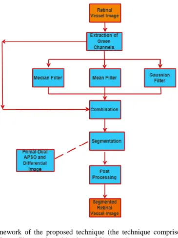
proposed technique is outlined below: (1) Removal of the green channels of
the coloured retina image.
(2) Filtering of the retinal image using median, mean, and Gaussian filtering methods.
(3) Producing the difference image. (4) Partitioning of the retinal vessels
from the difference image produced by the pdAPSO
algorithm.
(5) Execution of a post-processing stage using median filter for the elimination of miscategorisation. Figure 1 below depicts the framework
of the proposed system.
Figure 1: Framework of the proposed technique (the technique comprises of basically three steps using different filters and combinations of filters and a segmentation technique).
3.1 Filtering Techniques
The improvement of the green channel of the fundus retinal image is by achieved applying diverse filtering methods. The mean filter and Gaussian filter which are linear filters are employed to free the images from roughness and unevenness of the surface. These filters help to decrease image noise. However, they cannot
effectively protect the boundaries of an image. The median filter which is a non-linear filter is very effective in eliminating image noise in addition to safeguarding information at the borderline of images. It is very essential for us to state that the dimensions of the frame of the chosen image should not be too big to be able to proficiently handle the noise resulting from brightness
Emmanuel Gbenga Dada & Stephen Bassi Joseph CJICT (2018) 6(2) 64-78
deviation that usually characterises the retinal image. Being meticulous in the choice of window size that has adequate data points is very crucial to achieving for good enhancement. Choosing window sizes with adequate data points is crucial to attaining superior enhancement in the segmentation process. In this study, we employ the mean, Gaussian and median filters because of the complexity of the retinal image. The complexity of the retinal image is explained below:
R = F G (1)
R (i, j) = ∑ (x, y) ε F (i-x, j-y) ε G F (x, y) G (i-x,
j-y) (2) Given that R is the twisted retinal image, G represents the green channel of the retinal image and the twisted cover F is used to denote the filtering method used in this research work
3.2 Difference Image
The subtraction of the green channel of the coloured retinal image from the twisted retinal image produced the difference image. The formula for the difference image D(i, j) is as follows: D(i,j) = R(i,j) - G [i,j] (3) where D(i,j)= {Dq(i,j),DG(i,j),Dα(i,j)},
here Dq(i,j) is the difference image
created by median filter (DIMDF), DG(i,j) is the difference image created
by mean filter (DIMNF) and Dα(i,j) is
the difference image created by Gaussian filter (DIGF). We also experimented with using the combination of median and mean filters (DIMDMNF), median and Gaussian filters (DIMDGF), and mean and
Gaussian filters (DIMNGF). The possible permutations are
= DG(i,j) + Dq(i,j)
= Dq(i,j) + DG(i,j) (5)
= Dα(i,j) + DG(i,j) (6)
where represents the DIMDMNF, and is used to denote the DIMDGF, while represents the DIMNGF. The outputs of the equations (4) to (6) are regularised to the interval [0, 255].
3.3 Primal-Dual Asynchronous Particle Swarm Optimisation (pdAPSO) Algorithm
The Asynchronous PSO (APSO) is a variant of PSO proposed by Dada and Effirul (2015). The current personal best
(pbesti,m) and the global best (gbesti,m) of
a particle, its velocities, and positions of particles are instantly modified to be up to date after computing their fitness. As a result of this, the parameters are updated using partial or deficient information about the neighbourhood. This leads to diversities in the swarm of particles as the current swarm is a mixture of certain information from the earlier iteration and the ones from the current iteration. For detail information on the Primal-Dual PSO, the reader is referred to our previous work (Dada & Effirul, 2015). This present study adapted the pdAPSO algorithm for efficient segmentation of retinal images. In this approach, a single particle xi
denotes N cluster such that xi=(yi1,...,yij,…,yiN) where yij correspond
to the jth cluster centre of inertia trajectory of the ith particle.
Consequently, a swarm typifies a number of contending cluster centers.
The fitness of each group of cluster is computed based on the formula below: f(xi,mi) = w1distmax (mi,xi) + w2(mmax -
Emmanuel Gbenga Dada & Stephen Bassi Joseph CJICT (2018) 6(2) 64-78
distmin(xi)) (7)
where zmax = 2n-1 for an n-bit image; M
is a matrix denoting the allocation of pixels to clusters of particle i. Each element mijp specifies if the pixel mp is a
member of cluster Cij of particle i. The
coefficients w1and w2 are constants that
are specified by the user. Similarly, the highest value of the mean Euclidean distance of particles to their related clusters is
distmax (mi, xi) =
(8) and the distance between any set of clusters with the lowest Euclidean distance value is distmin (xi) =
(9) The flowchart of the pdAPSO algorithm is shown in figure 1 below.
Generate Initial Primal-Dual Parameters Generate Initial PSO Parameters Primal-Dual
Operators PSO operators Primal-Dual Method Evaluate Solutions Solution Feasible? Redefine Constraint Handling Method Solution Feasible? Yes No Display the Solutions Yes No Update Individual Particle's Position Evaluate Objective Function Update global best
position Compute Best
Function Velocity Update New set of solution
PSO method
Figure 2: Primal- Dual-APSO (Pdapso) Flowchart
The proposed algorithm for retinal vessel segmentation is outlined below: Algorithm 1 pdAPSO Segmentation algorithm
Step 1: Haphazardly assign an initial value to cluster centers for each particle. Step 2: On behalf of each particle, designate each pixel to a cluster with the shortest distance to its cluster center. Step 3: Compute the fitness function for each particle and obtain the global best solution.
Step 4: Save the best solution found so far as the pbest or personal best solution. Step 5: Use the equations below to update the cluster centers
) ( () ( () () , 2 2 ) ( , , 1 1 ) ( , ) 1 ( , * t m i m t m i m i t m i t m
i w v c rand pbest x c rand gbest x
v (10) ) 1 ( , ) ( , ) 1 ( , t m i t m i t m i x v x (11) Step 6: If the stopping condition is
fulfilled move to the subsequent step. Else, move to Step 3.
Emmanuel Gbenga Dada & Stephen Bassi Joseph CJICT (2018) 6(2) 64-78
The Primal-Dual Asynchronous Particle Swarm Optimisation method explained in Algorithm 1 is utilised in segmenting the retinal vessel network from the contextual tissue in the retinal images by utilising the outputs produced of equations (10) and (11).
3.4 Post-Processing
Median filter is utilised for the post-processing stage to ensure the reduction to the barest minimum of erroneously detected pixels in the vessel. We adopted a 2×2 median filter to remove the noisy pixels in the image to so that we can get the segmented vessel network. The median filter works by taking into consideration each pixel in the vessel image in turn and considers at its close neighbours to determine if it is a true exemplification of its surroundings. Rather than just merely changing the pixel values, it substitutes it with the median of these values. A 2x2 neighbourhood is considered here as larger neighbourhoods will produce better smoothing in the retinal vessel image. The median filter is very robust and it does not generate fresh impracticable pixel values when the filter overlaps a border. This makes it to be much better at maintaining sharp edges than some other filters.
3.5 Performance Measures
In this section, the experimental setup used to evaluate three algorithms proposed for retinal vessel segmentation. The sensitivity, specificity, and accuracy are the main performance metrics used in this work. The algorithms were experimented on the DRIVE database explained in section 3.3.
Sensitivity = TP/ (TP + FN) (12)
Specificity = TN/ (TN + FP) (13)
Accuracy = (TP + TN)/ (TP + TN + FP
+ FN) (14)
where TP = True Positive, TN = True Negative, FP = False Positive and FN = False Negative.
The amount of pixels that are rightly categorised as vessel pixels is the TP. The amount of pixels that are rightly categorised as non-vessel pixels is the TN, while FN is the number of pixels wrongly categorised as non-vessel pixels. The FP is the number of pixels wrongly categorised as vessel pixels. The sensitivity (SE) is the division of TP by the sum of vessel pixels in the ground truth segmentation, while the specificity (SP) is the division of TN by the sum of non-vessel pixels in the ground truth.
4. Results and Discussion
Some tests were carried out to ascertain the efficiency of our new technique. Our technique was tested using DRIVE
database on
http://www.isi.uu.nl/Research/Databases /DRIVE. Experiments were conducted using MATLAB 2015a on an AMD A 10-7300 Radeon R6, 10 Compute Cores 4C+6G, 1.90 GHz, 8.00GB of RAM. The optimal parameters for the filtering operation were discovered by inputting one of the images into the optimised median filter. The pdAPSO optimisation technique comprises of diverse arbitrary processes. It therefore means that using identical parameters for the filter does not guarantee getting the same result even when the program is restarted and executed.
4.1 Results
The results of our experiments indicate the performance of the diverse difference images hybridised with the pdAPSO algorithm. pdAPSO and
Emmanuel Gbenga Dada & Stephen Bassi Joseph CJICT (2018) 6(2) 64-78
DIMDF produced the most ideal segmentation result when compared to pdAPSO and DIMNF then pdAPSO and DIGF. pdAPSO and DIMDF generated the most superior accuracy rate 0.9559 and a specificity rate of 0.9855. In Figure 3, we provide a pictorial explanation that relates the results gotten from vessels segmented by pdAPSO and difference image using each of the filters.
4.2 Discussion
The fusion of DIMDF and pdAPSO
detected many of the big and tinny vessels, whereas some few gauzier vessels go on without detection. There are few cases of erroneous detection coupled with previous leftover close to the edge of the point where the optic nerve enters the retinal. The erroneous detection near the edge of the optic disc is nevertheless greatly reduced on the segmented vessels by DIMDF and
pdAPSO but much in number on
segmented vessels generated by the combination of pdAPSO with DIMNF,
and pdAPSO with DIGF. The reason for
this is because the median filter maintains boundary information of the vessels in the improved retinal image. Furthermore, the DIMDMNF and DIMDGF while hybridised with
pdAPSO produced good sensitivity of
0.7217 and 0.7205 respectively. The addition of DIMDF caused the increase in sensitivity of the retinal image. However, the accuracy of DIMDF is
better than the ones generated by DIMDMNF, DIMDGF, and DIMNGF while fused with the pdAPSO method. The statistical results generated by our approach are presented in table 1. In Fig. 4 there is a visual description of the result comparison of retina vessels segmented by the fusion of pdAPSO and DIMDF DIMDMNF, DIMDGF and DIMNGF. As illustrated in Fig. 3, a good number of the large and tinny vessels are detected, while the ones that remain undetected are very few. The erroneous detection near the edge of the optic nerve is greater on the outputs generated by DIMDMNF, DIMDGF and DIMNGF while fused with pdAPSO
but smaller on the outputs produced through pdAPSO fused with DIMDF. This explains the reason why a better accuracy rate was attained.
4.3 Performance comparison of pdAPSO with other segmentation techniques on DRIVE dataset
The performance comparison of some of the segmentation techniques on DRIVE database is presented in Table 2. The combinations of the proposed approach with the best performance yield higher and specificity rates, when compared to the earlier approaches. It is worth noting that the work of Ricci & Perfetti (2007) generated the highest accuracy rate among all the techniques compared, the value for the sensitivity and specificity rates are not specified.
Emmanuel Gbenga Dada & Stephen Bassi Joseph CJICT (2018) 6(2) 64-78
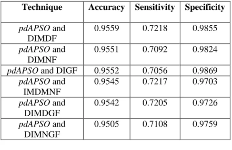
Table 1: Performances of Selected Segmentation Techniques on Drive
Technique Accuracy Sensitivity Specificity
pdAPSO and DIMDF 0.9559 0.7218 0.9855 pdAPSO and DIMNF 0.9551 0.7092 0.9824
pdAPSO and DIGF 0.9552 0.7056 0.9869
pdAPSO and IMDMNF 0.9545 0.7217 0.9703 pdAPSO and DIMDGF 0.9542 0.7205 0.9726 pdAPSO and DIMNGF 0.9505 0.7108 0.9759
Table 2: Performances of Selected Segmentation Techniques on Drive
Technique Accuracy Sensitivity Specificity
Human observer 0.9473 0.7761 0.9725 Mapayi et al., (2015) 0.9469 0.7477 0.9680 Saffarzadeh et al., (2014) 0.9387 N/A N/A Xiao et al., (2013) 0.9529 0.7513 0.9792 Oliveira et al. (2016) 0.9464 N/A N/A Meng et al. (2016) 0.9630 0.7680 0.9827 Mohsen et al. (2017) 0.9469 0.5147 0.9950 Memari et al. (2017) 0.9321 0.8124 0.9505 Sumathi, Vivekanandan & Ravikanth (2018) 0.9671 0.8139 0.9822 Proposed Technique 0.9559 0.7218 0.9855
The result below shows the images produced by the fusion of difference image and pdAPSO algorithm.
Emmanuel Gbenga Dada & Stephen Bassi Joseph CJICT (2018) 6(2) 64-78
(a) (b) (c) (d)
(e) (f) (g) (h)
Fig. 3: (a) & (e) DRIVE Database Gold Standard. (b) & (f) Segmented Vessels Using
pdAPSO and DIMDF. (c) & (g) Segmented Vessels Using pdAPSO and DIMNF. (d) & (h) Segmented Vessels Using pdAPSO and DIGF.
(a) (b) (c)
(d) (e) (f)
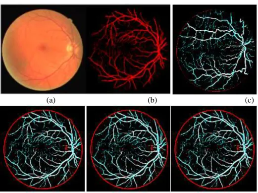
Fig. 4: (a) DRIVE Database Coloured Fundus Image (b) DRIVE Database Gold
Standard (c) Segmented Vessels Using pdAPSO and DIMDF (d) Segmented Vessels
Using pdAPSO and DIMNGF (e) Segmented Vessels Using pdAPSO and DIMDGF
Emmanuel Gbenga Dada & Stephen Bassi Joseph CJICT (2018) 6(2) 64-78
5. Conclusion
In this study, the hybrid of difference image and pdAPSO was used for the segmenting the retinal vessels. It was demonstrated that the proposed vessel segmentation technique is time efficient and yields a high accuracy, specificity rates, and sensitivity when compared to some segmentation methods on DRIVE database. The fusion of pdAPSO and difference image centered on median filter effectively segmented both large and tinny retinal vessels and also reduced erroneous detection near the edges of the optic disc. The results also proved the superiority of the hybrid of
pdAPSO and difference image centered
on median filter compared to difference image centered on mean filter and difference image centered on Gaussian filter fused with pdAPSO for the segmentation of retinal vessels. Moreover, the capacity of the median filter to preserve the boundaries of the
retinal image is the reason for the outstanding performance. This work also proved that the hybridisation of
pdAPSO with difference images
centered on linear filtering technique and difference image centered on the median filter generated an excellent vessel segmentation result. Finally, we deduced from our experiments that our proposed method that integrates difference image with pdAPSO
produced accuracy, specificity and sensitivity that are as good as that of other earlier methods on DRIVE database. Future work, will concentrate on developing more effective segmentation methods using Softcomputing algorithms such as Moth flame Optimisation and Grey Wolf Optimisation algorithms, and also exploit filters such as Gabor Wavelet filters, matched filter, and Frangi’s filters.
References
Akram, M.U. & Khan, S.A. (2013). Multilayered thresholding-based blood vessel segmentation for screening of diabetic retinopathy, Eng Comput, vol. 29, pp. 165-173.
Dada, E. G. & Ramlan, E. I. (2015). Primal-Dual Interior-Point Method Particle Swarm Optimization (pdipmPSO) Algorithm. In: 3rd Int'l Conference on Advances in Engineering Sciences & Applied Mathematics (ICAESAM’2015), London (UK), pp. 117-124. Gergely, K. & Gerinec, A. (2009).
Retinopathy of prematurity epidemics, incidence, prevalence, blindness, Bratislavske lekarske
listy, vol. 111 no. 9, pp. 514 - 517.
Gopi, R.N. & Nageswara, R. P. A. (2013). Particle Swarm Optimization Methods for Image Segmentation Applied In Mammography. Int. Journal of Engineering Research and Applications, vol. 3, Issue 6 pp. 1572-1579.
Jaemin, S., Sang, J. P. & Kyu-Hwan, J. (2017). Retinal Vessel Segmentation in Fundoscopic Images with Generative Adversarial Networks. arXiv:1706.09318v1 [cs.CV] 28 Jun 2017.
Jiang, X. & D. Mojon, D. (2003). Adaptive local thresholding by verification based
multi-Emmanuel Gbenga Dada & Stephen Bassi Joseph CJICT (2018) 6(2) 64-78
threshold probing with application to vessel detection in retinal images, IEEE Transactions on Pattern Analysis and Machine Intelligence, vol. 25 no. 1, pp. 131-137.
K.S. Sreejini, V.K. Govindan (2015). Improved multiscale matched filter for retina vessel segmentation using PSO algorithm, Egyptian Informatics Journal (2015) 16, 253–260. Kande, G. B. Subbaiah, P. V. &
Savithri, T. S. (2010). Unsupervised fuzzy based vessel segmentation in pathological digital fundus images, Journal of Medical Systems, vol. 34, no. 5, pp. 849–858. Kanski, J. J. (2007). Clinical Ophthalmology: A Systematic Approach. 6th Edition, Edinburgh: Butterworth-Heinemann/Elsevier, 491 pages. Klonoff, D. C. & Schwartz, D. M.
(2000). An economic analysis of interventions for diabetes”, Diabetes Care, vol. 23, no. 3, pp. 390-404.
Lupascu, C. A. & Tegolo, D. (2011). Automatic unsupervised segmentation of retinal vessels using self-organizing maps and k-means clustering, In Computational Intelligence Methods for Bioinformatics and Biostatistics. Springer Berlin Heidelberg. pp. 263-274.
Mahalakshmi, S. & Velmurugan, T. (2015). Detection of Brain Tumor by Particle Swarm Optimization, Image Segmentation. Indian Journal of Science and Technology, vol 8 issue 22, pp. 13-19. DOI: 10.17485/ijst/2015/v8i22/79092.
Mapayi, T., Tapamo, J. R & Viriri, S. (2015). Retinal Vessel Segmentation: A Comparative Study of Fuzzy C-means and Sum Entropy Information on Phase Congruency International Journal of Advanced Robotic Systems, vol. 12, no. 133, pp 1-11, doi: 10.5772/60581.
Mapayi, T., Viriri, S. & Tapamo, J. R. (2014). A New Adaptive Thresholding Technique for Retinal Vessel Segmentation Based on Local Homogeneity Information, In Image and Signal Processing. Springer International Publishing, Ser. Lecture Notes in Computer Science, pp. 558-567
Mapayi, T., Viriri, S. & Tapamo, J.R. (2015). Comparative study of retinal vessel segmentation based on global thresholding techniques, Computational and Mathematical Methods in Medicine, vol. 2015 Article ID 895267.
Marin, D., Aquino, A., Gegundez-Arias, M. E. & Bravo, J. M. (January 2011). A New Supervised Method for Blood Vessel Segmentation in Retinal Images by Using Gray-Level and Moment Invariants-Based Features, IEEE transaction on medical imaging, vol.30 no. 1, pp. 146-158.
Marrugo, A. G., Milln, M. S., Cristbal, G., Gabarda, S., Sorel, M. & Sroubek, F. (June, 2012). Image analysis in modern ophthalmology: from acquisition to computer-assisted diagnosis and telemedicine, In SPIE Photonics Europe, International Society for Optics and Photonics, pp. 84360C-84360C.
Emmanuel Gbenga Dada & Stephen Bassi Joseph CJICT (2018) 6(2) 64-78
Mendonca, M., & Campilho A.J. (2006). Segmentation of Retinal Blood Vessels by Combining the Detection of Centerlines and Morphological Reconstruction, IEEE Trans Med Imag., vol 25, pp. 1200-1213.
Meng, L., Zhenshen, M., Chao, L., Guang, Z., & Zhe, H. (2017). Robust Retinal Blood Vessel Segmentation Based on Reinforcement Local Descriptions. Hindawi BioMed Research International, Volume 2017, Article ID 2028946, 9 pages
https://doi.org/10.1155/2017/202 8946
Mohsen, H., Nader, K.S.M., Reza, S., Shadrokh, S. & Kayvan, N. (2017). Retinal blood vessel segmentation for macula detachment surgery monitoring instruments, Int J Circ Theor Appl. 2018;1–15. DOI: 10.1002/cta.2462.
Niemeijer, M. Staal, J. Van Ginneken, B. Loog, M. & Abramoff, M. D. (2004). Comparative study of retinal vessel segmentation methods on a new publicly available database, Proc SPIE Med Imaging, vol. 5370, pp. 648-656.
Oliveira, W. S., Teixeira, J. V., Ren, T. I., Cavalcanti, G. D. C., Sijbers, J. (2016). Unsupervised Retinal Vessel Segmentation Using Combined Filters. PLoSONE, vol. 11, issue 2, e0149943. DOI:10.1371/journal.pone.01499 43.
Qin, L. You, J., Zhang, D. & Bhattacharya, P. (2006). A Multiscale Approach to Retinal Vessel Segmentation Using Gabor Filters and Scale
Multiplication, IEEE International Conference on Systems, Man and Cybernetics (SMC ’06). vol.4, pp. 3521-3527.
Ramaswamy, M., Anitha, D., Priya Kuppamal, S., Sudha, R., Fepslin, S. A. M. (2011). A Study and Comparison of Automated Techniques for Exudate Detection Using Digital Fundus Images of Human Eye: A Review for Early Identification of Diabetic Retinopathy, Int. J. Comp. Tech. Appl., vol. 2 no. 5, pp. 15031516.
Research Section, Digital Retinal Image for Vessel Extraction (DRIVE) Database (2017). Utrecht, The Netherlands, Univ. Med. Center Utrecht, Image Sci. Inst. [Online]. Available: http://www.isi.uu.nl/Research/Da tabases/DRIVE.
Ricci, E. & Perfetti, R. (2007). Retinal blood vessel segmentation using line operators and support vector classification”, IEEE Transactions on Medical Imaging, vol. 26 pp. 1357-1365. Saatchi, S. & Cheng Hung, C.
(December 2007). Swarm Intelligence and Image Segmentation. Swarm Intelligence: Focus on Ant and Particle Swarm Optimization, Book edited by Felix T. S. Chan and Manoj Kumar Tiwari, pp. 532, Itech Education and Publishing, Vienna, Austria ISBN 978-3-902613-09-7. Saffarzadeh, V. M., Osareh, A., &
Shadgar, B. (2014). Vessel Segmentation in Retinal Images Using Multi-Scale Line Operator and K-Means Clustering”,
Emmanuel Gbenga Dada & Stephen Bassi Joseph CJICT (2018) 6(2) 64-78
Journal of medical signals and sensors, vol. 4 no. 2, pp. 1-22. Sumathi, T., Vivekanandan, P.,
Ravikanth, B. (2018). Retinal vessel segmentation using neural network. IET Image Process., 2018, Vol. 12 Iss. 5, pp. 669-678, doi: 10.1049/iet-ipr.2017.0284. Szpak, Z. L. & Tapamo, J. R. (2008).
Automatic and Interactive Retinal Vessel Segmentation, South African Computer Journal, vol. 40, pp. 23-30.
Varughese, S., Gilbert, C., Pieper, C. & Cook, C. (2008). Retinopathy of prematurity in South Africa: an assessment of needs, resources, and requirements for screening programmes, British Journal of Ophthalmology, vol. 92 no. 7, pp. 879- 882.
Wang, Y., Ji, G., Lin, P. & Trucco, E. (2013). Retinal vessel segmentation using multiwavelet kernels and multiscale hierarchical decomposition, Pattern Recognition vol. 46, pp. 2117-2133.
Wen, Y. H., Bainbridge-Smith, A., Morris, A.B. (2007). Automated Assessment of Diabetic Retinal Image Quality Based on Blood Vessel Detection, Proceedings of Image and Vision Computing,
Hamilton, New Zealand, pp. 132-136.
World Health Organization Prevention of Blindness and Visual
Impairment (2016).
http://www.who.int/blindness/ca uses/priority/en/index8.html. Xiao, Z., Adel, M. & Bourennane, S.
(2013). Bayesian Method with Spatial Constraint for Retinal Vessel Segmentation, Computational and mathematical methods in medicine vol. 2013, Article ID 260410.
Yin, Y., Adel, M. & Bourennane, S. (2013). Automatic Segmentation and Measurement of Vasculature in Retinal Fundus Images Using Probabilistic Formulation, Computational and mathematical methods in medicine 2013 Article ID260410.
Zhang, W., Zhou, C. & Bao, X. (2015). Investigation on digital media image processing algorithm based on asynchronous and inertia adaptive particle swarm optimization. International Journal of Signal Processing, Image Processing and Pattern Recognition, vol. 8, no. 2, pp. 65–76.