www.fm.viamedica.pl
Address for correspondence: O.E. Idowu, Neurological Surgery Unit, Department of Surgery, Lagos State University College of Medicine (LASUCOM), Ikeja, Lagos, Nigeria, e-mail: [email protected]
Dimensions, septation, and pattern of
pneumatization of the sphenoidal sinus
O.E. Idowu
1, B.O. Balogun
2, C.A. Okoli
31Neurosurgery Unit, Department of Surgery, Lagos State University College of Medicine (LASUCOM) and Lagos
State University Teaching Hospital (LASUTH), Ikeja, Lagos, Nigeria
2Department of Radiology, Lagos State University College of Medicine (LASUCOM) and Lagos State University
Teaching Hospital (LASUTH), Ikeja, Lagos, Nigeria
3Department of Radiology, JPI Diagnostic Centre, Ikeja, Lagos, Nigeria
[Received 2 August 2009; Accepted 13 October 2009]
The endoscopic endonasal transsphenoidal approach (EEA) to treat sellar, para-sellar, and suprasellar tumours continues to gain increased significance. Due to the close proximity of the sphenoid sinus to the carotid artery and the optic canal, it is very important for surgeons to know the anatomical features and variations of the sphenoid sinus as relevant to EEA.
A prospective study of the sphenoid sinus morphology was carried out on the cranial tomographic (CT) scan images of 60 Nigerian adult patients. The CTs were reviewed regarding the different anatomical variations of the sphenoid sinus: dimen-sions, septation, and pattern of pneumatisation.
There were 37 males and 23 females. The patients’ ages ranged from 18 years to 85 years, with a mean of 47.2 years. There was a main single intersphenoid septum in most patients (95%). The insertion of the septum was usually to the right posteriorly (38%) and in the midline anterior (65%). Although there is usually a main septum, the septa present were multiple in 29 of the sinuses studied. There was no gender difference with respect to the attachment of the main sphenoid sinus septum. The sphenoid anterior, posterior, and transverse dimensions were not significantly dependent on age, but they were longer in males than in females. Sellar pneumatization was present in the majority of the patients (83%), with 4 patients having postsellar pneumatization (6.7%) and 3 patients having presellar pneumatization (5%). There were no cases with conchal pneumatization or lateral pneumatization of the greater wing of the sphenoid.
The present study provides anatomical information about the sphenoid sinus dimensions morphology that is essential for avoiding complications in perform-ing an endoscopic sphenoidotomy. (Folia Morphol 2009; 68, 4: 228–232)
INTRODUCTION
In the last 15 years, the endoscopic endonasal transsphenoidal approach (EEA) to the sellar has been embraced by many neurosurgeons and otorhi-nolaryngologists. The endoscopic endonasal trans-sphenoidal approach is in widespread use for the treatment of sphenoid sinus, and sellar and supra-sellar tumours [9]. Due to the close proximity of the carotid artery, the optic canal, and the skull base to the sphenoid sinus, it is very important for surgeons to know the anatomical features and vari-ations of the sphenoid sinus. This has stimulated more detailed study of the surgical anatomy and relationships of the sphenoid sinus to vital intra-cranial structures.
The sphenoid sinus is situated within the body of the sphenoid bone, and is widely recognized to be an irregular cavity, with the degree of pneumati-zation varying from absent to extensive [9]. It is ex-tremely variable in size, shape, and relation to the sella. It is divided by one or more vertical septa that are often asymmetric.
The sphenoid septum is an important landmark during the endonasal endoscopic trans-sphenoid approach to important structures such as the carot-id artery, optic canal, and skull base [9, 10]. Although there are several reports about the sphenoid sinus, none has been carried out in the African continent south of the Sahara [4, 7, 8, 10, 15]. Due to the large variations in the size and shape of the sphe-noid sinus, it is crucial to know in detail about the dimensional anatomical features of the sphenoid sinus and its septation in order to safely perform the EEA, and even more so in an environment where intra-operative neuronavigation is absent.
The aim of this study is to evaluate the frequen-cy of the different anatomical variations of the sphe-noid sinus that are relevant to the EEA as detected by cranial tomographic scans, and to highlight the these variations in the adult Nigerian population.
MATERIAL AND METHODS
A prospective study of 60 patients was carried out. All the cranial tomographic scans examined in this study were adult Nigerians with a minimum age of 18 years. High-resolution spiral computerized tomographic (CT) images were taken of all the sub-jects at 3 mm thickness at the Radiologic Depart-ment of JPI, Ikeja, Lagos, Nigeria. Exclusion criteria were patients with skull base fractures or previous craniofacial trauma, sphenoid or parasphenoid tu-mours, and those with previous sinus surgery.
The following anatomical variations were not-ed, degree of pneumatization (Conchal, presellar, sellar, and postsellar) [2, 5] and septation type (the presence or absence of the main intersphenoid sep-tum with the side of insertion anteriorly and poste-riorly). The number of accessory septum was also noted. The dimensions were taken under the bone window (window width 4000; window level 400) in the CT images. Each dimension was measured twice and the mean recorded. The data collected was checked for errors prior to analysis.
The mean, median, and standard deviation (SD) of each dimension were computed. Right and left, and gender differences were analyzed. A compari-son was made of the means of the dimensions us-ing the Student’s t-test. The association between continuous variables was investigated by means of Pearson’s correlation coefficient. A probability (p) of less than 0.05 was considered statistically signi-ficant. Data analysis was performed with SPSS ver-sion 14.
RESULTS
Sixty CT images of the sphenoid sinuses of 60 parti-cipants (37 males and 23 females) were reviewed. The patients’ ages ranged from 18 years to 85 years, with a mean and median of 47.2 years (males: 45.6 years; females: 49.9 years) and 45.0 years (males: 43.0 years; females: 55.0 years), respectively. The dimensions measured are summarized in Table 1.
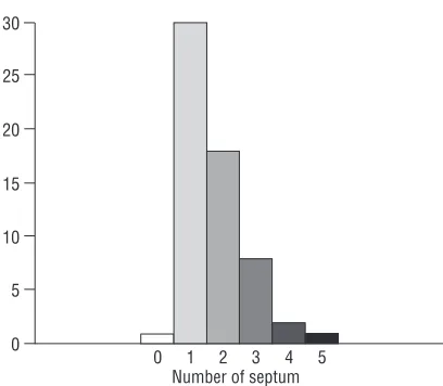
There was no septum in one patient, and a main single intersphenoid septum in most patients (95%). The insertion of the main septum was usually in the midline anteriorly (65%) but to the right posteriorly (38%) in most individuals (Fig. 1). Although there is usually a main septum, the septa present were multi-ple in 29 of the sinuses studied (Fig. 2). There was no gender difference with respect to the attachment of the main sphenoid sinus septum anteriorly (p = 0.753) or posteriorly (p = 0.261). The sphenoid anterior, posterior, and transverse dimensions were not de-pendent on age. Although they were longer in males compared to females, the dimensions were not sta-tistically significant (p was 0.154, 0.670, 0.430, and 0.826 for right anterioposterior, left anterior poste-rior, right transverse, and left transverse dimensions, respectively).
pneu-matization or lateral pneupneu-matization of the greater wing of the sphenoid.
DISCUSSION
The versatility of the trans-sphenoid approach is based on solid foundations: it is the least traumatic route to the sella turcica, it avoids brain retraction, and it provides excellent visualization of the pitu-itary gland and related lesions. It also offers a lower morbidity and mortality rate when compared with
a transcranial procedure [3]. Knowing the details of the dimensions of the sphenoid sinus and the ex-tent of pneumatization can guide the surgeon through difficult corners of the approach.
Table 1. Dimensions of the sphenoid sinus in all patients
Gender Statistic Age Right Left Right Left
(years) anterioposterior anterioposterior transverse transverse
[mm] [mm] [mm] [mm]
Male Mean 45.6 24.8 26.3 19.1 20.3
Median 43.0 23.0 25.0 18.0 19.0
Minimum 18.0 9.0 14.0 7.0 13.0
Maximum 85.0 41.0 50.0 35.0 40.0
SD 17.11 0.72 0.83 0.68 0.55
SEM 2.81 0.12 0.14 0.11 0.09
Female Mean 49.9 22.1 25.4 17.7 19.9
Median 55.0 22.0 24.0 16.0 20.0
Minimum 22.0 9.0 12.0 7.0 11.0
Maximum 78.0 36.0 42.0 34.0 36.0
SD 17.30 0.65 0.77 0.64 0.64
SEM 3.61 0.14 1.16 0.13 0.13
Total Mean 47.2 23.8 26.0 18.6 20.1
Median 45.0 23.0 25.0 17.8 19.0
Minimum 18.0 9.0 12.0 7.0 11.0
Maximum 85.0 41.0 50.0 35.0 40.0
SD 17.16 0.70 0.80 0.67 0.58
SEM 2.22 0.09 0.10 0.09 0.08
SD — standard deviation; SEM — standard error of mean
Figure 1. The location of the main sphenoid sinus septum
attachment posteriorly.
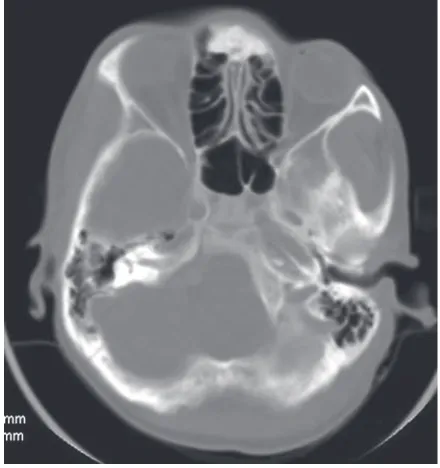
Figure 2. Computerized axial tomographic scan showing multiple
There is usually an intersphenoid septum within the sphenoid sinus. This septum is removed to ex-pose the floor of the sella. The septum usually devi-ates to one side, dividing the sinus into two unequal cavities, thereby resulting in an asymmetrical appear-ance of the sella turcica floor, of which the surgeon must be aware. In our series, we noted that although there is usually a single main septum, multiple sep-tation occurs frequently (Fig. 3).
The present data regarding the vertical and hori-zontal dimensions of the sphenoid sinus, as shown in Table 1, indicate that the dimensions of the sphe-noid sinus can vary tremendously. This might be due to large variations in the shape of the sphenoid si-nus: variations that often are encountered in daily clinical practice. As for differences between the sex-es, almost all the dimensions measured were great-er in males than in females. Although most of the differences were small, surgeons should at least keep this fact in mind when performing an EEA on fe-male patients.
It is wise to use extreme caution while removing the terminal septum as this is not usually in the midline.
The sella turcica is seen as a prominence in the roof of a well-pneumatized sphenoid sinus and is known as the sellar bulge [13]. This is considered one of the most important surgical landmarks to the sellar floor. High-resolution cranial tomographic scan may show pneumatization of the sphenoid si-nuses from as early as 2 years of age. Pneumatiza-tion progresses in an inferior and posterolateral di-rection. The pneumatized basisphenoid plate often extends to, but not past, the spheno-occipital syn-chondrosis in the mature sphenoid sinus. The sinus attains its mature size by the age of 14 years [14].
Figure 3. The number of septa within the sphenoid sinus.
The degree of pneumatization of the sphenoid si-nus varies considerably. The pattern of pneumatiza-tion of the sphenoid sinus significantly affects safe access to the sella. A highly pneumatized sphenoid sinus may distort the anatomic configuration, so in these cases it is extremely important to be aware of the midline when opening the sella to avoid acci-dental injury to the carotid and optic nerves. In the series by Hamid et al. [6], presellar, sellar type and postsellar pneumatization noted were 21%, 54.7%, and 22.3% respectively. The lateral pneumatization of the greater wing of the sphenoid, leading to a capacious sinus, was found in 15.9% of cases [6]. Our high number of sellar type pneumatizations may be due to our policy of excluding those with sellar diseases which might have invaded the sinus.
The conchal non-pneumatized sphenoid is con-sidered a relative contraindication to the trans-sphe-noid approach to the sella [12]. However, with the surgeon informed in advance, different tools can make such an approach feasible. The availability of intraoperative fluoroscopic imaging or intraopera-tive navigational devices can be used to confirm surgical landmarks, making it possible to access the sella through the sphenoid sinus safely even in poorly pneumatized cases [11]. In our series, conchal pneu-matization was absent but was noted in 2% of pa-tients by Hamid et al. [6]. The rarity of conchal pneu-matization agrees with different studies [1, 11, 16].
CONCLUSIONS
In conclusion, there are different anatomical con-figurations of the sphenoid sinus. These different anatomical variations should be kept in mind, and problems anticipated, in order to avoid difficulty during surgery. It is imperative that clinicians deter-mine the anatomic features of the sphenoid sinus and its septum whenever EEA is contemplated. The stated features in this study are important to keep in mind preoperatively while planning an EEA.
REFERENCES
1. Banna M, Olutola PS (1983) Patterns of pneumatiza-tion and septapneumatiza-tion of the sphenoidal sinus. J Can Assoc Radiol, 34: 291–293.
2. Batra PS, Citardi MJ, Gallivan RP, Roh HJ, Lanza DC (2004) Software-enabled computed tomography ana-lysis of the carotid artery and sphenoid sinus pneuma-tization patterns. Am J Rhinol, 18: 203–208.
4. Elwany S, Yacout YM, Talaat M, El-Nahass M, Gunied A, Talaat M (1983) Surgical anatomy of the sphenoid sinus. J Laryngol Otol, 97: 227–241.
5. Hamberger CA, Hammer G, Norlen G (1961) Trans-sphenoidal hypophysectomy. Arch Otolaryngol, 74: 2–8.
6. Hamid O, El Fiky L, Hassan O, Kotb A, El Fiky S (2008) Anatomic variations of the sphenoid sinus and their impact on trans-sphenoid pituitary surgery. Skull Base, 18: 9–16.
7. Hayakawa K, Yoshikawa H, Suzuki M (2003) Varia-tions in reciprocal distances between the ethmoid sinus, sphenoid sinus and posterior orbit: measure-ment on CT scans [in Japanese]. Juntendo Med J, 49: 89–96.
8. Ikeda T, Iinuma T (1992) Surgical anatomy of the sphe-noid sinus — development and intersinus septum [in Japanese]. Nippon Jibiinkoka Gakkai Kaiho, 95: 214– –223.
9. Kieff DA, Busaba N (2002) Treatment of isolated noid sinus inflammatory disease by endoscopic sphe-noidotomy without ethmoidectomy. Laryngoscope, 112: 2186–2188.
10. Kim HU, Kim SS, Kang SS (2001) Surgical anatomy of the natural ostium of the sphenoid sinus. Laryngoscope, 111: 1599–1602.
11. Liu S, Wang Z, Zhou B, Yang B, Fan E, Li Y (2002) Relat-ed structures of the lateral sphenoid wall anatomy stud-ies in CT and MRI [in Chinese]. Lin Chuang Er Bi Yan Hou Ke Za Zhi, 16: 407–409.
12. Massoud AF, Powell M, Williams RA, Hindmarsh PC, Brook CGD (1997) Trans-sphenoidal surgery for pitu-itary tumors. Arch Dis Child, 76: 398–404.
13. Romano A, Zuccarello M, Van Loveren HR, Keller JT (2001) Expanding the boundaries of the trans-sphenoidal ap-proach: a micro anatomic study. Clin Anat, 14: 1–9. 14. Scuderi AJ, Harnsberger HR, Boyer RS (1993)
Pneuma-tization of the paranasal sinuses: normal features of importance to the accurate interpretation of CT scans and MR images. Am J Roentgenol, 160: 1101–1104. 15. Smith WC, Boyd EM, Parsons DS (1996) Pediatric