This is an open access journal, and articles are distributed under the terms of the Creative Commons Attribution-Non Commercial-ShareAlike 4.0 License, which allows others to remix, tweak, and build upon the work non-commercially, as long as appropriate credit is given and the new creations are licensed under the identical terms.
© 2019 Journal of Advanced Pharmacy Education & Research | Published by SPER Publication
125
Nigella sativa protects Kidneys against Metabolic disorders
induced by high fat diet in Rats
Rasha Alshali
1,4, Aziza Alrafiah
2,4*, Ebtisam Al-ofi
3,4, Abeer Hanafy
5,6, Atif Hasan
7, Nora Bawaked
4,
Amina Fallata
41Department of Anatomy, Faculty of Medicine, King Abdulaziz University, Jeddah, Saudi Arabia. 2 Department of Laboratory Technology, Faculty of Applied Medical Sciences,
King Abdulaziz University, Jeddah, Saudi Arabia. 3 Department of Physiology, Faculty of Medicine, King Abdulaziz University, Branch of Sulaymaniyah, Jeddah, Saudi Arabia. 4Neuroscience Research Unit, Faculty of Medicine, King Abdulaziz University, Jeddah, Saudi Arabia. 5 Department of Pharmacology, Faculty of Pharmacy, King Abdulaziz
University, Jeddah 21589, Kingdom of Saudi Arabia. 6 Department of Pharmacology, Faculty of Veterinary Medicine, Kafrelsheikh University, Kafrelsheikh 33516, Egypt. 7
Department of Anatomy and Embryology, Faculty of Veterinary Medicine, Kafrelsheikh University, Kafrelsheikh 33516, Egypt.
Correspondence:
Aziza Alrafiah. P.O Box 80200, Jeddah 21589, Saudi Arabia. Email: [email protected].
ABSTRACT
Introduction: High fat diet (HFD) intake initiate variety of metabolic disorders as insulin resistance and type 2 diabetes. Adipose tissue secretes adiopocytokines, which cause lipid peroxidation in cell membrane of vital organs like kidneys. Nigella sativa (NS) is a famous condiment; it is a medicinal herb, which contains number of active contents and has no toxic effects. Methods: We applied 8 weeks HFD feeding protocol to induce metabolic disorders in rats which were divided randomly into three groups; control group, HFD group and NS group. Kidney function parameters (urea, uric acid and creatinine), Lipid peroxidation marker (Malondialdehyde MDA) and oxidative stress biomarkers (glutathione GSH – superoxide dismutase SOD) were measured. Paraffin sections were stained with different stains to examine histological architecture and collagen contents in renal tissues. Results: HFD feeding protocol resulted in disturbances in kidney function tests; there were rise in urea, uric acid and creatinine values; increased values of lipid peroxidation marker MDA and decreased values of oxidative stress markers GSH and SOD were observed. Histologically, there was batches of affected renal areas showing glomerulomegally, shortened epithelium of proximal convoluted tubules, dilatation of blood vessels (glomerular capillaries and rete mirabile). In NS group, kidney function tests were normal and plasma levels of biochemical markers MDA, GSH and SOD were around its normal values. NS preserved normal renal architecture. Conclusion: Administration of Nigella sativa with HFD ameliorated the adverse effects of HFD on kidneys through a mechanism utilizing NS antioxidant activity.
Keywords:High fat diet – kidney - metabolic disorders - Nigella sativa – antioxidant activity.
Introduction
High fat diet (HFD) consumption induces obesity in both humans and animals [1, 2]; it results in elevation of plasma level of free fatty
acids and production of adipocytokines and reactive oxygen species (ROS) which adversely affect non adipose tissues [3, 4];
excess free fatty acids interfere with the mechanism by which
insulin prevent output of hepatic glucose [5]. Obesity is
accompanied by low grade inflammation that finally leads to hepatic, renal, and cardiovascular disorders, in addition to type 2 diabetes [4, 6 7]. Previous studies showed that sustained HFD
intake causes a pre-diabetic state leading to renal dysfunction in lab model animals [8]; it was found that, consumption of HFD for
4, 8 and 16 weeks resulted in insulin resistance at 4 weeks, renal steatosis at 8 weeks and renal injury at 16 weeks in mice [9]. HFD
induced obesity and its subsequent metabolic syndrome come with a cluster of metabolic disorders (including type 2 diabetes) which increase the possibility of various renal problems [10];
moreover, recent studies proved that renal dysfunction is intimately related to uncontrolled diabetes [11]. Twelve weeks of
HFD feeding of female rats were enough to make them obese and show abnormal renal changes [12]. Previous studies stated that,
diet induced obesity is considered as a risk factor of development
Access this article online
Website: www.japer.in E-ISSN: 2249-3379
How to cite this article:Rasha Alshali, Aziza Alrafiah, Ebtisam Al-ofi, Abeer Hanafy, Atif Hasan, Nora Bawaked, Amina Fallata. Nigella sativa protects Kidneys against Metabolic disorders induced by high fat diet in Rats. J Adv Pharm Edu Res 2019;9(1):125-133.
of renal carcinoma [13, 14]. HFD long term feeding protocols cause
a variety of renal histological changes like dilatation of glomerular capillaries, shortened tubular epithelium, infiltration of inflammatory cells, thickening of glomerular basement membrane (GBM) and fibrosis [12, 15, 16]; however, short-term
HFD feeding protocol showed neither histological nor biochemical renal changes [17]. Biochemically, HFD long time
intake induces inflammatory cytokines production specially interleukin 6 (IL-6) and tumor necrosis factor–alpha (TNFα) which point to renal dysfunction [18, 19].
Nowadays, herbal medicine is a growing science and there is great interest in plants that have therapeutic effects [20]. Nigella
sativa (black seed) is a known plant that has valuable general health boosting effects [21], its oil contains more than 100
components like trace elements, aromatic oils and vitamins [22].
Many studies showed that N. sativa has divergent therapeutic effects such as antibacterial [23], antidiabetic [24], estrogenic [25],
diuretic, hypotensive [26], anti-tumor [27], anti-inflammatory [28],
antioxidant [29] and hepatic tissue protection [30, 31] effects. Several
studies showed that administration of various doses of N. sativa
has no toxic effects, and revealed that N. sativa has wide safety margin [32, 33]; in addition, normal plasma levels of urea and
creatinine beside normal renal architecture confirm that N. sativa
do not affect kidney function and structure [34].
In the current study, we induced obesity and metabolic syndrome in rats by using HFD feeding protocol (8 weeks); N. sativa powder was orally administrated in a daily dose-dependent manner; then kidney functions and structure were examined to elucidate the therapeutic and protective effect of Nigella sativa on metabolically affected kidneys.
Materials and Methods
Animals
For this study, eighteen adult Albino wester rats, aged 8 weeks, and weighing 160-200 g were purchased from King Fahd Medical Research Center, Jeddah, Saudi Arabia. The animals were accommodated at 25 °C with a 12 h light/12 h dark cycle, 55 ± 10% humidity. This study was conducted according to the guidelines of dealing with experimental animals that are followed in the KFMRC. The study ethical approval number (222-19) was obtained from the Medical Research Ethics Committee, Faculty of Medicine, King Abdulaziz University, Jeddah, KSA
Materials:
The black cumin (Nigella sativa -NS) seeds were purchased from the local market, Jeddah. KSA. NS powder was dissolved daily in distilled water and given to rats by oral gavage into the stomach. High fat diet composed of 20% saturated fat (butter; purchased from local markets in Jeddah), 2% cholesterol and 0.5% bile acid salts.
Experimental design
Rats were divided randomly into three groups including 6 rats each. Group 1: control rats which fed with standard diet for eight weeks, group 2: fed with high fat diet (HFD) at a dose of 6 ml/day for 8 weeks, group 3: fed high HFD for 4 weeks then
added Nigella sativa seed powder at a dose of 300 mg/kg/day for 4 weeks.
After 8 weeks, blood samples were obtained from the tail tip, and glucose levels were determined by the glucometer method (Accutrend Alpha; Boehringer, Mannheim, Germany). The animals were anesthetized and samples of blood were collected from the rats by cardiac puncture; blood samples were centrifuged to obtain serum for the assessment of oxidative biomarkers and kidney function tests.
Serum insulin level:
Serum insulin was assayed by ELISA (Millipore, Billerica, MA, USA) kit that uses a coated plate with anti-rat insulin monoclonal antibodies and an ELISA reader.
Kidney function tests:
Urea was measured according to the method of Berthelot reagent as described by [35]. Creatinine was measured as described by [36].
Uric acid was measured according to the method of Enzyme Colorimetric as described by [37].
Serum concentration of tumor necrosis factor
– alpha (TNF-α):
TNF-α serum concentration was determined using ELISA Kit (R&D), catalog number (RTA00). Instructions of the manufacturer were followed using ELISA reader to quantify the reaction.
Determination of Lipid Peroxide (MDA)
Malondaldehyde (MDA) was determined in serum spectrophotometrically as thiobarbituric acid reactive substances (TBARS) [38].
Determination of Glutathione (GSH)
GSH level was determined in serum as described by Ellman [39].
Serum Superoxide Dismutase (SOD) activity
SOD activity was estimated according to the method adopted by Marklund [40].
Statistical analysis
The results were represented as mean ± standard deviation (SD) and were analyzed by the Statistical Package of Social Sciences (SPSS) program version 16. We used one-way analysis of
variance (ANOVA). ANOVA was followed by Bonferroni’s
multiple comparison test to compare different groups [41].
P<0.05 was considered significant.
Histological Techniques
Samples from kidneys fixed in 10% formalin were routinely processed and impregnated in paraffin for sectioning [42]. 5 μm
thick paraffin sections were obtained for staining. We used hematoxylin and eosin (H&E) stain to examine histological architecture; Masson’s trichrome stain (MT) and periodic acid
Schiff (PAS) technique were used to detect collagen and proteoglycan in glomeruli and interstitial renal tissue [42]. It is
known that glomerular basement membrane (GBM) of the glomerular epithelial tuft is formed of three laminae namely lamina densa (collagen), laminae rara interna and externa (proteoglycan rich in heparan sulfate) of epithelial origin [42]. It is
also known that, collagen stained blue with Masson’s trichrome
Stained sections were mounted and examined under light microscope. Diameter of renal corpuscle (between vascular pole and urinary pole) and epithelial height of proximal convoluted tubules were measured by using image analyzer software (Leica Qwin 500).
Results
Effect of NS on serum insulin level
Serum insulin level was significantly elevated in rats on HFD but animals given HFD and NS showed nearly normal insulin level as shown in table (1).
Effect of NS on kidney function
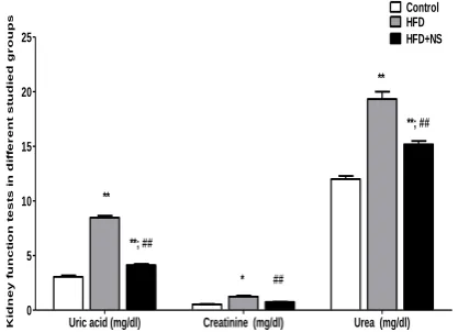
Urea, creatinine and uric acid serum levels were elevated significantly in rats fed on HFD. Adding NS to the HFD alleviated the deterioration in the kidney function produced by HFD significantly.
Effect of NS on serum concentration of
TNF-α
TNF-α serum concertation was elevated significantly in rats fed on HFD. On the other hand, rats fed on HFD showed significant decrease in TNF-α concentration in serum as shown in Table (1).
Effect of NS on MDA
A significant increase in MDA levels was shown in serum of HFD-treated group compared to the group fed on normal diet, while administration of NS significantly reduced MDA level compared to the group fed on HFD (Table 1).
Effect of NS on Glutathione (GSH)
Compared to the control group, HFD induced significant reduction in the GSH level. Oral NS treated group combined with HFD resulted in a significant increase of GSH level when compared to the HFD-treated group (Table 1).
Effect of NS on Superoxide Dismutase (SOD)
As shown in table 1, HFD resulted in increased SOD serum level when compared to control group, while NS increased the SOD level significantly in comparison with the HFD group.
Uric acid (mg/dl) Creatinine (mg/dl) Urea (mg/dl) 0 5 10 15 20 25 Control HFD HFD+NS * **; ## ** ## ** ## **; ## K id n e y f u n c ti o n t e s ts i n d if fe r e n t s tu d ie d g r o u p s
Figure 1:Comparison of serum levels of kidney function tests
in the different studied groups. Values are expressed as mean ±SD. *compared to control (*P <0.05; ** P <0.001);
##compared to HFD (P <0.001).
Table 1: Comparison of serum level of glucose, Insulin,
GSH, SOD, MDA and TNF-α in the different studied
groups.
Control HFD HFD + NS
Glucose (mg/dl) 101.2 ± 12.4 201.0 ± 8.8* 111.1 ± 18.6#
Insulin (µIU/ml) 3.97 ± 0.20 1.29 ± 0.17* 3.92 ± 0.46#
GSH (mol/l) 7.25 ± 0.36 1.32 ± 0.36* 9.76 ± 1.09#
SOD (u/ml) 167.67 ± 10.67 98.83 ± 5.41* 178.67 ± 16.02#
MDA (nmol/l) 0.755 ± 0.11 2.07 ± 0.35* 0.700 ± 0.15#
TNF-α (pg/ml) 10.7 ± 0.7 28.2 ± 3.1* 17.3 ± 0.9#
Values are represented as mean±SD, 𝑛 = 6. ∗compared to control group (P <0.05). #compared to HFD group (P <0.05).
Table 2: Comparison of renal corpuscle diameter and proximal convoluted tubules (P.C.T.) epithelial height in
the different studied groups.
Control HFD HFD + NS Renal corpuscle
diameter 32.94 ± 1.46 40.66 ± 1.21* 31.20 ± 1.19# P.C.T. epithelial
height 5.98 ± 0.62 2.56 ± 0.44* 4.37 ± 0.24#
Values are represented as mean±SD, 𝑛 = 6. ∗compared to control group (𝑃
< 0.05). # compared to HFD group (𝑃 < 0.05).
Histological findings
After eight weeks of high fat diet feeding of experimental rats (HFD group), several histopathological changes were recorded in renal tissues when compared with that in control rats.
In sections stained with H&E:
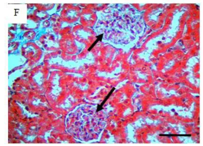
Affected areas of renal tissues in HFD group were batches in outline; in the cortex, marked abnormal changes occurred in glomeruli and proximal convoluted tubules; marked increase in glomerular size was recorded (fig 2D), glomerular capillaries appeared thin walled with wider lumens (fig 2D) than that of control group (fig 2B). proximal convoluted tubules in HFD group showed marked luminal dilatation (fig 2C, D) due to decreased epithelial height (fig 2D). In cortico-medullary and outer medullary zones, in HFD group, renal tubules showed marked luminal dilatation and thinning of their walls (fig 3 C, D) when compared with control rats (fig 3 A, B); In addition, rete mirabile in HFD group showed marked vasodilatation (Fig 3 E, F); this was not the same in control ones (Fig 3 A). In N. sativa
treated group (NS group–group3), the histological findings were nearly similar to that in control group (Fig 2 E, F).
In Masson’s trichrome and PAS stained sections:
In the renal cortex, due to presence of collagen and proteoglycan in glomerular basement membrane (GBM), glomeruli stained blue with Masson’s trichrome stain and magenta with PAS
staining technique.
In Masson’s trichrome stained sections, glomeruli of control
convoluted tubules, the brush border of their epithelial cells stained magenta in control (Fig 5 A) and NS groups (Fig 5C) but in HFD group, the magenta color of the epithelial brush border was markedly diminished (Fig 5 B). Distal convoluted tubules were PAS negative in all groups (Fig 5 A, B, C).
Taken together, In HFD group, abnormal renal tissue architecture was observed in many glomeruli and its related tubules but treatment with N. sativa protects kidneys against HFD adverse effects in NS group.
Figure 2:Photomicrographs of hematoxylin and eosin (H&E)
stained renal cortex. A, B: lower and higher magnifications of renal cortex of control group (Co-group) revealed normal tissue architecture. C, D: lower and higher magnifications of renal cortex of HFD group (group 2) showed large number of
dilated tubules (C, arrows), enlarged glomeruli (D - stars), Shortened epithelial cell in proximal convoluted tubules with lost nuclei (D- arrowheads) and thin walled dilated glomerular capillaries (D- arrows). E, F: lower and higher magnifications of renal cortex of NS group (group-3), tissue architecture is nearly
Figure 3:Photomicrographs of hematoxylin and eosin (H&E) stained renal medulla and cortico - medullary junction. A, B:
lower and higher magnifications of control group showing normal tubules (B-arrows) and ducts (B- stars). C, D: lower and higher magnifications of HFD group showing dilated thin walled tubules and ducts (C, D arrows). E, F: lower and higher
magnifications of cortico-medullary junction showing dilated blood vessels of rete mirabile HFD group (E, F arrows), notice normal sized rete mirabile in control group (A-arrows). Bars =
Figure 4:Photomicrographs of Masson’s trichrome (MT)
stained sections of renal cortex detecting collagen content. A, B: lower and higher magnifications of renal cortex of control
group, glomeruli stained blue (arrows) due to its normal collagen content in its basement membranes. C, D: lower and
higher magnifications of HFD group showing glomeruli (arrows) stained faint blue due to decreased collagen content.
E, F: lower and higher magnifications of NS group showing nearly normal collagen content in glomeruli (arrows) as in
control group. Bars = 40 µm.
Figure 5:Photomicrographs of Periodic acid Schiff (PAS)
stained sections of renal cortex detecting proteoglycan content in glomeruli. A: normal proteoglycan content stained magenta color in glomeruli of control group (arrows). B: glomeruli stained faint magenta color in HFD group indicating decreased
proteoglycan content (stars). C: NS group showed normal magenta color of glomeruli (arrows). In HFD group, luminal epithelium of proximal convoluted tubules showed sloughed brush borders and lost nuclei (B – arrows). Bars = 40 µm.
Discussion
In the current study, N. sativa oral administration ameliorated the adverse effects of HFD on the rat kidney. Biochemically, urea and creatinine are biochemical byproducts that mostly excreted through kidneys [43], they are commonly used in kidney function
tests [44, 45]; in the current study, there was increase in plasma urea
and creatinine levels in HFD treated group which returned back to its normal values after addition of N. sativa to the feeding protocol in NS group (group 3). TNF-α (tumor necrosis factor –
alpha) is a pro-inflammatory cytokine produced by macrophages and adipocytes [46]; it induces lipolysis and generates ROS
(reactive oxygen species) which might cause damage of glomeruli and tubules [47, 48]. TNF-α plasma level was markedly increased in
HFD group and preserved its normal level in group 3 (NS group). Malondialdehyde (MDA) is a lipid peroxidation end product [49],
it is used to detect oxidative stress and lipid peroxidation; its plasma level was elevated in HFD group indicating occurrence of lipid peroxidation process while in group 3 where N. sativa was co- administrated with HFD, it preserved its normal values. Oxidative stress biomarkers, glutathione (GSH) and superoxide dismutase (SOD) plasma levels were markedly decreased in HFD group, and similar results were obtained in a biochemical study on rats [50]; while in group 3, N. sativa maintained GSH and SOD
plasma levels indicating its powerful antioxidant effect. Previous studies confirmed the antioxidant activity of NS [51, 52], and it is
thought that NS performs its protective antioxidant effect through its thymoquinone basic component [51, 53].
Persistent HFD intake affects the body in time-dependent manner. It was found that short term HFD intake in rats produced neither marked metabolic disorders nor renal dysfunction [17]. On the other hand, long time HFD intake in rats
produced cluster of metabolic and histologic disorders [54].
tubular epithelium are marked abnormal morphological renal changes observed in HFD prone animal models [15 15, 55]; similar
abnormal histological findings were recorded in our study (8 weeks), in addition to dilated renal blood vessels (rete mirabile) in the corticomedullary zone. Severe morphological renal lesions like glomerulosclerosis, inflammatory cells infiltration, fibrosis, thickening of glomerular basement membrane (GBM) and nephron degradation were recorded in long term (11 months) HFD administration protocol [54] but not in our study. It is worthy
to state that, severe pathological changes due to obesity create microenvironment encourage carcinogenesis in affected tissues
[13, 14].
In the current study, Masson,s trichrome and PAS stains revealed
no thickening of GBM in which its basal lamina is formed of collagen and proteoglycan. In contrast, the faint color of the glomeruli of HFD group indicated marked decrease in collagen and proteoglycan content. Comparable results were obtained in earlier study where high fat diet prevented GBM thickening in diabetic rats and reduced its thickening in control ones [54]. In our
study, daily oral administration of N. sativa with HFD prevented the decrease of collagen and proteoglycan content of the GBM. Proximal convoluted tubules in HFD treated group showed reduced epithelial cells heights due to sloughing of its brush borders. Shortened tubular epithelium is a constant abnormality in HFD treated rats [12, 15]. However, addition of NS to HFD
feeding protocol ameliorated its effect on tubular epithelium in the examined rats.
In conclusion, N. sativa (NS) protects kidneys against the adverse effects of high fat diet (HFD) through its powerful antioxidant components. our study revealed direct protective effect of NS on kidneys. We recommend its use as food additive due to its safety and curative properties. Further studies are necessary to elucidate the effect of N. sativa on kidneys suffering from long term HFD complications and renal cell carcinoma.
Competing interests
The authors declare that they have no competing interests.
Acknowledgment
Authors would like to acknowledge Neuroscience research unit at King AbdulAziz University, Jeddah, Saudi Arabia
References
1. Rothwell NJ & Stock MJ (1984) The development of obesity in animals – the role of dietary factors. Clin Endocrinol Metab 13, 437–449
2. Warwick ZS & Schiffman SS (1992) Role of dietary fat in calorie intake and weight gain. Neurosci Biobehav Rev 16, 585–596
3. Unger R. H., Clark G. O., Scherer P. E., Orci L. (2010). Lipid homeostasis, lipotoxicity and the metabolic syndrome. Biochim. Biophys. Acta 1801, 209–214 4. Gregor M. F., Hotamisligil G. S. (2011). Inflammatory
mechanisms in obesity. Annu. Rev. Immunol. 29, 415–
445
5. Jensen MD: Role of body fat distribution and the metabolic complications of obesity. J Clin Endocrinol Metab 2008, 93:S57-63
6. Haslam D. W., James W. P. (2005). Obesity. Lancet 366, 1197–1209
7. Brunt E. M. (2010). Pathology of nonalcoholic fatty liver disease. Nat. Rev. Gastroenterol. Hepatol. 7, 195–203 8. Thethi T, Kamiyama M, Kobori H: The Link Between the
Renin-AngiotensinAldosterone System and Renal Injury in Obesity and the Metabolic Syndrome. Curr Hypertens Rep 2012, 14:160-169.
9. Kume S, Uzu T, Araki S, Sugimoto T, Isshiki K, Chin-Kanasaki M, Sakaguchi M, Kubota N, Terauchi Y, Kadowaki T, Haneda M, Kashiwagi A, Koya D: Role of altered renal lipid metabolism in the development of renal injury induced by a high-fat diet. J Am Soc Nephrol 2007, 18:2715-2723.
10. de Castro, Uberdan Guilherme Mendes, Dos Santos, Robson Augusto Souza, Silva ME, de Lima WG, Campagnole-Santos MJ, Alzamora AC: Age-dependent effect of high-fructose and high-fat diets on lipid metabolism and lipid accumulation in liver and kidney of rats. Lipids Health Dis 2013, 12:136.
11. Reutens A: Epidemiology of Diabetic Kidney Disease. Med Clin North Am 2013, 97:1-18.
12. Altunkaynak ME, Ozbek E, Altunkaynak BZ, Can I, Unal D, Unal B (2008): The effects of high-fat diet on the renal structure and morphometric parametric of kidneys in rats. J Anat, 212:845-852.
13. Calle E. E., Rodriguez C., Walker-Thurmond K., Thun M. J. (2003). Overweight, obesity, and mortality from cancer in a prospectively studied cohort of U.S. adults. N. Engl. J. Med. 348, 1625–1638
14. Renehan A. G., Tyson M., Egger M., Heller R. F., Zwahlen M. (2008). Body-mass index and incidence of cancer: a systematic review and meta-analysis of prospective observational studies. Lancet 371, 569–578 15. Deji N, Kume S, Araki S, Soumura M, Sugimoto T, Isshiki
K, Chin-kanasaki M, Sakaguchi M, Koya D, Haneda M, Kashiwagi A, Uzu T, Sakaguchi M, Koya D, Haneda M (2009): Structural and functional changes in the kidneys of high-fat diet-induced obese mice. Am J Physiol Renal Physiol, 118-126.
16. Amaral LSB, Silva JA, Trindade TM, Ribas WBD, Macedo CL, Coimbra TM, Belo NO, Magalhaes ACM, Soares TJ (2014) Renal Changes in the Early Stages of Diet-Induced Obesity in Ovariectomized Rats Physiol. Res. 63: 723-732 17. Crinigan C, Calhoun M, Sweazea KL (2015) Short-Term High Fat Intake Does Not Significantly Alter Markers of Renal Function or Inflammation in Young Male Sprague-Dawley Rats J Nutr Metab. 2015: 157520.
19. Ruster C, Wolf G: Adipokines promote chronic kidney disease (2013). Nephrol Dial Transplant, 28 Suppl 4:8-14. 20. Schwartsmann G, Ratain MJ, Cragg GM, Wong JE, Saijo N, Parkinson DR, Fujiwara Y, Pazdur R, Newman DJ, Dagher R, and Di Leone L. 2002. Anticancer Drug Discovery and Development throughout the World. J Clin Oncol, 20: 47s-59s.
21. Butt MS, Sultan MT (2010). Nigella sativa: Reduces the risk of various maladies. Crit Rev Food Sci Nutr;50: 654-65.
22. Ali BH and Blunden G. 2003. Pharmacological and toxicological peroperties of Nigella Sativa. J Phytotherapy Res, 17: 299-305.
23. Rakhshandeh H, Vahdati-Mashhadian N, and khajekaramadini M. 2011. In vitro and in vivo study of the antibacterial effects of Nigella sativa methanol extract in dairy cow mastitis, Avicenna J Phytomed, 1: 29-35. 24. Kanter M, Meral I, Yener Z, Ozbek H, Demir H. 2003.
Partial regeneration/proliferation of the beta-cells in the islets of Langerhans by Nigella sativa L. in streptozotocininduced diabetic rats. Tohoku J Exp Med, 201: 213-219.
25. Parhizkar S, Latiff L, Rahman S, Dollah MA. 2011. Assessing estrogenic activity of Nigella sativa in ovariectomized rats using vaginal cornification assay. Afr J Pharm Pharmacol, 5: 137-142.
26. Zaoui A, Cherrah Y, Lacaille-Dubois MA, Settaf A, Amarouch H, and Hassar M. 2000. Diuretic and hypotensive effects of Nigella sativa in the spontaneously hypertensive rat. Therapie, 55: 379-382.
27. Turkdogan MK, Agaoglu Z, Yener Z, Sekeroglu R, Akkan HA, and Avci ME. 2001. The role of antioxidant vitamins (C and E), selenium and Nigella sativa in the prevention of liver fibrosis and cirrhosis in rabbits: new hopes. Dtsch Tierarztl Wochenschr, 108: 71-73.
28. Houghton PJ, Zarka R, de las Heras B, Hoult JR (1995). Fixed oil of Nigella sativa and derived thymoquinone inhibit eicosanoid generation in leukocytes and membrane lipid peroxidation. Planta Med; 61:33-6.
29. Zineb Mammad, K. Mammad, T. Aqeil, A. Kribii, and K. Ounine (2017) Antibacterial and Antioxidant activity of Nigella Sativa. International Journal of Innovation and Scientific Research Vol. 31 No. 1 Jun. pp. 167-172 30. EL-Kholy WM, Hassan HA, Nour SE, Abe Elmageed ZE,
Matrougui K. 2009. Hepatoprotective effects of Nigella
sativa and bees’ honey on hepatotoxicity induced by
administration of sodium nitrite and sunset yellow. FASEB J, 23: 733
31. Ahmad A, Husain A, Mujeeb M, et al (2013). A review on therapeutic potential of Nigella sativa: A miracle herb. Asian Pac J Trop Biomed; 3:337-52.
32. Al-Ghamdi MS. 2003. Protective effect of Nigella sativa seeds against carbon tetrachloride-induced liver damage. Am J Chin Med, 31:721-728.
33. Al Ameen NM, Altubaigy F. Jahangir T. Mahday IA, Esmaeel Abdurrahman Mohammed EA and Musa OAA. 2011. Effect of Nigella sativa and bee honey on pulmonary, hepatic and renal function in Sudanese in Khartoum state. J Med Plant Res, 5: 6857-6863.
34. Mohammad Aziz Dollah, Saadat Parhizkar, Mohammad Izwan (2013). Effect of Nigella sativa on the kidney function in rats. Avicenna Journal of Phytomedicine Vol. 3, No. 2, 152-158
35. Hammes, S. R. (2003). The further redefining of steroid-mediated signaling. Proc Natl Acad Sci,USA 100(5): 21680–2170.
36. Agbafor. K. N., Engwa. A. G., Ude. C. M., Obiudu. I. K. and Festus. B. O. (2015). Effect of Aqueous Leave Extract of Ageratum Conyzoideson Blood Glucose, Creatinine and Calcium Ion Levels in Albino rats. Journal of pharmaceutical Chemical and Biological Sciences,3(3): 408-415.
37. Barham, T. (1972). Enzymatic Colorimetric Determination of Uric Acid. Journal of Clinical Chemistry, 97(2): 142 -144.
38. Preuss H., Jarrel S., Scheckenobach R., Lieberman S., and Anderson R. (1998), Comparative effects of chromium, vanadium and Gymnema sylvestre on sugar-induced blood pressure elevations in SHR. J. Am. Coll. Nutr. 17, 116 –
123.
39. Ellman G. L. (1970), SH groups determination in biological fluids. Anal. Biochem. 46, 237.
40. Marklund S. L. (1992), Regulation by cytokines of extracellular superoxide dismutase and other superoxide dismutase isoenzymes in fibroblasts. J. Biol. Chem. 267, 6696 – 6701.
41. Katz A., Nambi S. S., Mather K., Baron A. D., Follmann D. A., Sullivan G., and Quon M. J. (2000), Quantitative insulin sensitivity check index: a simple, accurate method for assessing insulin sensitivity in humans. J. Clin. Endocrinol. Metab. 85, 2402 – 2410.
42. Bancroft JD and A Stevens.1996. Theory and practice of histological techniques, 4th edition
43. Weber DK, Danielson K, Wright S, and Foley JE. 2002. Hematology and serum biochemistry values of dusky-footed wood rat (neotoma fuscipes). J Wildlife Dis, 38: 576–582.
44. Treasure J. 2003. Urtica semen reduces serum creatinine levels. J Am Herbal Guild, 4: 22-25
45. Mitchell HR and Kline W. Core curriculum in nephrology, Renal Function Testing. Am J Kidney Dis. 2006; 47:174–
183.
46. Ramos EJ, Xu Y, Romanova I, Middleton F, Chen C, Quinn R, Inui A, Das U, Meguid MM (2003). Is obesity an inflammatory disease? Surgery, 134:329-335.
48. Ahmadiasl N, Banaei S, Alihemmati A (2013). Combination antioxidant effect of erythropoietin and melatonin on renal ischemia reperfusion injury in rats. Iran J Basic Med Sci; 16:1209-16.
49. Noeman SA, Hamooda HE, Baalash AA (2011). Biochemical study of oxidative stress markers in the liver, kidney and heart of high fat diet induced obesity in rats. Diabetol Metab Syndr, 3:17-5996-3-17.
50. Burits M, Bucar F 2000. Antioxidant activity of Nigella sativa essential oil. Phytother Res. Aug;14(5):323-8. 51. Zineb Mammad, K. Mammad, T. Aqeil, A. Kribii, and K.
Ounine 2017.Antibacterial and Antioxidant activity of Nigella Sativa. International Journal of Innovation and Scientific Research Vol. 31 No. 1 Jun., pp. 167-172 52. Hanieh Shaterzadeh-Yazdi, Mohammad-Foad Noorbakhsh,
Saeed Samarghandian d Tahereh Farkhondeh (2018) An
Overview on Renoprotective Effects of Thymoquinone. Kidney Dis 4(2):74-82.
53. Kerstin stemmer, Diego Perez-Tilve, Gayathri Ananthakrishnan, Anja Bort, Randy J seeley, Matthias H Tschoep, Daniel R Dietrich and Paul T Pfluger (2012). High Fat Diet induced obesity causes an inflammatory and tumor promoting microenvironment in the rat kidney. Dis Model Mech. Sep 5(5): 627 – 635.
54. Dumler F, Schmidt R, Vera G, Cortes P, Bernstein J (1992). Effect of diet composition on renal morphology in diabetic rats. Miner Electrolyte Metab. 18(2-5):120-2 55. Walmsley SJ, Broeckling C, Hess A, Prenni J, Curthoys