Cite as
Wang SH, Li L-H, Zou DM, Zheng XM, Deng J. Roles of the mammalian target of rapamycin (mTOR) signaling pathway in the repair of hyperoxia-induced acute lung injury. Adv Clin Exp Med. 2020;29(1):13–23. doi:10.17219/acem/76170
DOI
10.17219/acem/76170
Copyright
© 2020 by Wroclaw Medical University This is an article distributed under the terms of the Creative Commons Attribution Non-Commercial License (http://creativecommons.org/licenses/by-nc-nd/4.0/)
Address for correspondence
Shao-Hua Wang
E-mail: [email protected]
Funding sources
This study was supported by the Science and Technology Committee, Shenzhen, China (45576779-3) and the Health and Family Planning Committee, Shenzhen, China (201505022).
Conflict of interest
None declared
Received on March 21, 2017 Reviewed on May 27, 2017 Accepted on July 31, 2017
Published online on November 26, 2019
Abstract
Background. Rapamycin inhibits the mammalian target of rapamycin (mTOR) activity and has been proven effective for the treatment of lung injury.
Objectives. The objective of this study was to investigate the roles of the mTOR pathway and its inhibitor rapamycin in the repair of hyperoxia-induced acute lung injury (ALI).
Material and methods. Firstly, premature rat lung fibroblast L929 cells were cultured under differ-ent oxygen concdiffer-entrations (40%, 60% and 90%). At day 3, 7 and 14 after exposure, MTT assay and flow cytometry were used to evaluate the effect of oxygen stress on cell viability and apoptosis of L929 cells, respectively. Secondly, microscopy, MTT assay and flow cytometry was used to investigate the effect of 10 nM rapamycin on 90% O2 exposed L929 cells. We also used small interfering RNAs (siRNAs) to abrogate the expression of mTOR in 90% O2 exposed L929 cells, and then evaluated the apoptosis and cell viability using flow cytometry and the MTT assay, respectively. In addition, western blot was used to detect the protein expression of Bcl-2, p53, TGF-β and connective tissue growth factor (CTGF). A hyperoxia-induced lung injury model was established in Sprague Dawley (SD) rats in order to evaluate the histopathological changes in lung tissues and expression of the mTOR pathway and fibrosis related factors.
Results. Exposure to 40%, 60% or 90% oxygen all significantly inhibited the growth of L929 cells. Application of 10 nM rapamycin was found to effectively promote apoptosis of 90% O2 exposed L929 cells. In addition, mTOR siRNA promoted the apoptosis and inhibited the growth of L929 cells. Rapamycin inhibited the activation of the mTOR signaling pathway, down-regulated the expression of downstream proteins p70S6K and 4EBP1, reduced the collagen deposition and the production of fibrosis-inducing factors, including TGF-β and CTGF in hyperoxia-induced lung injury rats.
Conclusions. Rapamycin may be useful for the treatment of hyperoxia-induced acute lung injury (ALI) by inhibiting the activation of mTOR signaling pathway.
Key words: mTOR, rapamycin, siRNA interference, hyperoxia-induced lung injury
Roles of the mammalian target of rapamycin (mTOR) signaling pathway
in the repair of hyperoxia-induced acute lung injury
Shao-Hua Wang
A,C,D,F, Long-Hui Li
A,D,F, Dong-Mei Zou
B,E,F, Xue-Mei Zheng
B,E,F, Jian Deng
C,E,FNeonatal Intensive Care Unit, Women and Children Health Institute Futian, University of South China, Shenzhen, China
A – research concept and design; B – collection and/or assembly of data; C – data analysis and interpretation; D – writing the article; E – critical revision of the article; F – final approval of the article
Introduction
Bronchopulmonary dysplasia (BPD), a form of chronic lung disease (CLD), is characterized by the abnormal formation of alveoli and chronic pulmonary vascular
changes, especially in preterm infants.1 BPD has
a re-ported incidence of >60% in preterm infants, and is one of the leading causes of death and disability in preterm
infants in China.2 Oxygen therapy is a common method
for the treatment of preterm infants, and appropriate oxygen therapy can effectively save the lives of preterm
infants suffering from hypoxia.3 However, sustained
oxy-gen inhalation at high concentrations can lead to exten-sive and non-specific inflammatory responses in lung tissue, followed by pulmonary stromal hyperplasia, and
pulmonary fibrosis, or even acute lung injury (ALI).4 Lung
fibroblasts (LFs) are the main cells involved in the repair
of lung injury.5 The accumulation of the abnormal
ex-tracellular matrix (ECM) that is produced by LFs plays
an important role in the pathogenesis of BPD fibrosis.6
Currently, one of the major problems in the prevention and early treatment of lung injury is the excessive prolif-eration of LFs in lung repair and reconstruction, which may end up replacing the normal terminal bronchioles and alveoli, resulting in irreversible damage of both the
structure and function of the lung.7,8
As a serine/threonine protein kinase, mammalian target of rapamycin (mTOR) is a member of the phosphoinositide-3-kinase-related kinase family (PI3K). The mTOR signal-ing pathway can phosphorylate multiple target proteins for the regulation of transcription, translation initiation, pro-tein synthesis, and degradation, mainly through the
activa-tion of p70S6K/S6 and the inhibiactiva-tion of 4EBPI/eIF4E.9–11
Mammalian target of rapamycin signaling pathway has been
shown to be able to regulate embryonic development,12,13
and lung development14, and is involved in a variety of lung
diseases, such as chronic obstructive pulmonary disease
(COPD),15 cystic fibrosis,16 and pulmonary fibrosis.17
Pulmonary fibrosis is characterized by the activation of the mTOR and its down-stream target, the ribosomal S6 kinase (p70S6K), pulmonary fibrosis generated fibrotic foci with abundant activated hepatic stellate cells and ex-cessive collagen deposition, it has been reported that ra-pamycin, an inhibitor of p70S6K phosphorylation, could inhibit hepatic stellate cell proliferation and limits
fibro-genesis.18–20 Blocking the mTOR signaling pathway has
been shown to suppress the proliferation of fibroblasts and the over-production of extracellular matrix (ECM) in liver
tissue, which may reverse pulmonary fibrosis.20
Rapamy-cin is a macrolide immunosuppressive agent, an inhibitor of mTOR, and it has been proven effective for the
treat-ment of pulmonary fibrosis.21 Nevertheless, the detailed
molecular mechanisms underlying the role of the mTOR signaling pathway in the repair of hyperoxia-induced lung injury have not yet been fully elucidated and require fur-ther investigation.
In this study, we aim to investigate the roles of mTOR signaling pathway in the repair of hyperoxia-induced acute lung injury in vivo and in vitro.
Material and methods
Ethics statement
All animal handling procedures were carried out in ac-cordance with the protocols approved by the Institutional Animal Care and Use Committee of University of South
China (No. 2014).11
Animals and cell lines
The mouse lung fibroblast cell line L929 was purchased from the American Type Culture Collection (ATCC) (Manassas, USA). The L929 cells were thawed at 37°C, centri-fuged at 1000 g for 5 min, and rinsed twice with RPMI-1640 medium containing 10% fetal bovine serum (Gibco, USA). Cells were harvested by centrifugation at 1000 g for 5 min, and cultured in RPMI-1640 medium containing 10% fetal bovine serum (Gibco, USA), which was replaced every day. Fifty-four specific-pathogen-free (SPF) SD rats (weighing 225–240 g and aged 8 weeks), including 36 females and 18 males, were obtained from the Experimental Animal Center of Nanhua University (Hunan, China). Female and male rats were co-housed at a ratio of 2:1 for mating. Fe-males were checked for the identification of vaginal plugs each morning. The day a vaginal plug was identified was
the 1st day of pregnancy. On the 21st day of pregnancy,
66 neonatal rats were obtained as the premature rats in this study. These 66 premature rats were used for 2 parts of the
in vivostudies. In the first part of animal study, 42
prema-ture rats were included and divided into 7 groups (n = 6 per
group): 1) control, 2) 90% O2 3d, 3) 90% O2 7d, 4) 90% O2 14d,
5) 90% O2 + rapamycin 3d, 6) 90% O2 + rapamycin 7d,
7) 90% O2 + rapamycin 14d. In the second part of animal
study, 24 premature rats were included and divided into
4 groups (n = 6 per group): 1) 90% O2, 2) 90% O2 + rapamycin
3d, 3) 90% O2 + rapamycin 7d, 4) 90% O2 + rapamycin 14d.
Oxygen exposure on L929 cells
L929 cells were cultured in 37°C with 5% of carbon di-oxide and in saturated humidity until moisture began ad-hering to the wall. After aspiration of the culture medium, the cells were rinsed twice with 37°C pre-warmed PBS, and cultured in fresh complete medium. The L929 cells were cultured in distinct media and were randomly
di-vided into 4 groups (control, 40% O2, 60% O2, and 90% O2).
chambers. Air, including the specific concentrations
of ox-ygen and nitrogen, as well as 5% CO2,was injected into the
culture chamber at a speed of 1 L/min for 30 min. The cul-ture chamber was sealed once the oxygen monitor showed expected oxygen concentrations, and placed in an incu-bator for culture. The 40% oxygen group was cultured
in 40% O2, 55% N2 and 5% CO2. The 60% oxygen group
was cultured in 60% O2, 35% N2 and 5% CO2. The 90%
oxygen group was cultured in 90% O2, 5% N2 and 5% CO2.
The corresponding concentration air was injected to the culture chambers every 12 h to confirm that the oxygen concentration was as expected. After being cultured for 3, 7, or 14 days, the cells of each group were harvested for follow-up experiments.
Rapamycin intervention
The 90% oxygen group of L929 cells was cultured in 37°C with 5% carbon dioxide and saturated humid-ity (until moisture adhered to the wall). After aspiration of the culture medium, the cells were rinsed twice with 37°C pre-warmed PBS, and were then cultured in fresh complete medium with 10 nM rapamycin in independent culture chambers. In order to keep the oxygen concentra-tion in the culture consistent, mixed air of the specific
oxygen and nitrogen concentrations, and 5% CO2, was
injected into the culture chambers every 12 h. After be-ing cultured for 3 days under 90% concentration of oxy-gen concentration, the cells were harvested for follow-up experiments.
Flow cytometry for apoptosis
To evaluate the apoptosis of L929 cells, cells were double stained with annexin V-FITC and PI, and analyzed by flow cytometry (BD Biosciences, New York, USA). The L929
cells (105/mL) cultured under different conditions were
in-cubated at 37°C and 5% CO2 for 24 h before being digested
by trypsin without EDTA. The cells were then rinsed with pcooled PBS, centrifuged at 1500 g for 5 min, and re-suspended in 300 μL of ×1 binding buffer. After incubation with Annexin V-FITC (5 µL) for 15 min and PI (10 µL) for 5 min, the L929 cells were analyzed by FACSCalibur flow cytometry (BD Biosciences).
MTT assay for cell viability
To evaluate the cell viability of L929 cells, the cell
con-centration of each group was adjusted to 3 × 10⁴ cells/mL.
The L929 cells were inoculated into a 96-well culture plate and 15 μL MTT was added to each well. Cells were
incubat-ed at 37°C in a humidifiincubat-ed chamber and 5% CO2 for 3–4 h.
After aspiration, 200 μL DMSO was added to each well and incubated at room temperature in a shaker for 10 min; the absorbance of each well was measured at 492 nm. All samples were assayed in duplicate.
Inverted phase contrast microscopy
L929 cells exposed to 90% O2 concentrations of oxygen,
with rapamycin intervention, were harvested after 3 days. The morphology and number of L929 cells were evaluated under an inverted phase contrast microscope to investi-gate the effects of rapamycin on the morphology of oxygen exposed L929 cells.
Inhibition of mTOR expression using siRNA
The cells were diluted to a concentration of 1 × 10⁵cells/mL
and inoculated in a 6-well plate (3 mL/well). They were
then cultured at 37°C and 5% CO2 for 24 h. To prepare
siRNA-Lipofectamine for (Invitrogen, Carlsbad, USA) L929 cell transfection, 75 pmol synthesized mTOR siRNA diluted in 100 μL serum-free RPMI1640 medium (Invit-rogen Life Technologies, New York, USA), and 5 μL lipo-fectamine 2000 diluted in 100 μL serum-free RPMI1640 medium were mixed and incubated at room temperature for 20 min. After cells were washed with PBS, 200 μL siRNA-Lipofectamine 2000 was added to each well and
cells were incubated at 37°C and 5% CO2 for 4–6 h.
The su-pernatant was removed and 3 mL RPMI1640 was added to each well. The siRNA-transfected L929 cells were
di-vided into 4 groups: control, 90% O2, 90% O2 + rapamycin
(10 nM), and the mTOR siRNA groups.
Quantitative real-time PCR
The mTOR siRNA transfected cells were harvested for RNA isolation using the Trizol reagent (Takara, Japan). The Takara kits were used for cDNA synthesis and qPCR, which was conducted in a 20 μL reaction system with 10 μL 2 × Mix buffer (Aidlab Biotechnologies, Beijing, China), forward and reverse primers (Table 1) (0.4 μL each), 1 μL DNA, and 15.4 μL double distilled water. The primers used in this study were shown in Table 1. The qPCR was con-ducted according to the following program: 94°C for 5 min, 25 cycles of denaturation at 94°C for 1 min, annealing at 55°C for 1 min, and extension at 72°C for 40 s. Glyceral-dehyde 3-phosphate dehydrogenase (GAPDH) was used as the internal reference. The relative mRNA level was
Table 1. The primers for real-time quantitative PCR
Gene names Primers sequences (5’-3’)
GAPDH F: CCGAGAATGGGAAGCTTGTC
R: AAGCACCAACAGAGGAGAA
mTORC1 F: TCAACTGGGGAAGAAGTACC
R:TCATGGGTCCTGTCTCAACT
p70S6K F: AAATCTCCATGGCTTTGGGG
R: AGGGGCCATGTATTCTATTG
4EBP1 F: CCAGGATTATCTATGACCGG
calculated according to the 2–ΔΔCt method with ABI Prism 7500 SDS software (Applied Biosystems, Foster City, USA).
Western blot
Harvested cells were mixed with 500 μL RIPA lysate with PMSF (Sigma-Aldrich, St. Louis, USA) and incubated on ice for 2 h, before being centrifuged at 12,000 g at 4°C for 10 min to obtain the supernatant. Protein quantification was performed using the BCA method. Western blotting was used to evaluate the protein expressions of p70S6K and 4EBP1 proteins in the control and mTOR siRNA groups. The expressions of Bcl-2, p53, TGF-β, and connective
tis-sue growth factor (CTGF) in the control, 90% O2, 90% O2 +
rapamycin, and mTOR siRNA groups were also evaluated using western blot. Relative density of western blot band was analyzed by using Image J software (National Institutes of Health, Bethesda, USA; http://imagej.nih.gov/ij/) and protein expres sion level of each group was normalized with β-actin.
Establishment of a model
of hyperoxia-induced lung injury in SD rats
and rapamycin intervention
On the 21st day of pregnancy, the rats were sacrificed
in order to obtain premature neonatal rats. After 3, 7, and 14 days, the premature rats were sacrificed for follow-up experiments (n = 6 rats per group). The hyperoxia-induced lung injury rat model was established according to the
study conducted by Zhang, et al.22 Briefly, the premature
rats were raised in 90% oxygen (5–6 L/min), at 22–25°C, and 65–75% humidity with unlimited food and water.
The CO2 level in the rat house was set to 5%, using the
appropriate amount of barium hydroxide. Sun et al.23
reported that mTOR signaling was blocked by intraperi-toneal injection of rapamycin (1.5 mg/kg) in mice, thus 1.5 mg/kg dose of rapamycin was chosen in the animal
study. Next, the 90% O2 + rapamycin group received
intra-peritoneal injection of rapamycin (1.5 mg/kg) once a day. To avoid hyperoxicosis, the maternal rats of the control and hyperoxia groups were interchanged every 24 h.
Histopathological examination
Six rats were randomly selected from each group and sacrificed for lung dissection. The left lung was fixed in 4% paraformaldehyde to prepare paraffin sections for his-topathological examination. The paraffin sections were stained with hematoxylin and eosin and examined un-der light microscopy (XDS-1A; Supore, Shanghai, China). Other lung tissue was washed with PBS at 4°C for 3 min and frozen in liquid nitrogen for follow-up experiments. In order to evaluate the effects of 90% oxygen and intra-peritoneal injection of 1.5 mg/kg rapamycin on lung tissues of rats, histopathological scores were evaluated according
to the protocol developed by Murakami et al.24
Enzyme-linked immunosorbent assay
Five lung tissue samples (10 mg per sample) randomly selected from each group were homogenized in cold PBS (pH = 7.4). The homogenate was centrifuged at 10,000 g for 20 min at 4°C. Total protein concentration of each sample was assayed using a BCA protein quantification kit (Sangon, Shanghai, China). The contents of collagen I, collagen III, and FN in ECM were measured using an ELISA kit (Cos-mo Bio, Tokyo, Japan) according to the manufacturer’s instructions. The absorbance was measured at 450 nm on a microtiter reader (Thermo Fisher Scientific, Waltham, USA). In addition, the levels (ng/mg) of TGF-β, CTGF, and collagen I in the lung tissue of premature rats were also determined with an ELISA kit (Sangon, Shanghai, China) according to the manufacturer’s instructions. Absorbance values were normalized to the standard curve.
Western blot analysis for the expression
of mTORC1, p70S6K, and 4EBP1 in lung
tissues
The lung tissue was washed with pre-cooled saline to re-move residual blood and was then dried. One gram of lung tissue was used to prepare a 10% lung homogenate. After centrifugation at a low temperature, the supernatant from the lung homogenate was used for western blot to quantity the protein levels of mTORC1, p70S6K, and 4EBP1.
Statistical analyses
The data was expressed as mean ± standard devia-tion (x ±SD) and analyzed using SPSS V. 19.0 software (IMB Corp., Armonk, USA). One way analysis of variance (ANOVA) for repeated measures and post-hoc Tukey’s test for pairwise comparison were performed. A p-value less than 0.05 was considered to be statistically significant.
Results
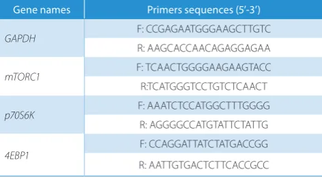
Oxygen exposure induced apoptosis and inhibited proliferation in L929 cells in a time and concentration-dependent manner.
Effects of 10 nM rapamycin on morphology
of L929 cells exposed to 90% O
2According to the results of above, 90% oxygen caused more severe damage to the L929 cells than 40% and 60% oxygen. Therefore, 90% oxygen was used in the follow-up experiments. We added 10 nM rapamycin to L929 cells
exposed to 90% O2 and cultured for 3 days, to investigate
the effects of rapamycin on the morphology of L929 cells. As shown in Fig. 2, the majority of L929 cells cultured with
regular air grew well and had bright, translucent, and
com-pact cell bodies. With exposed to 90% O2, cells cultured
with rapamycin exhibited a reduced refractive index and had increased numbers of granules in the cytoplasm, while most cells were still viable and they exhibited a slightly different morphology (Fig. 2).
Rapamycin inhibited the growth and promoted
apopto-sis of L929 cells exposed to 90% O2.
The L929 cells cultured with 10 nM rapamycin and
ex-posed to 90% O2 exhibited significantly higher apoptosis
Fig. 1. A,B – flow cytometry analysis for apoptosis of L929 cells exposed to 40%, 60% and 90% of oxygen; C – MTT assay for cell viability of L929 cells exposed to 40%, 60% and 90% of oxygen
* p < 0.05; ** p < 0.01 vs control group.
Fig. 2. Inverted phase contrast microscopy. L929 cells exposed to 90% O2 with 10 nM rapamycin intervention were harvested after 3 days. The morphology
and number of cells were evaluated under an inverted phase contrast microscope
101 101 102 103 104 105 106 107.2
102103 AV-FITC-A 3 d control A B C PI -A
104105106 107.2
101 101 102 103 104 105 106 107.2
102103 AV-FITC-A
40% O2
60% O2
90% O2
PI
-A
104105106 107.2
101 101 102 103 104 105 106 107.2
102103 AV-FITC-A PI -A 101 102 103 104 105 106 107.2 PI -A
104105106 107.2
101102103
AV-FITC-A10
4105106 107.2
101 101 102 103 104 105 106 107.2
102103 AV-FITC-A
7 d
PI
-A
104105106 107.2
101 101 102 103 104 105 106 107.2
102103 AV-FITC-A
PI
-A
104105106 107.2
101 101 102 103 104 105 106 107.2
102103 AV-FITC-A PI -A 101 102 103 104 105 106 107.2 PI -A
104105106 107.2
101 102103
AV-FITC-A10
4105106 107.2
101 101 102 103 104 105 106 107.2
102103 AV-FITC-A 14 d 3 d control 40% O2 60% O2 90% O2 control 40% O2 60% O2 90% O2
7 d 14 d
3 d 2.5 2.0 1.5 1.0 0.5 0.0 7 d ce ll viabilit y (4 92 nm ) 80 60 40 20 0 ap op
tosis of L9
29 [%
]
14 d
PI
-A
104105106107.2
101 101 102 103 104 105 106 107.2
102103 AV-FITC-A
PI
-A
104105106107.2
101 101 102 103 104 105 106 107.2
102103 AV-FITC-A PI -A 101 102 103 104 105 106 107.2 PI -A
104105106107.2
101102103
AV-FITC-A10
4105106107.2
A (20 µm)
control
90% O2
90% O2 + rapamycin
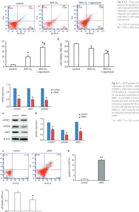
Fig. 4. A – qPCR analysis for the mRNA expression of mTORC1, 4EBP1 and p70S6K in L929 cells transfected with mTOR siRNA; B – western blot analysis for the protein expressions of mTORC1, 4EBP1 and p70S6K in L929 cells transfected with mTOR siRNA; C – flow cytometry analysis for the apoptosis of L929 cells transfected with mTOR siRNA; D – MTT assay for cell viability of L929 cells transfected with mTOR siRNA
* p < 0.05, ** p < 0.01 vs control group.
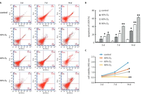
Fig. 3. A,B – Flow cytometry analysis for apoptosis of L929 cells exposed to 90% O2 with
rapamycin intervention; C – MTT assay for cell viability of L929 cells 90% O2 with rapamycin
intervention
* p < 0.05 vs control group; #p < 0.05 vs 90% O2 group.
101
101
102
103
104
105
106
107.2
102 103
AV-FIFC-A siRNA
siRNA
control
siRNA control
siRNA
control control
siRNA control
siRNA control
mTORC1 p70S6K 4EBP1 mTORC1
p70S6K
4EBP1
β-actin
mTORC1 4EBP1 p70S6K
C
D B A
a b
a b
PI-A
104 105 106 107.2
101
101
102
103
104
105
106
107.2
102 103
AV-FIFC-A
PI-A
104 105 106 107.2
25
20
15
10
5
0 1.6
1.2
0.8
0.4
0.0
apoptosis of L
92
9 [
%]
prot
ein expressio
n
1.0
0.5
0.0
cel
l v
iabilit
y (
49
2 n
m)
1.2
0.8
0.4
0.0
mRNA expressio
n
101
101
102
103
104
105
106
107.2
102 103
AV-FITC-A control
control
A
B C
PI
-A
104 105 106 107.2 10101 1
102
103
104
105
106
107.2
102 103
AV-FITC-A
90% O2 90% O2 + rapamycin
90% O2 90% O2
+ rapamycin control
1.0 0.8 0.6 0.4 0.2 0.0
cell v
iabilit
y
(4
92
nm
)
25 20 15 10 5 0
apoptosis [%
]
90% O2 90% O2
+ rapamycin
PI
-A
104 105 106 107.2 10101 1
102
103
104
105
106
107.2
102 103
AV-FITC-A
PI
-A
rates than the control group (*p < 0.05, Fig. 3A,B), and
90% O2 exposure significantly inhibited the cell viability
of L929 cells (*p < 0.05, Fig. 3C). In addition, the apoptosis ratio was increased and the cell viability was inhibited
in the 90% O2 + rapamycin group compared with 90% O2
group (#p < 0.05) (Fig. 3A–C).
Validation of mTOR siRNA
After transfected with mTOR siRNA in L929 cells, we found that the mRNA and protein expression levels of mTORC1, 4EBP1 and p70S6K in L929 cells transfected with mTOR siRNA were significantly lower than in control cells (*p < 0.05, Fig. 4A,B). In addition, the rates of apop-totic cells transfected with mTOR siRNA (1.2%) were sig-nificantly increased than in the control (20.1%) (**p < 0.01, Fig. 4C), and the cell viability of cells transfected with mTOR siRNA was significantly decreased compared with control group (*p < 0.05, Fig. 4D). These results suggest that mTOR siRNA could down-regulate the expression of mTORC1, 4EBP1 and p70S6K, inhibit the cell viability and promote apoptosis of L929 cells.
Effects of rapamycin and mTOR siRNA
on the protein expressions of Bcl-2, p53,
TGF-β, and CTGF in L929 cells
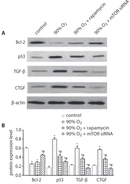
We evaluated the protein expressions of apoptosis-related genes, including p53 and Bcl-2, as well as fibrosis-related genes, including TGF-β and CTGF, in L929 cells which were transfected with mTOR siRNA, treated with 10 nM
rapamycin, and exposed to 90% O2. As shown in Fig. 5,
the expression of Bcl-2 in cells exposed to 90% oxygen and cultured with 10 nM rapamycin or transfected with mTOR siRNA was significantly lower than in cells exposed to regular air (*p < 0.05), and mTOR siRNA significantly
increased the expression of Bcl-2 compared with 90% O2
group (#p < 0.05). The expression level of p53 in L929 cells exposed to 90% oxygen and cultured with rapamycin or
those transfected with mTOR siRNA was significantly higher than in cells exposed to regular air (*p < 0.05); how-ever, the application of rapamycin or mTOR siRNA groups all exhibited lower expression level of p53 compared with
90% O2 group (#p < 0.05). The expression levels of TGF-β
and CTGF in L929 cells exposed to 90% oxygen was signifi-cantly higher than in the control group (*p < 0.05); however, the application of 10 nM rapamycin or mTOR siRNA groups
Fig. 5. A – western blot analysis of the protein expressions of Bcl-2, p53, TGF-β, and CTGF in the control, 90% O2, 90% O2 + rapamycin, and 90%
O2 + mTOR-siRNA L929 cell groups; B – relative density of western blot
bands was analyzed using ImageJ software
* p < 0.05 vs control group; #p < 0.05 vs 90% O2 group.
Fig. 6. The contents of col I (A), col III (B) and FN (C) in ECM of L929 cells from each group were determined with ELISA
col I – collagen I; col III – collagen III; FN – fibronectin; ECM – extracellular matrix; * p < 0.05 vs control group; #p < 0.05 vs 90% O2 group.
90% O2 +
rapam ycin
cont rol
90% O2
90% O2 +
mTOR siRN
A
90% O2 + rapamycin control
A
B 90% O2
90% O2 + mTOR siRNA
Bcl-2 p53 TGF-β CTGF
Bcl-2
p53
CTGF TGF-β
β-actin
1.0 0.8 0.6 0.4 0.2 0.0
prot
ei
n e
xpressio
n l
evel
90% O
2 + rapa
mycin contro
l
90% O
2
90% O
2 + m
TOR siR NA
90% O
2 + rapa
mycin contro
l 90% O
2
90% O
2 + m
TOR siR NA
90% O
2 + rapa
mycin contro
l
90% O
2
90% O
2 + m
TOR siR NA 30
25
20
15
10
5
0
A B C
co
l I content
[n
g/
mL
]
30
25
20
15
10
5
0
co
l III content
[n
g/
mL
]
35
30
25
20
15
10
5
0
FN co
ntent
[n
g/
mL
all exhibited lower expression levels of TGF-β and CTGF
compared with 90% O2 groups. These results suggest that
90% O2 exposure could promote L929 cell apoptosis and
successfully inhibit the expression of CTGF and TGF-β.
The contents of collagen I, III
and fibronectin in extracellular matrix
The contents of collagen I, collagen III (col I, col III), and fibronectin in extracellular matrix (ECM) of L929 cells
ex-posed to 90% O2 was significantly higher than those in cells
exposed to regular air (*p < 0.05), suggesting that 90% O2
could induce the production of FN and collagen. However, the contents of col I, col III, and FN in L929 cells treated with rapamycin or transfected with mTOR siRNA were
signifi-cantly lower than that in 90% O2 (#p < 0.05), suggesting that
blocking the mTOR signaling pathway suppressed collagen deposition and decreased the production of FN (Fig. 6).
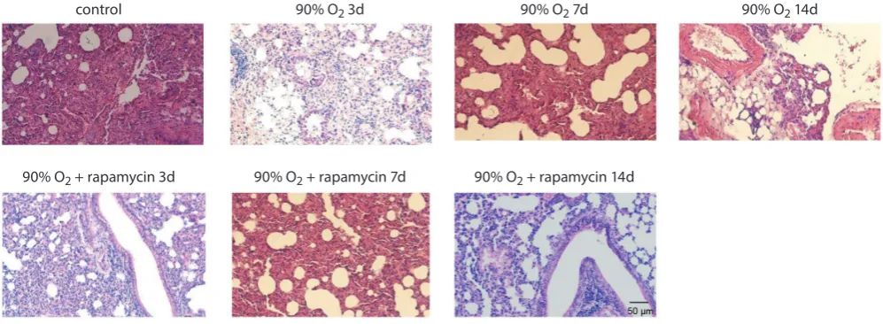
Histopathological changes
in the lung tissues
The lung tissues of control group rats exhibited normal histopathology, without exudation of inflammatory cells
(Fig. 7). Exudation of a few inflammatory cells and red cells
was observed in the lungs exposed to 90% O2 for 3, 7 and
14 days, and in a time-dependent manner. After 7 days of exposure to 90% oxygen, we observed alveolar rupture and fusion, and disordered distribution of cells along the tracheal arteries. After 14 days of exposure to 90% oxygen, extensive exudation of inflammatory cells and thickening blood vessels and airway walls were observed. The lungs of rats that had received intraperitoneal rapamycin injec-tions and were exposed to 90% oxygen for 3 days exhibited normal histopathology. Alveolar rupture and fusion, as well as thickening blood vessels and airway walls, were observed in the lungs of rats that had received intraperitoneal injec-tions of rapamycin and were exposed to 90% oxygen for 7 days. After 14 days of exposure to 90% oxygen, we observed more severe alveolar rupture and fusion in the lungs of rats that had received intraperitoneal injection of rapamycin.
The pathological scores of lung injury are shown in Ta-ble 2. The pathological scores of lung injury in the rats ex-posed to 90% oxygen for 3, 7, and 14 days were significantly
higher than those of the rats from the control (a p < 0.05).
After 3 and 7 days, the pathological lung injury scores
of the 90% O2 + rapamycin group were (3.50 ±0.84) and
(9.67 ±1.97), respectively, which were significantly lower
than those of the 90% O2 group for 3 (6.33 ±2.34) and 7 days
(14.0 ±2.45), respectively (b p < 0.05).
Dynamic changes of col I, TGF-β
and CTGF in lung tissues
The col I concentration in the lung tissues of rats exposed to 90% oxygen for 3, 7, and 14 days were (471.87 ±5.72 ng/mg), (529.72 ±6.97 ng/mg), and (556.44 ±8.52 ng/mg), which were significantly higher than those of the air control group
(414.43 ±8.97 ng/mg) (ap < 0.05). The TGF-β concentration
in the lung tissues of rats exposed to 90% oxygen for 3, 7 and 14 days were (33.74 ±2.84 ng/mg), (58.65 ±3.10 ng/mg), and
Fig. 7. Histopathological changes of the lung tissues from hyperoxia-induced lung injury rats Hematoxylin and eosin stain; bar 50 μm; magnification ×200.
Table 2. The pathological scores of lung injury in rat model (n = 6)
Group Pathological scores
Control 1.95 ±0.34
90% O2 3d 6.33 ±2.34a
90% O2 + rapamycin 3d 3.50 ±0.84a,b
90% O2 7d 14.0 ±2.45a
90% O2 + rapamycin 7d 9.67 ±1.97a,b
90% O2 14d 23.83 ±3.71a
90% O2 + rapamycin 14d 24.0 ±3.74a
a compared with control group, p < 0.05; b compared with 90% O 2 groups
at the same time, p < 0.05.
control 90% O2 3d
90% O2 + rapamycin 3d 90% O2 + rapamycin 7d 90% O2 + rapamycin 14d
(98.81 ±1.55 ng/mg), which were significantly higher than
those of the control group (25.50 ±1.86 ng/mg) (a p < 0.05).
The CTGF concentration in the lung tissues of rats ex-posed to 90% oxygen for 3, 7, and 14 days were (50.72 ±1.80 ng/mg), (68.65 ±2.24 ng/mg), and (94.39 ±2.48 ng/mg), which were significantly higher than those of the control
group (41.23 ±1.08 ng/mg) (ap < 0.05). These results
sug-gest that the production of pulmonary fibrosis factors was induced by 90% oxygen. Compared with the 90% oxygen group, rapamycin significantly reduced the concentra-tions of col I, TGF-β1, and CTGF in the lung tissues of rats
exposed to 90% oxygen for 3, 7, and 14 days (bp < 0.05)
(Table 3).
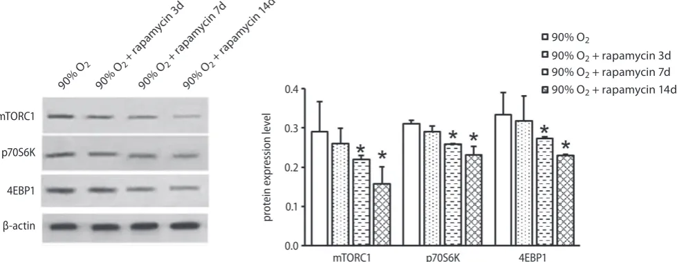
Rapamycin inhibits the mTOR pathway
in the lung tissue of rats
The above results indicated that rapamycin attenuates acute lung injury induced by 90% oxygen. The objective of western blot was to determine whether the mTOR path-way was involved in the repair of lung injury. As shown in Fig. 8, the expression levels of mTORC1, p70S6K, and
4EBP1 in the lung tissue of rats in the 90% O2 + rapamycin
group were significantly decreased compared with control animal at day 7 and 14 (*p < 0.05), which suggested that the application of rapamycin could inhibit the activation of mTOR signaling pathway and inhibit the expression of downstream proteins p70S6K and 4EBP1.
Discussion
In the present study, we found that exposure to oxy-gen concentrations of 40%, 60%, and 90% all promoted the apoptosis of L929 cells in a time and concentration-dependent manner. Application of 10 nM rapamycin or transfected with mTOR siRNA could inhibit the activation of mTOR signaling pathway and promote the apoptosis
of L929 cells exposed to 90% O2, and down-regulate the
ex-pression of the downstream proteins 4EBP1, and p70S6K. Exposure to 90% oxygen increased the production of col I, col III, and FN in ECM of L929 cells; however, inhibition of the mTOR signaling pathway decreased the production of col I, III and FN, which could reduce hyperoxia-induced pulmonary fibrosis. In lung tissues of hyperoxia-induced lung injury rats, we found that rapamycin inhibited
Table 3.The concentrations of TGF-β, CTGF and col I in the lung tissue determined with ELISA (n = 6)
Group TGF-β [ng/mg] CTGF [ng/mg] Col I [ng/mg]
Control 25.50 ±1.86 41.23 ±1.08 414.43 ±8.97
90% O2 3d 33.74 ±2.84a 50.72 ±1.80a 471.87 ±5.72a
90% O2 + rapamycin 3d 25.72 ±1.44b 43.45 ±1.71b 425.11 ±3.50b
90% O2 7d 58.65 ±3.10a 68.65 ±2.24a 529.72 ±6.97a
90% O2 + rapamycin 7d 34.39 ±3.32b 50.92 ±2.42b 466.17 ±10.60b
90% O2 14d 98.81 ±1.55a 94.39 ±2.48a 556.44 ±8.52a
90% O2 + apamycin 14d 54.31 ±4.25b 63.64 ±2.78b 511.72 ±13.72b
a compared with control group, p < 0.05; b compared with 90% O
2 groups at the same time, p < 0.05; col I – collagen I.
Fig. 8. The protein expression of mTORC1, p70S6K and 4EBP1 in the lung tissues of rats of all experimental groups were detected with western blot * p < 0.05 vs 90% O2 group.
90% O2 +
rapam ycin
3d
90% O2 + rapamycin 3d
90% O2
90% O2
90% O2 +
rapam ycin
7d
90% O2 + rapamycin 7d
90% O2 +
rapam ycin
14d
90% O2 + rapamycin 14d
mTORC1 p70S6K 4EBP1
mTORC1
p70S6K
4EBP1
β-actin
0.4
0.3
0.2
0.1
0.0
prot
the activation of the mTOR pathway, which protected the lung from the oxidative injury and fibrosis induced by high concentration of oxygen.
Lung fibroblasts play an important role in the repair of lung injury. Yang et al. found that high oxygen concen-trations inhibited the proliferation of fibroblasts isolated
from embryonic lungs.25 The mTOR signaling pathway
is essential for cell proliferation, survival, and migration by regulating the transcription and translation
of pro-teins.26 Rapamycin is a specific inhibitor of the mTOR
pathway.27 Sun et al. found that 0.01–10 nM rapamycin
could inhibit the proliferation of lung cancer cells; how-ever, only >1 nM rapamycin quickly inhibited the phos-phorylation of downstream proteins and the growth
of lung cancer cells.28 In the present study, mTOR siRNA
down-regulated the expression of mTORC1, 4EBP1, and p70S6K, suggesting that mTOR can specifically regulate the expression of 4EBP1 and p70S6K. The extracellular matrix is synthesized and secreted by animal cells to form a network structure between cells, which consists
of pro-tein and polysaccharide.29 In the present study, we found
that blocking the mTOR signaling pathway inhibited col-lagen deposition in L929 cells, which prevented the lung fibrosis caused by oxidative injury. Collagen deposition in the lung is one of the most important causes and major
characteristics of pulmonary fibrosis.30 In the pathology
of pulmonary fibrosis, apoptosis-related proteins, such as Bcl-2, P53, and caspase-3, as well as other cytokines, interplay to promote the proliferation of lung fibroblasts, which replace alveolar type II epithelial cells (AT II),
subse-quently causing pulmonary fibrosis.31–33 TGF-β and CTGF
could activate the PI3K/Akt/mTOR signaling pathway, promoting the proliferation, collagen synthesis, and anti-apoptotic ability of lung fibroblasts. TGF-β, which is a key component in a number of cytokine networks, is currently recognized to be one of the most powerful factors causing
fibrosis.34 Lee and Kim reported that CTGF was closely
associated with TGF-β in pulmonary fibrosis.35 Connective
tissue growth factoris a newly identified cytokine closely
involved in pulmonary fibrosis and is widely distributed in human tissues and organs; it plays important roles in cell
adhesion, fibroblast proliferation, and ECM synthesis.36
In the present study, we observed increased expression of CTGF and TGF-β, which was induced by 90% oxygen. Additionally, we found that blocking the mTOR pathway could down-regulate the expression of CTGF and TGF-β, which suggests that rapamycin could decrease the expres-sion CTGF and TGF-β through inhibiting the activation of mTOR signaling pathway. In other words, the mTOR pathway is involved in lung fibrosis by regulating the ex-pression of CTGF and TGF-β.
The mammalian target of rapamycin is widely found in mammals in 2 complex forms: mTORC1 and mTORC2. As the target protein of rapamycin, mTORC1 is sensitive
to rapamycin; however, mTORC2 is not.27 The
mamma-lian target of rapamycin complex 1 regulates cell growth,
proliferation, and metabolism by phosphorylating 2 major downstream proteins, S6K1 and eukaryotic initiation
fac-tor 4E-BP1. In the present study, we found that 90% O2
induced lung injury in premature rats in a time-dependent manner. We also observed increased expression of fibro-sis-related proteins, including TGF-β, CTGF, col II and col III, in the lung tissue of rats, also in a time-dependent manner. These results suggest that lung injury induced by a high concentration of oxygen is consistent with lung fibrosis. High concentrations of oxygen induced changes in the expression of mTORC1, p70S6K, and 4EBP1, which are key components of the mTOR pathway, suggesting that the mTOR pathway is activated by high concentrations of inhaled oxygen. It has been reported that the mTOR pathway is activated in lipopolysaccharide (LPS)-induced acute lung injury (ALI) in mice and rapamycin reduced the expression of inflammatory factors in bronchoalveolar
lavage fluid (BLF).37 Lorne et al. reported that the
phos-phorylation of rpS6 and 4EBP1, 2 effector proteins of the mTOR signaling pathway, induced the over-production of inflammatory cytokines by neutrophils in ALI, which
could be inhibited by rapamycin.38 It has been reported
that rapamycin administration causes a significant re-duction of p70S6K phosphorylation, increased autoph-agy, decreases neuronal cells apoptosis and significantly reduces brain damage in neonatal rats, which testifies to the neuroprotective effect of rapamycin in neonatal
hypoxia-ischemia.39 Furthermore, it was found that the
inhibition of the mTOR signaling pathway by rapamy-cin prevented the development and progression of lung fibrosis in a rat pulmonary fibrosis model that was
in-duced by TGF-α.21 Tulek et al. also found that rapamycin
could inhibit the progress of lung fibrosis in the early
stage of bleomycin-induced pulmonary fibrosis in rats.40
Therefore, we speculate that the mTOR signaling pathway plays an important role in the repair of hyperoxia-induced lung injury by regulating the expression of TGF-β and CTGF. Rapamycin can effectively inhibit the activation of the mTOR signaling pathway and downregulate down-stream target proteins P70S6K and 4EBP1, and reduce the production of TGF-β and CTGF, inhibiting the develop-ment of pulmonary fibrosis induced by high concentration oxygen exposure.
Conclusions
References
1. Mirza H, Ziegler J, Ford S, Padbury J, Tucker R, Laptook A. Pulmonary hypertension in preterm infants: Prevalence and association with bronchopulmonary dysplasia. J Pediatr. 2014;165(5):909–914. 2. Zhou W, Zhang X, Feng Z. Risk factors, incidence and severity
of bron-chopulmonary dysplasia (BPD) in the world’s largest neonatal inten-sive care units (NICUs) in China. Klin Padiatr. 2012;224(7):2095–2099. 3. Greenspan JS, Goldsmith JP. Oxygen therapy in preterm infants:
Hit-ting the target. Pediatrics. 2006;118(4):1740–1741.
4. Garat C, Jayr C, Eddahibi S, Laffon M, Meignan M, Adnot S. Effects of inhaled nitric oxide or inhibition of endogenous nitric oxide forma-tion on hyperoxic lung injury. Am J Respir Crit Care Med. 1997;155(6): 1957–1964.
5. Dubaybo BA, Rubeiz GJ, Fligiel SE. Dynamic changes in the function-al characteristics of the interstitifunction-al fibroblast during lung repair. Exp Lung Res. 1992;18(4):461–477.
6. Rocco PR, Negri EM, Kurtz PM, et al. Lung tissue mechanics and extra-cellular matrix remodeling in acute lung injury. Am J Respir Crit Care Med. 2001;164(6):1067–1071.
7. Xia H, Diebold D, Nho R, et al. Pathological integrin signaling enhanc-es proliferation of primary lung fibroblasts from patients with idio-pathic pulmonary fibrosis. J Exp Med. 2008;205(7):1659–1672. 8. Uhal BD, Joshi I, True AL, et al. Fibroblasts isolated after fibrotic lung
injury induce apoptosis of alveolar epithelial cells in vitro. Am J Physiol. 1995;269(1):819–828.
9. Hay N, Sonenberg N. Upstream and downstream of mTOR. Genes Dev. 2004;18(16):1926–1945.
10. Zoncu R, Efeyan A, Sabatini DM. mTOR: From growth signal integra-tion to cancer, diabetes and ageing. Nat Rev Mol Cell Biol. 2010;12(1): 21–35.
11. Thomas GV, Tran C, Mellinghoff IK, et al. Hypoxia-inducible factor determines sensitivity to inhibitors of mTOR in kidney cancer. Nat Med. 2006;12(1):122–127.
12. Hentges KE, Sirry B, Gingeras AC, et al. FRAP/mTOR is required for proliferation and patterning during embryonic development in the mouse. Proc Natl Acad Sci USA. 2001;98(24):13796–13801.
13. Hwang M, Perez CA, Moretti L, Lu B. The mTOR signaling network: Insights from its role during embryonic development. Curr Med Chem. 2008;15(12):1192–1208.
14. Land SC, Scott CL, Walker D. mTOR signalling, embryogenesis and the control of lung development. Semin Cell Dev Biol. 2014;36:68–78. 15. Costes F, Gosker H, Feasson L, et al. Impaired exercise training-induced muscle fiber hypertrophy and Akt/mTOR pathway activation in hypoxemic patients with COPD. J Appl Physiol. 2015;118(8):1040–1049. 16. Makam M, Diaz D, Laval J, et al. Activation of critical, host-induced, metabolic and stress pathways marks neutrophil entry into cystic fibrosis lungs. Proc Natl Acad Sci USA. 2009;106(14):5779–5783. 17. Gui YS, Wang L, Tian X, et al. mTOR overactivation and compromised
autophagy in the pathogenesis of pulmonary fibrosis. PLos One. 2015; 10(9):e0138625. doi: 10.1371/journal.pone.0138625
18. Gäbele E, Reif S, Tsukada S, et al. The role of p70S6K in hepatic stel-late cell collagen gene expression and cell proliferation. J Biol Chem.
2005;280(14):13374–13382.
19. Park JS, Park HJ, Park YS, et al. Clinical significance of mTOR, ZEB1, ROCK1 expression in lung tissues of pulmonary fibrosis patients. BMC Pulm Med. 2014;14:168.
20. Chung EJ, Sowers AL, Thetford A, Mckay-Corkum G, Mitchell JB, Citrin DE. mTOR inhibition with rapamycin mitigates radiation-induced pul-monary fibrosis in a murine model. Int J Radiat Oncol Biol Phys. 2016; 96(4):857–866.
21. Korfhagen TR, Le CT, Davidson CR, et al. Rapamycin prevents trans-forming growth factor-alpha-induced pulmonary fibrosis. Am J Respir Cell Mol Biol. 2009;41(5):562–572.
22. Zhang L, Zhao S, Yuan L, et al. Knockdown of placental growth fac-tor (PLGF) mitigates hyperoxia-induced acute lung injury in neona-tal rats: Suppressive effects on NFκB signaling pathway. Int Immu-nopharmacol. 2016;38:167–174.
23. Sun X, Threadgill D, Jobin C. Campylobacter jejuni induces colitis through activation of mammalian target of rapamycin signaling.
Gastroenterology. 2012;142(1):86–95.
24. Murakami K, Mcguire R, Cox RA, et al. Heparin nebulization attenu-ates acute lung injury with sepsis after smoke inhalation in sheep.
Shock. 2002;18(3):236–241.
25. Yang P, He XQ, Peng L, et al. The role of oxidative stress in hormesis induced by sodium arsenite in human embryo lung fibroblast (HELF) cellular proliferation model. J Toxicol Environ Health A. 2007;70(11): 976–983.
26. Sarbassov DD, Ali SM, Sabatini DM. Growing roles for the mTOR pathway. Curr Opin Cell Biol. 2005;17(6):596–603. doi: 10.1371/jour-nal.pbio.1000038
27. Feldman ME, Apsel B, Uotila A, et al. Active-site inhibitors of mTOR target rapamycin-resistant outputs of mTORC1 and mTORC2. PLoS Biol. 2009;7(2):e38.
28. Sun SY, Rosenberg LM, Wang X, et al. Activation of Akt and eIF4E sur-vival pathways by rapamycin-mediated mammalian target of rapamy-cin inhibition. Cancer Res. 2005;65(16):7052–7058.
29. Pizzo AM, Kokini K, Vaughn LC, Waisner BZ, Voytik-Harbin SL. Extra-cellular matrix (ECM) microstructural composition regulates local cell-ECM biomechanics and fundamental fibroblast behavior: A mul-tidimensional perspective. J Appl Physiol. 2005;98(5):1909–1921. 30. Mercer RR, Scabilloni J, Wang L, et al. Alteration of deposition
pat-tern and pulmonary response as a result of improved dispersion of aspirated single-walled carbon nanotubes in a mouse model. Am J Physiol Lung Cell Mol Physiol. 2008;294(1):L87–97.
31. Wu SH, Wu XH, Lu C, Dong L, Chen ZQ. Lipoxin A4 inhibits prolifera-tion of human lung fibroblasts induced by connective tissue growth factor. Am J Respir Cell Mol Biol. 2006;34(1):65–72.
32. Choi JE, Lee SS, Sunde DA, et al. Insulin-like growth factor-I recep-tor blockade improves outcome in mouse model of lung injury. Am J Respir Crit Care Med. 2009;179(3):212–219.
33. Vittal R, Horowitz JC, Moore BB, et al. Modulation of prosurvival sig-naling in fibroblasts by a protein kinase inhibitor protects against fibrotic tissue injury. Am J Pathol. 2005;166(2):367–375.
34. Moussad EE, Brigstock DR. Connective tissue growth factor: What’s in a name? Mol Genet Metab. 2000;71(1–2):276–292.
35. Lee HS, Kim CK. Effect of recombinant IL-10 on cultured fetal rat alve-olar type II cells exposed to 65%-hyperoxia. Respir Res. 2011;12:68. doi: 10.1186/1465-9921-12-68
36. Chen S, Rong M, Platteau A, et al. CTGF disrupts alveolarization and induces pulmonary hypertension in neonatal mice: Implication in the pathogenesis of severe bronchopulmonary dysplasia. Am J Physiol Lung Cell Mol Physiol. 2011;300(3):330–340.
37. Wang L, Gui YS, Tian X, et al. Inactivation of mammalian target of rapamycin (mTOR) by rapamycin in a murine model of lipopoly-saccharide-induced acute lung injury. Chin Med J (Engl). 2011;124(19): 3112–3117.
38. Lorne E, Zhao X, Zmijewski JW, et al. Participation of mammalian tar-get of rapamycin complex 1 in toll-like receptor 2- and 4-induced neutrophil activation and acute lung injury. Am J Respir Cell Mol Bio.
2009;41(2):237–245.
39. Carloni S, Girelli S, Scopa C, Buonocore G, Longini M, Balduini W. Acti-vation of autophagy and Akt/CREB signaling play an equivalent role in the neuroprotective effect of rapamycin in neonatal hypoxia-isch-emia. Autophagy. 2010;6(3):366–377.