Anna Brzozowska
1, 2, A–D, Anna Toruń
3, G, Maria Mazurkiewicz
1, 2, E, FThe Impact of Surgery on the Efficacy
of Adjuvant Therapy in Glioblastoma Multiforme
1 Chair and Department of Oncology, Medical University of Lublin, Poland2 St. John’s Cancer Center, Lublin, Poland
3 Department of Mathematics and Medical Biostatistics, Medical University of Lublin, Poland
A – research concept and design; B – collection and/or assembly of data; C – data analysis and interpretation; D – writing the article; E – critical revision of the article; F – final approval of article; G – other
Abstract
Background. Glioblastoma multiforme (GBM) is the most malignant brain tumor. Surgery still remains a funda-mental part of treatment in GBM, followed by radiotherapy and chemotherapy.
Objectives. The aim of the study was to assess the impact of surgery on the efficacy of adjuvant therapy in patients with glioblastoma multiforme.
Material and Methods. The study involved 181 patients: 50 treated with adjuvant radiotherapy (RTH) only (60 Gy in daily 2Gy fractions) and 131 treated with postoperative radiochemotherapy (RTH-CHT) (60 Gy, 2Gy/d) + 75 mg/m2 temozolomide for 42 days of radiotherapy, followed by 6 courses every 28 days; the first course was
150 mg/m2 for 1–5 days, and the subsequent courses were 200 mg/m2 for 1–5 days). Overall survival (OS) and
disease-free survival (DFS) were assessed. The statistical analysis entailed the log-rank test, Kaplan-Meier curves and Cox proportional hazards regression; the threshold of statistical significance was set at p = 0.05.
Results. Median OS and DFS were significantly increased (p = 0.001) in the RTH-CHT group compared with the RTH group: 9.77 months vs 6.38 months for OS, and 8.4 months vs 4.33 months for DFS. After radical surgery, RTH-CHT extended OS by 5.3 months and DFS by 4.5 months in comparison to RTH. In patients who underwent non-radical surgery, the type of adjuvant therapy made no difference in either OS or DFS. In the RTH-CHT group, OS and DFS depended on the extent of the surgery, and were significantly longer in patients who underwent radical surgery (OS: p = 0.03128; DFS: p = 0.01206). In the RTH group, the type of surgery had no effect on survival. Conclusions. Radiochemotherapy significantly prolongs OS and DFS compared with radiotherapy alone in GBM patients who have undergone radical removal of the tumor.Among patients who had non-radical surgery, the type of adjuvant treatment has no effect on OS or DFS (Adv Clin Exp Med 2015, 24, 2, 279–287).
Key words: glioblastoma multiforme, adjuvant treatment, surgery.
Glioblastoma multiforme (GBM) is a brain tu-mor with an especially adverse prognosis. Despite the use of modern methods of surgical treatment supplemented with radical postsurgical radiothera-py combined with the administration of temozolo-mide, the average overall survival time still does not exceed 2 years [1]. The main prognostic factors de-scribed in the literature are the patient’s age, the patient’s general condition, the adjuvant therapy method and the tumor resection margin [2, 3].
According to the most recent studies, the ex-tent of surgery in GBM patients affects not only the patient’s neurological condition and the sur-vival time, but also the efficacy of the postsurgical
systemic treatment [26]. The published data show that the more radical the surgery is, the more ef-fective postsurgical radiochemotherapy combined with temozolomide will be [27].
The aim of this study was to evaluate and com-pare the effectiveness of postsurgical radiotherapy and radiochemotherapy as adjuvant treatment of patients with glioblastoma multiforme and the in-fluence of the extent of surgery on the treatment outcome. The prognostic value of such factors as age, general condition, the waiting time for the ini-tiation of adjuvant therapy and the length of time
before disease progression or relapse were also
evaluated.
Adv Clin Exp Med 2015, 24, 2, 279–287
Material and Methods
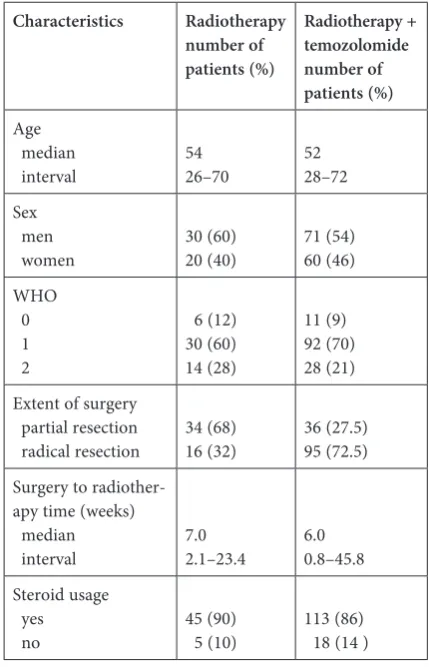
The study involved 181 patients radically treat-ed for GBM at St. John’s Cancer Center in Lublin, Poland, from 2004 to 2010. Among them, 50 pa-tients underwent only postsurgical radiotherapy in the years 2004–2006, while 131 patients treated be-tween 2006 and 2010 were treated with radiothera-py combined with temozolomide. The analysis did not include patients with a relapse of GBM who underwent only tumor biopsy or were undergoing palliative treatment (Table 1).
All the patients underwent surgery and the clin-ical diagnosis of GMB was confirmed by histopath-ological examination. The evaluation of the extent of surgery was based on the description of the sur-gery and the results of computer tomography exam-inations conducted after the surgery; on these bas-es, the operations were categorized as total or partial resections. Radical radiotherapy was performed on all patients. A total dose of 60 Gy in 30 fractions of 2 Gy was applied. In the course of radiotherapy, the
group of 131 patients additionally received 75 mg/m2
temozolomide daily, 7 days a week, for 42 days com-mencing from the first day of radiotherapy. Next, upon the completion of radiotherapy, 1 to 6 cours-es of temozolomide were administered at a
dos-age of 150 mg/m2 (1–5 days) in the first course and
200 mg/m2 (1–5 days) in subsequent courses
ev-ery 28 days. The number of administered courses depended on the response to treatment. Observed progression or relapse of the neoplasm disqualified patients from further treatment. Almost all the pa-tients were additionally treated, depending on the indications, with decongestants, anti-emetic and anti-convulsant medicines.
Progression or relapse of GBM was diagnosed on the basis of clinical symptoms and confirmed with computer tomography. Tumor relapse was treated surgically or symptomatically. Disease-free survival (DFS) time and overall survival (OS) time were calculated from the beginning of radiothera-py to progression/relapse (DFS) or death (OS). Da-ta concerning the progression/relapse date or time of death were based on medical records and death registries, respectively.
In the first part of the statistical analysis, the differences between OS and DFS were examined using the log-rank test. Kaplan-Meier curves were used to illustrate the differences. The Cox propor-tional hazards regression model was used to eval-uate the influence of quantitative factors on over-all survival and disease-free survival, as well as to calculate the relative risk for qualitative variables. The threshold of statistical significance adopted was p = 0.05. The statistical analysis of the data was carried out using STATISTICA 8.0 software.
Results
Patients’ Characteristics
Both of the groups studied consisted main-ly of men of about 50 years of age with a WHO performance level of 1. A significant majority of the patients in the group treated with radioche-motherapy had had radical surgery with total tu-mor excision, while in the group receiving only radiotherapy most of the patients had undergone only partial tumor excision. The median waiting time for radiotherapy, measured from surgery to the commencement of irradiation, was 7 weeks for the patients undergoing radiotherapy with te-mozolomide, and 6 weeks for the patients receiv-ing radiotherapy alone. In course of radiothera-py, steroids were administered to most patients (Table 1).
Treatment
In the group that underwent only radiother-apy, the radiation was discontinued in 4 patients (8%) due to disease progression. The applied dos-es in those 4 patients were 50 Gy, 36 Gy, 48 Gy
Table 1. Patient characteristics
Characteristics Radiotherapy
number of patients (%)
Radiotherapy + temozolomide number of patients (%)
Age median
interval 5426–70 5228–72 Sex
men
women 30 (60)20 (40) 71 (54)60 (46) WHO
0 1 2
6 (12) 30 (60) 14 (28)
11 (9) 92 (70) 28 (21) Extent of surgery
partial resection
radical resection 34 (68)16 (32) 36 (27.5)95 (72.5) Surgery to
radiother-apy time (weeks) median
interval 7.02.1–23.4 6.00.8–45.8 Steroid usage
yes
and 28 Gy, respectively. In the group of 131 pa-tients, the concurrent radiochemotherapy course was completed by 122 patients (93%) and the full treatment was completed by 64 patients (49%). The reasons for discontinuing radiochemotherapy were disease progression and hematological
toxic-ity of the 3rd or 4th degree due to the
administra-tion of temozolomide.
Overall Survival
and Disease-Free Survival
in the Study Subjects
During follow-up periods lasting from 1 to
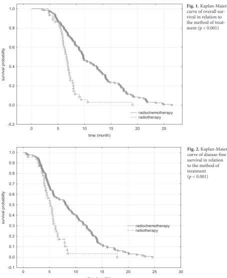
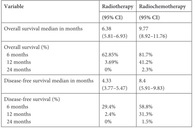
26 months all the study subjects died. Both the OS (Fig. 1) and DFS (Fig. 2) of the patients who un-derwent radiochemotherapy were significantly longer (p < 0.001) than in patients who were treat-ed solely with radiotherapy (Table 2).
Fig. 1. Kaplan-Maier curve of overall sur-vival in relation to the method of treat-ment (p < 0.001)
0 5 10 15 20 25
time (month) -0.2
0.0 0.2 0.4 0.6 0.8 1.0
su
rv
iv
al
pr
obab
ilit
y
radiochemotherapy radiotherapy
Fig. 2. Kaplan-Maier curve of disease-free survival in relation to the method of treatment (p < 0.001)
0 5 10 15 20 25 30
time (month) -0.1
0.0 0.1 0.2 0.3 0.4 0.5 0.6 0.7 0.8 0.9 1.0
su
rv
iv
al
pr
obab
ilit
y
Prognostic Factors
in the Study Subjects
Unifactorial Analysis
In both study groups the patients’ age, sex, general condition according to WHO criteria and waiting time for radiotherapy had no significant effect on OS and DFS. The only factor with signifi-cant influence on OS (p = 0.03) and DFS (p = 0.008) was the extent of surgery, but only in patients who underwent postsurgical radiochemotherapy.
Multifactorial Analysis
In the group of patients who underwent radio-chemotherapy, as many as 3 factors influenced the mortality risk value. Patients with time to relapse or progression equal to or longer than the medi-an (8.4 months) had a 19.29% lower risk of death compared to other patients from this group. Al-so, a waiting time for radiotherapy equal to or lon-ger than the median (6 weeks) increased the risk of death by 5.5% in these subjects. Furthermore, a less than radical surgical excision of the tumor
resulted in an increase in the mortality risk in-crease by as much as 61.9% (Table 4). Among the patients who underwent postsurgical radiotherapy only, without the administration of temozolomide, only the length of the time to relapse/progression was a significant factor affecting the mortality risk: patients whose time to relapse/progression was equal to or longer than the median (5.18 months) had a 17.9% lower mortality risk than the remain-ing patients (Table 3).
Survival Time in Relation
to the Extent of Surgery
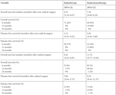
Among the patients who underwent non-rad-ical surgery, the OS and DFS medians were com-parable regardless of the method of postsurgical treatment. Administration of temozolomide dur-ing and/or after radiation therapy did not prolong these patients’ survival time (Table 4). However, the patients who underwent radical surgery ben-efited from the administration of temozolomide combined with postsurgical radiotherapy. Among the patients treated with the combined method, Table 2. The patients’ overall survival and disease-free survival
Variable Radiotherapy Radiochemotherapy
(95% CI) (95% CI)
Overall survival median in months 6.38
(5.81–6.93) 9.77(8.92–11.76) Overall survival (%)
6 months 12 months 24 months
62.85% 3.69% 0%
81.7% 41.2% 2.3% Disease-free survival median in months 4.33
(3.77–5.47) 8.4(5.91–9.83) Disease-free survival (%)
6 months 12 months 24 months
29.4% 2.4% 0%
58.8% 31.3% 1.5%
Table 3. Multifactorial analysis of the influence of the evaluated factors on the patients’ overall survival
Treatment method Prognostic factors Relative risk P-value 95% confidence interval
Radiochemotherapy time to relapse 0.8071 0.0000 0.777–0.837 surgery to radiotherapy time 0.945 0.007 0.906–0.985 extent of surgery 1.000
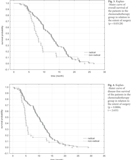
the OS median increased by 5.3 months and the DFS median increased by 4.5 months compared to patients who underwent postsurgical radiotherapy only (Table 4). The analysis of the survival time of patients who underwent only postoperative ra-diotherapy showed that the extent of surgery did not affect the OS and DFS (p > 0.05); the surviv-al times of surviv-all the patients were similar regardless of the extent of surgery. The probability of surviv-ing 6 months without relapse in patients treated radically and non-radically was 30% and 28%, re-spectively. The probability of 6-month overall sur-vival in these groups was 50% and 48%, respec-tively. The probability of 12-month and 24-month OS or DFS was 0%. Significant differences in OS and DFS were observed in the group of patients who underwent radiation therapy combined with the administration of temozolomide. Patients who had undergone radical resection were character-ized by significantly longer OS and DFS than pa-tients who had had non-radical surgery. The prob-ability of 6-month and 12-month overall survival was almost 40% higher in radically treated patients (Fig. 3). Similarly, in patients who had had radical
tumor resections there was an almost 50% low-er probability of relapse within 6 months, and about a 30% lower probability of relapse within 12 months (Fig. 4).
Discussion
Because there is no highly effective method of treating GBM patients, one of the main aims of the current study was to find factors that can modify existing therapeutic methods. This study has proven that the presence of a tumor after non- -radical surgical treatment is a factor significantly affecting the efficacy of adjuvant treatment using temozolomide.
In the study group, the median overall survival was lower than in most available studies, in which the median OS was 7.7 to 12.1 months in cases of radiation therapy alone and 12.0 to 14.6 months in cases of combined treatment [8–11, 20]. Howev-er, the disease-free survival of the subjects of the current study was comparable with published da-ta [8]. The reason for the shorter relapse-to-death
Variable Radiotherapy Radiochemotherapy
(95% CI) (95% CI)
Overall survival median (months) after non-radical surgery 6.35
(5.34–6.67) 7.36(6.48–8.24) Overall survival (%)
6 months 12 months 24 months
71.42% 0% 0%
69.43% 19.48% 0% Disease-free survival (months) after non-radical surgery 5.12
(4.34–5.62) 4.81(4.56–5.66) Disease-free survival (%)
6 months 12 months 24 months
28.57% 0% 0%
22.56% 13.60% 0% Overall survival median (months) after radical surgery 6.43
(5.63–6.85) 11.72(9.77–13.04) Overall survival (%)
6 months 12 months 24 months
55.8% 3.5% 0%
85.3% 49.4% 3.2% Disease-free survival (months) after radical surgery 5.06
(3.64–5.75) 9.53(8.42–11.37) Disease-free survival (%)
6 months 12 months 24 months
31.9% 3.5% 0%
Fig. 3. Kaplan- -Maier curve of overall survival of the patients in the chemoradiotherapy group in relation to the extent of surgery (p = 0.03128)
0 5 10 15 20 25 30
time (month) -0.1
0.0 0.1 0.2 0.3 0.4 0.5 0.6 0.7 0.8 0.9 1.0
su
rv
iv
al
pr
obab
ilit
y
radical non-radical
Fig. 4. Kaplan- -Maier curve of disease-free survival of the patients in the chemoradiotherapy group in relation to the extent of surgery (p = 0.0084, t = 2.635)
0 5 10 15 20 25 30 35
time (month) -0.1
0.0 0.1 0.2 0.3 0.4 0.5 0.6 0.7 0.8 0.9 1.0
su
rv
iv
al
pr
obab
ilit
y
radical non-radical
time in the current study compared to literature data may be the different relapse treatment scheme used in this study. In the present study, subjects with tumor relapse were surgically treated, but sec-ond-line chemotherapy was not used, while in the EORTC/NCIC study second-line chemotherapy was used in 72% of the patients treated with ra-diotherapy and 58% patients treated with radio-chemotherapy [8]. Among the evaluated prognos-tic factors, only the time to relapse, the waiting
times longer than 3 weeks significantly (p = 0.01) shorten survival time, and over 10 weeks the statis-tical significance is high (p = 0.0072) [21].
The radicality of surgery in GBM patients has been long considered one of the most impor-tant prognostic factors [8, 12–15], which was con-firmed by the present analysis. However, there are still many controversies concerning the role of the radicality of surgery as prognostic factor due to the recognized distribution bias. Many patients qual-ified for surgical procedures are younger and of-ten in better general condition than others [3]. The tumor location is also prognostically impor-tant. It is well known that small tumors are locat-ed superficially and can be removlocat-ed more radical-ly [23]. Moreover, in most studies, the evaluation of the extent of surgery was made intraoperative-ly by the surgeon, rather than on the basis of post- -surgical imaging. The current analysis showed that post-surgical radiochemotherapy successfully prolonged survival time only in patients who had undergone radical tumor resection. Among the patients with residual tumor, the efficacy of radio-chemotherapy combined with temozolomide was comparable with that of radiotherapy alone.
The results obtained in the present study dem-onstrate the importance of the extent of surgery not only as a factor improving the overall survival of GBM patients treated with the combined meth-od, but also influencing the efficacy of treatment using temozolomide. The concept that the efficacy of post-surgical treatment may be strictly connect-ed with the type of surgery performconnect-ed in GBM pa-tients is a new one. So far, similar correlations have been presented in just a few reports [22, 24–26]. Those studies described not only the influence of the extent of surgery on the patients’ survival, but also the efficiency of the adjuvant treatment: te-mozolomide and intraventricular chemotherapy using carmustine (BCNU). In a study conduct-ed by Westphal et al., the mconduct-edian overall surviv-al of patients who underwent radicsurviv-al surgery was 14.5 months in the BCNU group and 12.4 months in the placebo group (p = 0.02); in patients who had undergone non-radical surgery, the me-dians were 11.7 and 10.6 months, respectively (p = 0.98) [22]. In the EORTC-NCIC study the pro-longation of survival time in the group receiving temozolomide compared to the group receiving only radiotherapy was 4.1 months among radical-ly treated patients, 1.8 months in patients who had had non-radical resections and 1.5 months in pa-tients who only had biopsies [24]. In a prospective study published in 2012 involving 143 GBM pa-tients, Stummer et al. also found a statistically
sig-nificant correlation (p < 0.001) between the extent
of surgery and the response to radiochemotherapy.
The radicality of surgery was evaluated on the ba-sis of postsurgical MRIs. Median survival time was 16.9 months (95% CI: 13.3–20.5, p = 0.039) for pa-tients with residual tumors up to 1.5 cm in diame-ter and 13.9 months (95% CI: 10.3–17.5, p < 0.001) for those with residual tumors larger than 1.5 cm in diameter [25].
The mechanism responsible for this pattern is unknown. One theory is that the effect of hypox-ia characterizing the peripheral tumor fragments probably plays the main role in the phenomenon described. Hypoxia causes the activation of associ-ated genes, which leads to an aggressive course of the disease, metastasis and a worse response to sys-temic treatment [27]. The peripheral tumor frag-ments are also characterized by augmented
angio-genesis [28], and the cells in this region have more
migratory capabilities, resistance to apoptosis and
to chemical treatment [29]. Removal of these parts
of the tumor decreases the number of the most re-sistant tumor cells and facilitates better penetra-tion of medicine. A second theory explaining this phenomenon is associated with changes in the in-terstitial fluid pressure in GBM patients. Increased pressure leads to better penetration of medicine in the necrotic tissues and the tissues surrounding the tumor. Following surgery there is a short spell of pressure increase caused by edema. After detumes-cence, the interstitial fluid pressure decreases and the administered medicines probably persist lon-ger in the tissues surrounding the postsurgical tu-mor bed [30].
Evaluating the impact of the extent of surgery in GBM patients on the postsurgical combined treatment is a very important issue because radi-cal surgery is always risky and is associated with many complications. Everyday practice would significantly benefit from definite confirmation that in non-radically treated patients the prog-nosis is unfavorable even when temozolomide is administered.
This study has confirmed that the degree of surgery radicality in GBM patients plays a much more important role than was previously believed. Postsurgical radiochemotherapy is much more efficient in patients who have undergone radi-cal tumor excision than in patients with residual tumors. This means that surgery should be as rad-ical as possible, not only in order to eliminate tu-mor symptoms but also – and importantly – due to its significant impact on the effect of subsequent treatment and the prognosis.
radiotherapy alone, and that the efficacy of post- -surgical treatment depends considerably on the extent of surgery and the degree of tumor resec-tion. The presence of residual tumor is probably
a factor significantly modifying the activity of te-mozolomide. Thus, the quality of the surgical pro-cedure is a key element in the treatment of patients with GBM.
References
[1] Stupp R, Gander M, Leyvraz S: Current and future developments in the use of temozolomide for the treatment of brain tumours. Lancet Oncol 2001, 2, 552–560.
[2] Ohgaki H, Kleihues P: Epidemiology and etiology of gliomas. Acta Neuropathol 2005, 109, 93–108.
[3] Laws ER, Parney IF, Huang W: Survival following surgery and prognostic factors for recently diagnosed malig-nant glioma: data from the glioma outcomes project. J Neurosurg 2003, 99, 467–473.
[4] Buckner JC: Factors influencing survival in high-grade gliomas. Semin Oncol 2003, 30, Suppl 19, 10–14.
[5] Curran WJ, Scott CB, Horton J: Recursive partitioning analysis of prognostic factors in three Radiation Therapy Oncology Group malignant glioma trials. J Natl Cancer Inst 1993, 85, 704–710.
[6] DeAngelis LM: Brain tumors. N Engl J Med 2001, 344, 114–123.
[7] Tolcher AW, Gerson SL, Denis L: Marked inactivation of O6-alkylguanine-DNA alkyltransferase activity with protracted temozolomide schedules. Br J Cancer 2003, 88, 1004–1011.
[8] Stupp R, Mason WP, Bent MJ: Radiotherapy plus concomitant and adjuvant temozolomide for glioblastoma. N Engl J Med 2005, 352, 987–996.
[9] Athanassiou H, Synodinou M, Maragoudakis E: Randomized phase II study of temozolomide and radiotherapy compared with radiotherapy alone in newly diagnosed glioblastoma multiforme. J Clin Oncol 2005, 23, 2372–2377. [10] Van Genugten J, Leffers P, Baumert B: Effectiveness of temozolomide for primary glioblastoma multiforme in
routine clinical practice. J Neurooncol 2010, 96, 249–257.
[11] Helseth R, Helseth E, Johannesen TB: Overall survival, prognostic factors, and repeated surgery in a consecutive series of 516 patients with glioblastoma multiforme. Acta Neurol Scand 2010, 122, 159–167.
[12] Laws ER, Parney IF, Huang W: Survival following surgery and prognostic factors for recently diagnosed malig-nant glioma: data from the glioma outcomes project. J Neurosurg 2003, 99, 467–473.
[13] Lacroix M, Abi-Said D, Fourney DR: A multivariate analysis of 416 patients with glioblastoma multiforme: prog-nosis, extent of resection, and survival. J Neurosurg 2001, 95, 190–198.
[14] Stummer W, Reulen HJ, Meinel T: Extent of resection and survival in glioblastoma multiforme: identification of and adjustment for bias. Neurosurgery 2008, 62, 564–576.
[15] Gorlia T, Bent MJ, Hegi ME: Nomograms for predicting survival of patients with newly diagnosed glioblastoma: prognostic factor analysis of EORTC and NCIC trial 26981–22981/CE.3. Lancet Oncol 2008, 9, 29–38.
[16] Irwin C, Hunn M, Purdie G: Delay in radiotherapy shortens survival in patients with high grade glioma. J Neurooncol 2007, 5, 339–343.
[17] Do V, Gebski V, Barton MB: The effect of waiting for radiotherapy for grade III/IV gliomas. Radiother Oncol 2000, 57, 131–136.
[18] Burnet NG, Jena R, Jefferies SJ: Mathematical modelling of survival of glioblastoma patients suggests a role for radiotherapy (R Coll Radiol) dose escalation and predicts poorer outcome after delay to start treatment. Clin Oncol 2006, 18, 93–103.
[19] Lutterbach J: Live fast, die early? The deleterious effects of waiting time in patients with glioblastoma. Anticancer Drugs 2003, 14, 447–448.
[20] Rock K, McArdle O, Forde P: A clinical review of treatment outcomes in glioblastoma multiforme – the valida-tion in a non-trial populavalida-tion of the results of a randomised Phase III clinical trial: has a more radical approach improved survival? Br J Radiol 2012, 85, 729–733.
[21] Caloglu M, Yurut-Caloglu V, Karagol Het: Prognostic factors other than the performance status and age for glioblastoma multiforme: a single-institution experience. J BUON 2009, 14, 211–218.
[22] Westphal M, Hilt D.C, Bortey E: A phase 3 trial of local chemotherapy with biodegradable carmustine (BCNU) wafers (Gliadel wafers) in patients with primary malignant glioma. Neuro Oncol 2003, 5, 79–88.
[23] Steltzer KJ, Sauvé KI, Spence AM: Corpus callosum involvement as a prognostic factor for patients with high-grade astrocytoma. Int J Radiat Oncol Biol Phys 1997, 38, 27–30.
[24] Van den Bent MJ, Stupp R, Mason W: Impact of extent of resection on overall survival in newly-diagnosed glio-blastoma after chemo-irradiation with temozolomide: further analysis of EORTC study 26981. Eur J Cancer 2005, Suppl. 2, 134.
[25] Stummer W, Meinel T, Ewelt C: Prospective cohort study of radiotherapy with concomitant and adjuvant temo-zolomide chemotherapy for glioblastoma patients with no or minimal residual enhancing tumor load after surgery. J Neurooncol 2012, 108, 89–97.
[26] Stummer W, Van den Bent M, Westphal M: Cytoreductive surgery of glioblastoma as the key to successful adju-vant therapies: new arguments in an old discussion. Acta Neurochir 2011, 153, 1211–1218.
[27] Koritzinsky M, Seigneuric R, Magagnin MG: The hypoxic proteome is influenced by gene-specific changes in mRNA translation. Radiother Oncol 2005, 76, 177–186.
J Clin Oncol 2003, 21, 1624–1636.
[30] Navalitloha Y, Schwartz ES, Groothuis EN: Therapeutic implications of tumor interstitial fluid pressure in sub-cutaneous RG-2 tumors. Neuro Oncol 2006, 8, 227–233.
Address for correspondence:
Anna Brzozowska Department of Oncology Medical University of Lublin Jaczewskiego 7
20-090 Lublin Poland
Tel.: +48 81 747 56 82
E-mail: [email protected]
Conflict of interest: None declared