Paulina Kołodziejczyk, Jadwiga Kuciel-Lewandowska,
Małgorzata Paprocka-Borowicz, Sławomir Jarząb, Krzysztof Aleksandrowicz
A Photographic Method of Recording Movement
Trajectory to Evaluate the Effectiveness of Physiotherapy
in Temporomandibular Joint Dysfunctions
– A Preliminary Study
Fotograficzna ocena toru ruchu jako ocena skuteczności fizjoterapii
w zaburzeniach czynności stawów skroniowo-żuchwowych
– badania wstępne
Department of Health Care Sciences, Institute of Physiotherapy, Wroclaw Medical University, Wrocław, Poland
Abstract
Background. Dysfunctions of temporomandibular joint become a more frequent disorder of the stomatognathic system. Despite that masticatory organ performs very important role in nutrition of human, it’s diseases and dis-orders are notoriously by society ignored and considered as minor.
Objectives. The aim of the study was to elaborate a method of assessment useful in physiotherapy practice to evaluate progress in temporomandibular dysfunctions therapy. In this paper effectiveness’ evaluation of physio-therapeutic treatment in temporomandibular joint’s dysfunctions is presented.
Material and Methods. Research was conducted in a group of students who complained about disorder of mas-ticatory organ. Research comprises 15 people (12 women, 3 men) with confirmed temporomandibular joint dys-functions. Functional evaluation of the temporomandibular joint was performed by measuring the jaw’s range of motion (ROM) in the sagittal plane and by using a photographic method of recording the jaw movement trajec-tory.
Results. In 60% of the patients, the jaw’s active ROM increased after therapy. The method used in the research was a simple, cheap and non-invasive form of evaluation of the masticatory organ’s functional state: a photographic method of recording the jaw movement trajectory. Deviations from the normal jaw movement trajectory are evi-dence of temporomandibular joints dysfunctions observed in 100% of the patients in the study.
Conclusions. Physical therapy performed using readily available home-use apparatus can be an ideal supplement to stomatognathic treatment, and the outlined photographic method of recording the jaw movement trajectory can be a non-invasive, simple, cheap and fast diagnostic tool for practicing physiotherapists to use when dealing with temporomandibular joints dysfunctions. Further research on this method is warranted to confirm these results, with a larger study group and an appropriate control group (Adv Clin Exp Med 2011, 20, 1, 79–85).
Key words: physiotherapy, temporomandibular joint dysfunctions, photographic evaluation.
Streszczenie
Wprowadzenie. Zaburzenia czynności stawów skroniowo-żuchwowych stają się coraz częstszą dolegliwością ze strony układu stomatognatycznego. Mimo że narząd żucia pełni istotną rolę w odżywianiu człowieka, to choroby i zaburzenia, które go dotykają są notorycznie ignorowane przez społeczeństwo i uznawane za mało ważne.
Cel pracy. Opracowanie metody badań przydatnej w praktyce fizjoterapeuty do oceny postępów terapii w zaburzeniach czynności stawów skroniowo-żuchwowych. W niniejszej pracy przedstawiono ocenę skuteczności postępowania fizjoterapeutycznego w zaburzeniach czynności stawu skroniowo-żuchwowego.
Materiał i metody. Badania przeprowadzono na grupie studentów skarżących się na dolegliwości narządu żucia. Badaniami objęto 15 osób (12 kobiet, 3 mężczyzn) ze stwierdzonymi dysfunkcjami stawów skroniowo-żuchwowych. Oceny czynnościowej stawów skroniowo-żuchwowych dokonano na podstawie badania zakresu ruchomości żuchwy w płaszczyźnie strzałkowej oraz fotograficznej metody rejestracji toru ruchu.
Adv Clin Exp Med 2011, 20, 1, 79–85 ISSN 1230-025X
ORIgINAL PAPERS
The study group comprised 15 people (12 wo- men, 3 men) with confirmed temporomandibu-lar joint dysfunctions. The average age in the study group was 23.9 years (SD = 2.6 years). Among the women the average age was 22.9 years (SD = 1.8 years), and among the men it was 27.7 years (SD = 2.1 years). A histogram of the age distribution in the study group is shown in Fi-gure 1.
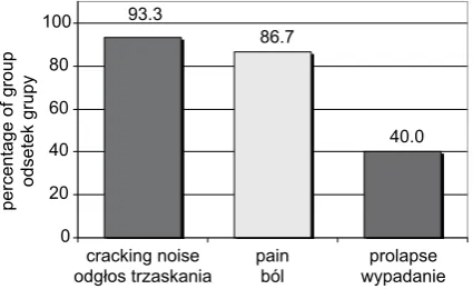
Participants were qualified to the study group on the basis of the occurrence of at least one of the following symptoms: cracking noises, pain in the vicinity of the temporomandibular joint or tem-poromandibular joint prolapse [9, 14, 18]. The
fre-in Figure 2.
Each participant in the research was submitted to two physical therapy treatments: TENS (Trans-cutaneous Electrical Nerve Stimulation) and
infra-Fig. 1. Age distribution in the study group
Ryc. 1. Rozkład wieku grupy badanej
0 5 10 15 20 25 30 35
20 21 22 23 24 25 26 27 28 29 30
age – years wiek – lata
percentage of group
odsetek grup
y
Fig. 2. Distribution of symptoms
Ryc. 2. Rozkład objawów
40.0 93.3
86.7
0 20 40 60 80 100
cracking noise
odgłos trzaskania painból wypadanieprolapse
percentage of group odsetek grupy
Fig. 3. Infrared lamp
Ryc. 3. Lampa na podczerwień
Fig. 4. Portable TENS apparatus
red radiation, which were conducted in accordance with accepted methods [2, 5, 10, 15–17]. Both of these treatments were performed using read-ily available home-use apparatus (Fig. 3, 4). The treatment area was chosen on the basis of physi-cal examinations and the patients’ descriptions of the discomfort [6]. The scheme of electrode ar-rangement during the TENS treatment is shown in Figure 5 [5, 16]. Each TENS treatment lasted 20 minutes and the current intensity was adjusted to each patient’s comfort level. Infrared radiation was performed from a distance of 20–30 cm; the dura-tion of each treatment was 15 minutes.
Functional evaluation of the temporoman-dibular joint was performed by measuring the jaw’s range of motion (ROM) in the sagittal plane and by using a photographic method of recording the jaw movement trajectory. Active and passive
ROM were measured three times and then aver-aged; the average values were compared before and after therapy. The jaw’s ROM was defined as the distance between the midline of the top and bot-tom incisors measured by slide calliper (Figure 6). Active movement was measured by having the pa-tients open their mouths maximally while the re-searcher measured the distance between the edges of the incisors. Passive movement was measured in a similar way, but instead of using the jaw muscles to open their mouths, the patients put their thumbs on the edge of their upper teeth and second finger-tips on the edge of their lower teeth to increase the mouth opening [6, 8, 14, 24].
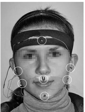
A special system was used in the study to evaluate the jaw movement trajectory. The system consists of a series of connected miniature light-emitting diodes (LEDs), powered by external bat-tery and located on moving (red diodes) and im-mobile (green diodes) parts of the patient’s head at selected characteristic points (Figure 7). A digital camera was used to take photos with a long expo-sure time (2 seconds) during abduction and ad-duction of the jaw.
Results
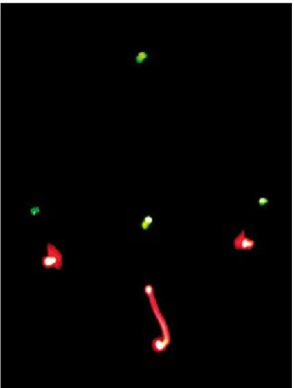
Figures 9–12 show deviations from the normal movement trajectory while the mouth is opening and closing. Differences can be observed between the shapes of the movement trajectories while
low-Fig. 5. Electrode arrangement
Ryc. 5. Rozmieszczenie elektrod
Fig. 6. Measurement of the jaw’s ROM in the sagittal plane [8]
Ryc. 6. Pomiar zakresu ruchomości żuchwy (z.r.ż.) w płaszczyźnie strzałkowej [8]
Fig. 7. Positions of LED markers on the angles of the jaw and protuberantia mentalis
Fig. 7. Pozycja wskaźników LED w kątach żuchwy
Fig. 8. Movement trajectory recorded during abduc-tion of the jaw (sagittal plane)
Ryc.8. Trajektoria ruchu zarejestrowana podczas odwodzenia żuchwy (płaszczyzna strzałkowa)
Fig. 9. Movement trajectory recorded during adduc-tion of the jaw (sagittal plane)
Ryc.9. Trajektoria ruchu zarejestrowana podczas przy-wodzenia żuchwy (płaszczyzna strzałkowa)
Fig. 10. Movement trajectory recorded during abduc-tion of the jaw (frontal plane)
Ryc.10. Trajektoria ruchu zarejestrowana podczas odwodzenia żuchwy (płaszczyzna czołowa)
Fig. 11. Movement trajectory recorded during adduc-tion of the jaw (frontal plane)
ering and raising the jaw. These differences may result from asymmetry in the temporomandibular joint structure and condyle-disc disorders [6].
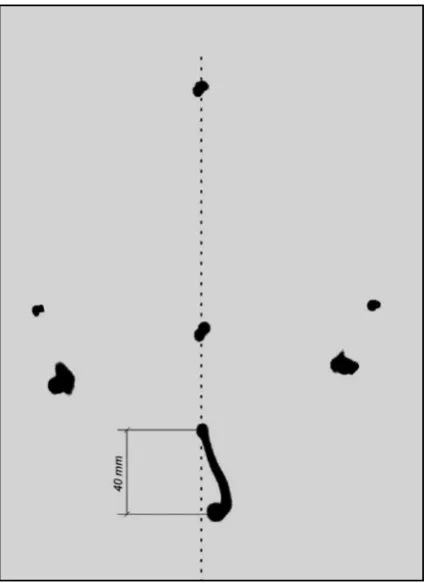
Figures 12–13 present an example analysis of the recorded mandible movement trajectory in the sagittal plain. Differences can be observed between the normal trajectory (shown a dotted line) and the recorded trajectory. No improvement in the mandible movement trajectory was observed in any of the patients after the therapy.
In 60% of the patients, the jaw’s active ROM increased after therapy. In these people the average value of the jaw’s ROM in the sagittal plane was 43.1 mm (SD = 8.4 mm) before therapy, while after physiotherapy the ROM was as high as 46.0 mm (SD = 6.3 mm).
Discussion
In an era when lifestyles are undergoing con-stant acceleration and the technology of medical diagnostics is improving, people still forget how to take care of their own health. This problem con-cerns spine or hip ailments as well as dysfunctions of the masticatory organ. It is rare for a person to consult a doctor only because of pain in the jaw area that appears while biting nails or chewing
gum, or when the third molars are growing. Epi-demiologic investigations indicate that dysfunc-tions of the temporomandibular joint affect about 80% of the human population, which makes them the third most frequent dental disorders after car-ies and parodontopathy [26]. Temporomandibu-lar dysfunction affects more women than men [9], which was confirmed in the current study by a pro-portion of 4:1. Although degenerative changes in temporomandibular joints are connected with long-term use, disease more and more frequently affects young people who are exposed to stress-related risk factors. The period of university stud-ies is a phase of life characterized by a particularly high susceptibility to stress. Because of this, uni-versity students are a high-risk group for diseases of the masticatory organ [20]. These facts were a crucial factor in deciding on the profile of the study group. People were qualified as participants in the research on the basis of common diagnostic criteria: the presence of acoustic symptoms, pain in the vicinity of temporomandibular joint or jaw prolapse [6, 9, 11, 13, 14, 18, 19].
Following the example of other clinicians [13, 14, 21], TENS was used in the current study (due to its remarkable analgesic effect) along with infra-red radiation (which infra-reduces muscle hypertonia).
Fig. 12. Example analysis of one patient’s movement trajectory during abduction of the jaw (frontal plane)
Ryc. 12. Analiza trajektorii ruchu żuchwy na przykładzie wybranego pacjenta podczas odwodzenia (płaszczyzna czołowa)
Fig. 13. Example analysis of one patient’s movement trajectory during adduction of the jaw (frontal plane)
Ryc. 13. Analiza trajektorii ruchu żuchwy na
References
[1] Bahnof R: Physiotherapy for Severe Restriction of Mouth Opening following Transtemporal Neurosurgery. Physiotherapy 1998, 84 (10), 501–505.
[2] Bauer A, Wiecheć M: Przewodnik metodyczny po wybranych zabiegach fizykalnych, Markmed Rehabilitacja s.c., Ostrowiec Świętokrzyski, 2005.
[3] Blanco RC et al.: Changes in active mouth opening following a single treatment of latent myofascial trigger points in the masseter muscle involving post-isometric relaxation or strain/counterstrain. J Body Mov Ther 2006, 10, 197–205.
[4] DeLany PJ: Temporomandibular dysfunction: neuromuscular therapy. J Bod Mov Ther 1997, 1(4), 199–203.
[5] Dobrogowski J, Wordliczek J (red.): Medycyna bólu. PZWL, Warszawa 2004.
[6] Dupas PH: Dysfunkcja czaszkowo-żuchwowa. PZWL, Warszawa 2009.
[7] Garcia IJ et al.: Changes in masseter muscle trigger points following strain-counterstrain or neuro-muscular tech-nique. J Body Mov Ther 2009, 13, 2–10.
[8] Gray M, Davies SJ, Quayle AA: Patologia układu mięśniowo-stawowego narządu żucia w ujęciu klinicznym. Wydawnictwo Medyczne Sanmedica, Warszawa 1996.
[9] Grey M et al: Temporomandibular pain dysfunction: can electrotherapy help? Physiotherapy 1995, 81 (1), 47–51.
[10] Kahn J: Elektroterapia. Zasady i zastosowanie. PZWL, Warszawa, 1996.
[11] Kalamir A et al.: TMD and the problem of bruxism. A review. J Bod Mov Ther 2007, 11, 183–193.
[12] Kleinrok M: Bólowe i bezbólowe objawy związane z zaburzeniem czynności układu ruchowego narządu żucia oraz zasady rozpoznawania i leczenia tych zaburzeń. Terapia 2004, 10 (157), 19–27.
[13] Kogut G, Kwolek A: Zaburzenia czynnościowe układu ruchowego narządu żucia – diagnostyka i leczenie. Rehabil Med 2006, 10 (1), 41–48.
[14] Maślanka T (red.): Zaburzenia czynnościowe narządu żucia. Urban&Partner, Wrocław 1997.
[15] Melzack R, Wall PD: Tajemnica bólu. WAM, Kraków 2006.
[16] Mieszkowski P, Kleinrok J: Ocena skuteczności leczenia chorych z bólowym zespołem dysfunkcji narządu żucia wyłącznie elektrostymulacją oraz w skojarzeniu z leczeniem repozycyjną szyną zgryzową. Czas Stomatol 2003, 56 (1), 56–61.
[17] Mika T, Kasprzak W: Fizykoterapia. PZWL, Warszawa 2006.
[18] Panek H et al.: Epidemiology of temporomandibular dysfunctions in young adult populations studied in Department of Prosthodontics Silesian Piast’s University of Medicine in Wrocław, Poland. Dent Med Probl 2007, 44 (1), 55–59.
[19] Panek H, Mankiewicz M: Zaburzenia funkcji stawów skroniowo-żuchwowych w obecności wyrżniętych trzecich zębów trzonowych.Dent Med Prob 2005, 42 (2), 311–315.
[20] Rasławska J, Dawid K, Janiszewska-Olszowska J: Występowanie bruksizmu u przyszłych stomatologów. Mag Stomatol 2008, 7–8, 72–76.
[21] Rzepka R: Leczenie bruksizmu cz. 2. Mag Stomatol 2009, 7–8, 30–34.
[22] Sawicki J et al.: Kompleksowa ocena stanu funkcjonalnego narządu żucia z użyciem różnych technik pomiaro-wych. Inż Biomed 2008, 14 (4), 336–339.
[23] Skaggs C: Temporomandibular dysfunction: chiropractic rehabilitation. J Body Mov Ther 1997, 1 (4), 208–213.
[24] Skolimowski T: Badania czynnościowe narządu ruchu w fizjoterapii. Wydawnictwo AWF we Wrocławiu, Wrocław 2009.
[25] Tilley L: Temporomandibular dysfunction: holistic dentistry. J Body Mov Ther 1997, 1 (4), 203–207.
[26] Wigdorowicz-Makowerowa N et al.: Zaburzenia czynnościowe narządu żucia. PZWL, Warszawa, 1984.
Address for correspondence:
Jadwiga Kuciel-Lewandowska Department of Health Care Sciences Institute of Physiotherapy
Wroclaw Medical University grunwaldzka 2
50-355 Wrocław Poland
Tel. (+48 71) 784 01 83; +48 603 672 374 E-mail: [email protected]
Conflict of interest: None declared