Beata Sobieszczańska
1, A, C, D, F, Anna Duda-Madej
1, B, C, Michał Turniak
1, B, C,
Roman Franiczek
1, B, C, Urszula Kasprzykowska
1, B, C, Anna K. Duda
1, B, C,
Marta Rzeszutko
2, B, C, Barbara Iwańczak
3, EInvasive Properties, Adhesion Patterns and Phylogroup
Profiles Among Escherichia Coli Strains Isolated from
Children with Inflammatory Bowel Disease*
Zdolności adhezyjne i inwazyjne oraz przynależność do grup
filogenetycznych szczepów E. coli izolowanych od dzieci
z nieswoistymi zapaleniami jelit
1 Department of Microbiology, Wroclaw Medical University, Wrocław, Poland 2 Department of Patomorphology, Wroclaw Medical University, Wrocław, Poland
3 2nd Department and Clinic of Pediatrics and Gastroenterology, Wroclaw Medical University, Wrocław, Poland
A – research concept and design; B – collection and/or assembly of data; C – data analysis and interpretation;
D – writing the article; E – critical revision of the article; F – final approval of article; G – other
Abstract
Background. A great deal of evidence indicates a link between Escherichia coli (E. coli) and Crohn’s disease in adult patients, but there is lack of information on the association of these bacilli with inflammatory bowel disease (IBD) among children.
Objectives. The study was carried out to determine the distribution of phylogenetic group, the adherence patterns and invasive properties of E. coli isolated from children with IBD and non-IBD chronic bowel diseases.
Material and Methods. A total of 22 E. coli isolated from biopsy specimens from children with IBD and 21 E. coli
strains obtained from children with indeterminate colitis and intestinal polyps were examined for adherence and internalization to the Int407 cell line. Genes involved in epithelial cell invasion and genes specific to E. coli phylo-groups were determined by polymerase chain reaction (PCR).
Results. The undefined adherence pattern predominated among the isolated E. coli, although most of them demon-strated the afaD and aggB genes encoding invasions of diffusely adhering and enteroaggregative E. coli. Regardless of the clinical entity, most E. coli were internalized by Int407 epithelial cells and belonged to the B2 and D phylo-groups.
Conclusions. The wide distribution of adhesive E. coli capable of entering Int407 cells but also having genes encod-ing adhesins and invasins characteristic to pathogenic E. coli strains seems to indicate that these E. coli may rep-resent a large group of pathogenic E. coli strains contributing to chronic intestinal disorders (Adv Clin Exp Med 2012, 21, 5, 591–599).
Key words: inflammatory bowel disease, invasion, E. coli, adherence pattern, invasion genes, phylogroups.
Streszczenie
Wprowadzenie. Wiele wyników badań wskazuje na związekEscherichia coli z chorobą Leśniowskiego-Crohna u osób dorosłych, jednak nadal brakuje danych na temat roli tych pałeczek w nieswoistych zapaleniach jelit (IBD) u dzieci.
Adv Clin Exp Med 2012, 21, 5, 591–599 ISSN 1899–5276
oRIGINAL PAPERS
© Copyright by Wroclaw Medical University
There have been several reports on increased numbers of Escherichia coli (E. coli) colonizing the intestinal mucosa of patients with inflammatory bowel disease (IBD) [1, 2]. Bacterial adherence to the intestinal mucosa is an essential step in the initiation of colonization and infection involving a wide variety of distinct bacterial adhesins. Fim-brial and nonfimFim-brial adhesins that mediate at-tachment to host cells are produced by most path-ogenic and non-pathpath-ogenic E. coli strains. Many of these adhesins are associated with specific adher-ence patterns to epithelial cells, i.e. the diffuse ad-herence pattern, aggregative adad-herence pattern or localized aggregative adherence pattern, which are characteristic to particular E. coli pathotypes [3]. Recently a new category has been described: ad-herent-invasive E. coli (AIEC), associated with ileal mucosa in Crohn’s disease [4–6]. AIEC strains are able to adhere to and invade intestinal epithelial cells, and are also able to survive in macrophages. Moreover, many AIEC have shown a mannose-re-sistant diffuse adherence pattern to intestinal epi-thelial cells, characteristic of the DAEC (diffusely adhering Escherichia coli) pathotype.
About 25% of all IBD patients will present before the age of 20 years. Childhood IBD, like adult IBD, comprises two phenotypes: Crohn’s disease (CD) and ulcerative colitis (UC). Although the pathophysiology of IBD is unknown, several studies have provided evidences that IBD may be a result of a genetic predisposition associated with defects of the innate response of the intestinal mu-cosa, conferring susceptibility to undefined envi-ronmental factors such as luminal bacteria [7, 8]. There is a lot of evidence linking E. coli and CD in adult patients, but there is lack of information on the association of these bacilli with inflammatory bowel disease IBD among children.
The present study was carried out to deter-mine the adherence patterns and invasive prop-erties as well as the distribution of phylogenetic
groups among of E. coli strains isolated from bi-opsy specimens obtained from children with IBD and non-IBD chronic bowel diseases.
Material and Methods
Biopsy Specimens and E. coli
Strains
Biopsy specimens from a total of 43 consecu-tive untreated children diagnosed on the basis of clinical, endoscopic and histological criteria of IBD at Wroclaw Medical University’s 2nd De-partment and Clinic of Pediatrics and Gastroen-terology (Wrocław, Poland) were examined. CD was diagnosed in 11 children (mean age 12 years, ranging from 3 to 18 years) and UC was diagnosed in 11 children (mean age 12 years, ranging from 2 to 15 years). Eleven E. coli isolates were obtained from 11 children (mean age 11 years, ranging from 3 to 13 years) with indeterminate colitis; ten E. coli
strains were from 10 otherwise healthy children (mean age 10 years, ranging from 2 to 17 years) with intestinal polyps. In the study these 21 E. coli
were categorized as non-IBD group. Biopsy sam-ples were taken during endoscopy from sites with visible macroscopic inflamed ileal mucosa. Imme-diately after being obtained, the biopsy specimens were plated onto MacConkey agar. After overnight incubation, five lactose-positive colonies were iso-lated from every sample and defined as E. coli by standard biochemical testing. All the isolated E. coli were tested in an adherence assay to determine their adherence types. A total of 43 E. coli strains representing 43 patients were included for further analysis. The following strains containing genes encoding invasins were used as controls in the polymerase chain reaction (PCR) and internaliza-tion assays: ipaH-positive Shigella flexneri strain (ATCCTM12022), invE-positive E. coli o29:NM
Cel pracy. Charakterystyka zdolności adhezyjnych i inwazyjnych oraz przynależność do grup filogenetycznych szczepów E. coli izolowanych od dzieci z nieswoistymi zapaleniami jelit oraz innymi przewlekłymi chorobami prze-wodu pokarmowego.
Materiał i metody. Test adhezji i inwazji in vitro do komórek nabłonka jelita linii Int407 wykonano dla 43 szczepów
E. coli izolowanych z bioptatów błony śluzowej jelita 22 dzieci z rozpoznaną chorobą Leśniowskiego-Crohna i wrzo-dziejącym zapaleniem jelita grubego oraz 21 dzieci z nieokreślonym zapaleniem jelita i polipami jelit. Geny kodujące inwazyny oraz geny klasyfikujące badane szczepy do konkretnej grupy filogenetycznej wykrywano testem PCR.
Wyniki. Wśród badanych izolatów E. coli przeważał nieokreślony typ adhezji, chociaż większość szczepów posiadała geny kodujące inwazyny, tj. afaD i aggB typowe dla szczepów o rozsianym i agregacyjnym typie adhezji. Niezależnie od jednostki chorobowej, z której izolowano badane szczepy, większość z nich była internalizowana przez komórki nabłonka jelita i należała do grup filogenetycznych B2 i D.
Wnioski. Z uwagi na to, że większość badanych szczepów E. coli wykazała zdolność adhezji i inwazji do komórek nabłonka jelita oraz prezentowała geny kodujące inwazyny typowe dla patogennych szczepów E. coli, prawdopo-dobnie badane izolaty mogą reprezentować większą grupę szczepów związanych z przewlekłymi chorobami prze-wodu pokarmowego (Adv Clin Exp Med 2012, 21, 5, 591–599).
(ATCCTM43892), and afaA, afaC and afaD
-posi-tive, but ipaH-, tia-, and aggB-negative wild E. coli
strain 48-2 of diffuse adherence pattern (DAEC) isolated from a child with diarrhea, enterotoxi-genic tia-positive E. coli (ATCCTM 35401) and E. coli 17-2 reference strain of aggregative adherence pattern was used as a source of the aggB gene. The
E. coli K-12 C600 strain was used as a noninvasive
control and negative control in all the PCR experi-ments. All bacterial strains were stored in Luria broth (LB) with glycerol 15% v/v at –80o C and
were cultured routinely in LB overnight at 37oC.
Ethical Considerations
The Ethical Committee of Wroclaw Medical University (Wrocław, Poland) approved the study, and the written consents of children’s parents were obtained in every case in the study.
Adherence Assay
The in vitro adherence assay to embryonic human epithelial cell line Int407 (ATCC CCL6) was performed as described by Cravioto et al. [9]. A chamber slide was seeded with cell suspension at a density ca. 4 × 10⁵ that was grown overnight to
a near confluent monolayer at 37oC in an
atmos-phere with 5% Co2 in Dulbecco modified Eagle’s
medium (DMEM; Invitrogen) supplemented with 10% fetal bovine serum (FBS, Lonza; Germany) and antibiotic-antimycotic solution (penicillin 100 U, streptomycin 100 U, amphotericin B 0.25 μg per ml; Invitrogen). Before the test was performed, the cells were washed twice with phosphate-buff-ered saline (PBS; pH 7.2; Sigma-Aldrich) and the cell culture medium in the chamber slides was replaced with 0.5 ml DMEM without antibiotics but supplemented with 2% FBS and 1% methyl α-D-mannopyranoside (Sigma-Aldrich). The bac-terial strains to be tested were grown overnight at 37oC in LB medium. Twenty-five μl of bacterial
culture was added to each chamber and incubated for 3 or 6 hours at 37oC in an atmosphere with
5% Co2. Then the slides were washed three times
with PBS, fixed with 70% methanol for 10 minutes and stained with 10% Giemsa (Sigma-Aldrich) for 30 minutes. After staining the slides were washed, mounted on glass slides and examined under the oil immersion objective of an olympus BX51 light microscope. The assay was repeated twice for each
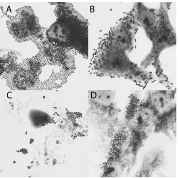
E. coli strain, with incubation periods of 3 hours and 6 hours. The sample results are presented in Figure 1.
Bacterial Internalization Assay
The invasive ability of E. coli isolates to the Int407 cells were evaluated by the standard gen-tamycin protection assay with some modifica-tions. This test is based on the principle that the gentamycin has limited capacity to penetrate to eukaryotic cells and internalized bacteria are thus protected. The minimal inhibitory concentration (MIC) of the gentamycin for all strains examined was determined by an agar dilution technique on Mueller-Hinton agar according to the Clini-cal Laboratory and Starndards Institute (CLSI) recommendations and the antibiotic was used at 100-fold the MIC value. The Int407 cells were grown to a confluence in minimal essential me-dium (MEM) with 10% FBS and antibiotic-anti-mycotic solution (penicillin 100U/l, streptomycin 100 μg/ml, amphotericin B 0.25 μg/ml) at 37oC ina humidified atmosphere containing 5% Co2. The
cells were harvested by trypsinization and seeded at a density of 4 × 10⁵ cells/ml into a 24-well
tis-sue culture plate, then incubated overnight to near (80%) confluence. Before infection, the cells were washed three times with pre-warmed PBS (pH 7.4) and fresh culture medium supplemented with FBS 1% v/v, and methyl α-D-mannopyranoside 0.5% v/v without antibiotics was added. The E. coli iso-lates were harvested from overnight cultures in LB and resuspended in PBS to a density of 9 × 10⁸cfu/
ml. The Int407 cells were then infected at a multi-plicity of infection (MoI) of 100 bacteria per cell. After centrifugation at 1200 rpm for 10 minutes to obtain efficient invasion, the cells were incubated at 37oC in a 5% Co
2 atmosphere. After a 4-hour
in-cubation period the cells were washed three times with PBS, and fresh culture medium containing 100 μg/ml gentamycin was added to kill extracel-lular bacteria. After a 1-hour incubation period the cells were washed three times and lysed by vigor-ous pipetting and shaking for 10 minutes at room temperature, after the addition of 1% Triton X-100 in deionized water. The cellular lysates were seri-ally diluted in PBS, placed onto MacConkey agar and incubated overnight at 37oC to determine the
number of bacterial colony forming units (CFU). The number of bacteria internalized was expressed as the mean percentage of the initial inoculum that was internalized. Each assay was run in duplicate and repeated three times.
PCR Assay
Genes involved in the invasiveness of E. coli
Table 1. Primer sequences and amplification conditions used in the study
Tabela 1. Sekwencje starterów oraz warunki reakcji zastosowane w badaniach Gene
(Gen) PCR primers (Startery PCR) PCR conditions(Warunki PCR) Product size – bp(Wielkość pro-duktu – bp)
References (Piśmiennictwo)
aggB (F) GCATATTACCGATGTCCTGCG 95oC 1 min, 42oC 1 min, 421 this study
(R) CCTCTTGATATTAGACATTCA 72oC 2 min (29 cycles)
afaD (F) GTCACCTGCGGGATGTTACT 94oC 1 min, 60oC 45 s, 392 this study
(R) GCTCCGCCAACCGAAAAC 72oC 45 s (34 cycles)
ipaH (F) GCTGGAAAAACTCAGTGCCT 94oC 1 min, 56oC 2 min, 424 [29]
(R) CCAGTCCGTAAATTCATTCT 72oC 1 min (29 cycles)
tia (F) ACCAGCGCTTCCGTCAGG 94oC 1 min, 56oC 1 min, 382 [4]
(R) GCCAGATTCATTCCAGGAGG 72oC 1 min (30 cycles)
invE (F) ATATCTCTATTTCCAATCGCGT 94oC 30 s, 47oC 1 min, 382 [30]
(R) GATGGCGAGAAATTATATCCCG 72oC 1.5 min (25 cycles)
Fig. 1. Adherence patterns of the E. coli strains examined. A) aggregative adherence (AA); B) diffuse adherence (DA); C) cell-detaching pattern (CDT); D) undefined adherence pattern (UD). Giemsa stain. Magnification 100×
were synthesized on the basis of published nucleo-tide sequences. The aggB anad afaD genes were amplified using primers designed in the authors’ laboratory (aggB Forward: positions 3460 to 3480 and Reverse: positions 3857 to 3880 Gen-Bank ac-cession no. U12894; afaD Forward: 7479 to 7498 and Reverse: positions 7853 to 7870 Gen-Bank accession no. X76688). The sequences of primers used, PCR conditions and the expected amplicon sizes are presented in Table 1. Triplex PCR with primers for chuA, yjaA and anonymous cryptic DNA fragment TSPE4.C2 for the phylogenetic groups was performed as described by Clermont et al. [10]. All PCR amplifications were performed in a DNA-Engine PT200 thermal cycler (MJ Re-search Waltham, MA, USA). The PCR products were visualized after electrophoresis on 2%
aga-rose gel in Tris-acetate-EDTA buffer by staining with SYBR Green I Nucleic Acid Gel Stain. The distribution of genes identified among the E. coli
isolates is shown in Table 2.
Statistical Analysis
The significance of differences observed in the prevalence of genetic determinants of invasiveness and phylogenetic groups among the E. coli exam-ined was assessed using the Pearson chi-square test (Excel 2007) with p ≤ 0.05 considered statistically significant. The data are reported as mean values with standard deviation (SD).
Table 2. Distribution of the genes identified in the study and the adherence patterns of E. coli strains from children with IBD and non-IBD groups
Tabela 2. Występowanie genów oznaczanych w badaniach oraz typy adhezji szczepów E. coli od dzieci z grup IBD i nie-IBD Characteristic/E.
coli group (Cecha/grupa E. coli)
№ of strains (Liczba szczepów) %
CD (n = 11) UC (n = 11) IC (n = 11) Po (n = 10) total (n = 43)
Adherence pattern (Typ adhezji)
UD 7 (63.6) 5 (45.4) 6 (54.5) 4 (40) 22 (51.2)
AA 2 (18.2) 4 (36.4) 2 (18.2) 0 8 (18.6)
DA 1 (9.1) 1 (9.1) 1 (9.1) 2 (20) 5 (11.6)
CDT 1 (9.1) 1 (9.1) 2 (18.2) 4 (40) 8 (18.6)
Invasion genes (Geny inwazji)
aggB 8 (72.7) 4 (36.4) 3 (27.3) 2 (20) 17 (39.5)
afaD 5 (45.4) 5 (45.4) 4 (36.4) 4 (40) 18 (41.9)
tia 1 (9.1) 3 (27.3) 0 1 (10) 5 (11.6)
ipaH 1 (9.1) 1 (9.1) 1 (9.1) 0 3 (7.0)
invE 0 0 0 0 0
Phylogroups (Grupy filogenetyczne)
A 0 0 1 (9.1) 2 (20) 3 (7.0)
B1 0 0 0 0 0
B2 7 (63.6) 8 (72.7) 6 (54.5) 5 (50) 26 (60.5)
D 4 (36.4) 3 (27.3) 4 (36.4) 3 (30) 14 (32.6)
Adherence patterns: UD: undefined; AA: aggregative; DA: diffuse; CDT: cell-detaching; CD: Crohn’s disease; UC: ulcerative colitis; IC: indeterminate colitis; Po: polyps.
Results
Adherence Assay
The distribution of the E. coli isolates’ adher-ence patterns is shown in Table 2. Undetermined adherence (UD), in which adhering bacteria do not form any characteristic pattern (Fig. 1), was the most prevalent, associated with 22 (51.2%) of the
E. coli isolates (p = 0.0015). This adherence pattern was also the one most frequently associated with
E. coli from CD children, although there was no statistical difference in the distribution of the E. coli
presenting the UD pattern among the strains iso-lated from CD, UC, IC and Po groups (p ≥ 0.05).
E. coli with aggregative adherence (AA) and cell-detaching (CDT) strains were isolated with equal frequency, i.e. 8 (18.6%); whereas diffusely adher-ing E. coli were isolated less frequently: 5 (11.6%) strains. There was no statistical difference in the frequency of isolation of E. coli strains presenting AA, CDT and DA adherence patterns among the 43 E. coli strains examined (p ≥ 0.05).
Invasion Assay
All the E. coli strains examined in the in vitro
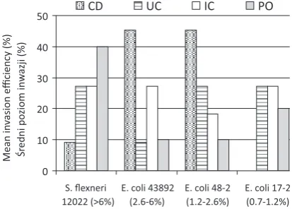
invasion assay were susceptible to gentamycin (MIC ≤ 2 μg per ml). The invasion assay demonstrated that 42 (97.7%) of the 43 E. coli isolates were inter-nalized by epithelial cells Int407 at the level ≥ 0.1%, although there were differences in the invasion ef-ficiency among isolates (Figure 2). E. coli isolates from CD and UC patients demonstrated similar invasion efficiency (3.85% and 3.39%, respectively), but lower than the mean invasion levels of E. coli
strains from patients with IC and Po (4.09% and 5.25%, respectively). Interestingly, E. coli isolated from children with Po, who were considered an otherwise healthy control group, showed the high-est invasion efficiency (5.25%) as compared with the other E. coli strains examined (p < 0.05). An invasion efficiency (> 6%) comparable to the ipaH -positive Shigella flexneri 12022 reference strain was demonstrated by 4 (40%) E. coli isolates from the Po group, 3 (27.3%) E. coli from the UC and IC groups, and only 1 (9.1%) strain from the CD patients (p > 0.05). As many as 5 (45.5%) E. coli from the CD group showed mean invasion levels ranging from 2% to 6%, comparable to the mean invasion level of the invE-positive E. coli 43892 reference strain. In contrast, mean invasion levels ranging from 2% to 6% were demonstrated by 3 (27.3%) E. coli isolates from the IC group and only one isolate from the UC group and the Po group (p > 0.05). Similarly, E. coli
from CD patients most frequently showed mean in-vasion levels ranging from 1% to 2%, comparable
with afaD-positive E. coli 48-2 strain, although this was not statistically significant (p > 0.05) in com-parison with E. coli from UC, IC and Po patients. Mean invasion levels comparable with aggB -posi-tive E. coli 17-2 strain, ranging from 0.7% to 1.2%, were shown by 3 (27.3%) E. coli from the UC and IC groups, but by none of the E. coli strain from CD patients. The lowest internalization levels < 0.06% to Int407 epithelial cells were demonstrated by single
E. coli strains isolated from the UC and Po groups. Generally, all E. coli isolated from CD patients dem-onstrated mean invasion levels ranging from 1% to > 6%. In contrast, E. coli isolated from UC and Po patients showed mean invasion levels ranging from < 0.7% to > 6%, whereas E. coli from IC group were internalized by Int407 cells at mean levels from > 0.7% to > 6%. However, these differences were not statistically significant (p > 0.05).
Distribution of Genes Encoding
for Invasins
The analysis of the invasion gene distribution demonstrated that afaD gene encoding the outer membrane protein involved in the internalization
0 10 20 30 40 50 S. flexneri 12022 (>6%)
E. coli 43892 (2.6-6%)
E. coli 48-2 (1.2-2.6%)
E. coli 17-2 (0.7-1.2%) Mean in va si on e ffi ci en cy (% ) Śr edni po ziom inw azji (% )
CD UC IC PO
Fig. 2. The mean invasion efficiency of E. coli strains isolated from children with CD (Crohn’s disease), UC (ulcerative colitis), IC (indeterminate colitis) and Po (polyps) in comparison with reference strains used in the study: S.flexneri 12022 ipaH-positive, E. coli 43892
invE-positive, E. coli 48-2 afaD-positive, E. coli 17-2
aggB-positive
Ryc. 2. Średni poziom inwazji szczepów E. coli
izolowanych od dzieci z CD (chorobą Crohna), UC (wrzodziejącym zapaleniem jelita grubego), IC (nieokreślonym zapaleniem jelit) oraz Po (polipami jelit) w porównaniu z referencyjnymi szczepami zasto-sowanymi w badaniach: S.flexneri 12022 ipaH-dodatni,
E. coli 43892 invE-dodatni, E. coli 48-2 afaD-dodatni,
of diffusely adhering E. coli (DAEC) strains [11, 12] and aggB gene encoding the outer membrane protein AggB considered as an invasion protein of enteroaggregative E. coli (EAEC) strains [13] were most frequently detected among E. coli isolates associated with 18 (41.9%) and 17 (39.5%) E. coli
strains, respectively (Table 2). Moreover, the aggB
gene was associated statistically more frequently with E. coli from CD patients than with IC (p = 0.03) and Po (p = 0.01) E. coli strains, although there was no significant difference (p > 0.05) in the distribution of the gene among E. coli from CD and UC patients. The ipaH invasion plasmid antigen associated with the invasiveness of Shigella
and enteroinvasive E. coli (EIEC) strains [14, 15], and the tia gene encoding the outer membrane protein engaged in the invasion of ETEC strains [16] were detected in a minority of E. coli isolates (Table 2). There was no statistical difference (p > 0.05) in the distribution of these genes among the
E. coli strains examined. None of E. coli examined showed the presence of invE regulator for cell in-vasion of enteroinvasive E. coli (EIEC). It is also worth noting that in some E. coli strains isolated in the study, the authors were unable to detect any of the invasion genes examined, although these iso-lates demonstrated invasion capability. This may indicate that these isolates may present other in-vasins not identified in this study.
Phylogroups Distribution
The E. coli species has been divided into four main phylogenetic groups based on an anony-mous DNA fragment designated the TSPE4.C2,
chuA gene, required for heme transport in EHEC o157:H7 E. coli, and the yjaA gene of unknown function present in nonpathogenic E. coli K12 strain. The A or B1 phylogroups comprise most commensal E. coli strains, whereas isolates asso-ciated with extraintestinal infections (ExPEC) de-rive predominantly from the B2 phylogroup and to a lesser extent the D phylogroup. Pathogenic E. coli responsible for acute and severe diarrhea are distributed between the A and B1 phylogroups, but isolates causing mild and chronic diarrhea are dis-tributed across all four phylogroups [10, 17]. In the present study, overall, 26 (60.5%) E. coli examined belonged to phylogenetic group B2 and 14 (32.5%) to D phylogroup. only 3 (7%) of the strains stud-ied belonged to phylogroup A, and none of the ex-amined isolate belonged to phylogenetic group B1 (Table 2). E. coli isolates from CD and UC patients derived from the B2 and D phylogroups, whereas
E. coli from IC and Po patients, comprised isolates from the A, B2 and D groups. There was no statisti-cally significant difference in the phylogroup
distri-bution among E. coli strains isolated from CD, UC, IC and Po patients (p > 0.05).
Discussion
In biopsy specimens from patients with IBD a high concentration of bacteria invading intes-tinal mucosa have been demonstrated [18–20]. Bacterial invasion of host cells induces a cascade of events to up-regulate innate immune defences, recruiting inflammatory cells responsible for epi-thelial barrier disruption and mucosal inflamma-tion through the released cytokines [21]. A new category of E. coli,i.e. AIEC,associated with the ileal mucosa of adult patients with Crohn’s dis-ease has been thoroughly investigated [4–6]. AIEC strains efficiently invade a wide range of human epithelial cell lines and have a unique capability to survive and replicate within macrophages [5, 22]. Moreover, AIEC strains present diffuse adherence pattern to intestinal epithelial cells, although only some of these strains harbor virulence genes char-acteristic of the DAEC pathotype, which confers this type of adhesion. AIEC strains have been iso-lated from 36.4% of adult patients with CD vs. 6% of controls [4, 23].
Most of the E. coli strains isolated in the cur-rent study, regardless of the clinical diagnosis, showed undefined adherence patterns and moder-ate levels of invasion into Int407 epithelial cells, most probably associated with invasion genes characteristic to diffusely adhering E. coli DAEC and/or EAEC. only some of the E. coli strains in the study presented defined adherence patterns – i.e. aggregative or diffuse patterns – and invasion levels corresponding to Shigella bacilli. Generally, when comparing E. coli isolates from children with IBD and other chronic bowel diseases (indeter-minate colitis or polyps), the results of the study showed no striking differences among these iso-lates. None of the adherence patterns was signifi-cantly associated with any of the disorders. More-over, all but one of the E. coli isolates in the study showed some degree of invasion capability, even the strains from children with polyps. In spite of the fact that the aggB and afaD genes were more frequently detected among E. coli from IBD than among E. coli isolated from children with inde-terminate colitis or polyps, generally none of the genes examined was significantly associated with any of these clinical entities. Although they dem-onstrated adhesive and invasive capabilities, E. coli
isolates from children with IBD and other chronic bowel diseases seem to be different from E. coli
CD patients were internalized by non-polarized, non-differentiated Int407 cells, which is a contrast to the results demonstrated by Darfeuille-Michaud et al. [4] for adult patients with UC. According to their study AIEC were associated with none of co-lonic specimens from patients with UC and 1.9% of the specimens from healthy controls. However, an increased number of mucosa-associated E. coli
in adult patients with UC was found in studies by Swidsinski et al. [24] and Kotlowski et al. [25]. Next, AIEC strains present diffuse adherence and, even more importantly, they lack known invasive determinants. In contrast, most of the E. coli iso-lates in the current study demonstrated at least one of the well-characterized invasion genes. Thus, it seems that the only similarity to AIEC shown by the E. coli isolated in the study from children IBD is the affiliation to phylogroups: Most of the E. coli
from children with IBD belonged to phylogropus B2 and D, which is similar to E. coli from adults IBD. However, both these phylogroups include E. coli associated with extraintestinal infections (Ex-PEC). Martinez-Medina et al. [26] showed that AIEC strains carry many virulence-associated genes characteristic of ExPEC, suggesting a close relationship between these both E. coli subgroups. Interestingly, Abe et al. [27] detected among some ExPEC isolates several virulence genes character-istic of the EAEC pathotype, e.g. the aatA gene encoding the dispersin transporter. The dispersin protein is responsible for dispersing EAEC across the intestinal epithelium, whereas the aatA gene is required for the proper function of aggregative
adherence fimbriae of EAEC [28]. This may indi-cate that at least some ExPEC strains arose from the EAEC pathotype. In the current study the aggB
gene, characteristic of the EAEC pathotype, was the second most common invasion gene detected in the E. coli isolated.
The results of the study indicate that E. coli
from IBD children do not differ significantly from
E. coli isolated from other chronic bowel diseases, but do differ from adult IBS E. coli. Taking into consideration that children become infected with many intestinal pathogens more frequently than adults, the authors hypothesize that the differences among the E. coli colonizing children’s and adults bowels depend on immunological, behavioral and nutritional divergences associated with these age groups. on the other hand, AIEC as well as the E. coli isolated in the study seem to be well adapted to colonize human intestinal tissue.
In summary, the study characterized the ad-herence patterns and invasion capabilities of E. coli
isolated from children with IBD and other chronic intestinal diseases. However, on the basis of the re-sults obtained, the comparison of these strains with AIEC associated with IBD in adults is too prelimi-nary and needs further investigation. Nevertheless, taking into consideration the fact that invasive E. coli of undefined adherence patterns were isolated in the study not only from children with IBD but also from children with other intestinal problems, e.g. polyps, it can be assumed that these strains may represent a larger group of pathogenic E. coli strains contributing to chronic intestinal disorders.
References
[1] Kim S, Ferry G: Inflammatory bowel disease in children. Curr Probl Pediatr Adolesc Health Care 2002, 32, 108–132.
[2] Masseret E, Boudeau J, Colombel JF: Genetically related Escherichia coli strains associated with Crohn’s disease. Gut 2001, 48, 320–325.
[3] Le Bouguenec C: Adhesins and invasions of pathogenic Escherichia coli. Int J Med Microbiol 2005, 295, 471–478.
[4] Darfeuille-Michaud A, Boudeau J, Bulois P, Neut C, Glasser AL, Barnich N, Bringer MA, Swidsinski A, Beaugerie L, Colombel JF: High prevalence of adherent-invasive Escherichia coli associated with ileal mucosa in Crohn’s disease. Gastroenterology 2004, 127, 412–421.
[5] Darfeuille-Michaud A, Neut C, Barnich N, Lederman E, Di Martino P, Desreumaux P, Gambiez L, Joly B, Cortot A, Colombel JF: Presence of adherent Escherichia coli strains in ileal mucosa of patients with Crohn’s disease. Gastroenterology 1998, 115, 1405–1413.
[6] Boudeau J, Glasser AL, Masseret E, Joly B, Darfeuille-Michaud A: Invasive ability of an Escherichia coli strain isolated from the ileal mucosa of a patient with Crohn’s disease. Infect Immun 1999, 67, 4499–4509.
[7] Moyer SM: A collaborative effort to define the epidemiology of pediatric inflammatory bowel disease: what can we learn from children with early-onset disease? J Pediatr 2005, 146, 7–8.
[8] Abdel-Hady M, Bunn SK: Inflammatory bowel disease. Curr Pediatr 2004, 14, 598–604.
[9] Cravioto A, Tello A, Navarro A, Ruiz J, Villafán H, Uribe F, Eslava C: Association of Escherichia coli HEp-2 adherence patterns with type and duration of diarrhoea. Lancet 1991, 337, 262–264.
[10] Clermont O, Bonacrosi S, Bingen E: Rapid and simple determination of the Escherichia coli phylogenetic groups. Appl Environ Microbiol 2000, 66, 4555–4558.
[11] Jouve M, Garcia MI, Courcoux P, Labigne A, Gounon P, Le Bouguénec C: Adhesion to and invasion of HeLa cells by pathogenic Escherichia coli carrying the afa-3 gene cluster are mediated by the AfaE and AfaD proteins, respectively. Infect Immun 1997, 65, 4082–4089.
[13] Garcia MI, Jouve M, Nataro JP, Gounon P, Le Bouguénec C: Characterization of the AfaD-like family of invasins encoded by pathogenic Escherichia coli associated with intestinal and extra-intestinal infections. FEBS Lett 2000, 479, 111–117.
[14] Oberhelman RA, Kopecko DJ, Venkatesan MM, Salazar-Lindo E, Gotuzzo E, Yi A, Chea-Woo E, Ruiz R, Fernandez-Prada C, León-Barúa R: Evaluation of alkaline phosphate-labeled ipaH probe for diagnosis of Shigella
infections. J Clin Microbiol 1993, 31, 2101–2104.
[15] Demers B, Sansonetti PJ, Parsot C: Induction of type III secretion in Shigella flexneri is associated with differen-tial control of transcription of genes encoding secreted proteins. EMBo J 1998, 17, 2894–2903.
[16] Fleckenstein JM, Kopecko DJ, Warren RL, Elsinghorst EA: Molecular characterization of the tia invasion locus from enterotoxigenic Escherichia coli. Infect Immun 1996, 64, 2256–2265.
[17] Duriez P, Clermont O, Bonacorsi S, Bingen E, Chaventré A, Elion J, Picard B, Denamur E: Commensal
Escherichia coli isolates are phylogenetically distributed among geographically distinct human populations. Microbiology 2001, 147, 1671–1676.
[18] Kleessen B, Kroesen AJ, Buhr HJ, Blaut M: Mucosal and invading bacteria in patients with inflammatory bowel disease compared with controls. Scand J Gastroenterol 2002, 37, 1034–1041.
[19] Swidsinski A, Loening-Baucke V, Theissig F, Engelhardt H, Bengmark S, Koch S, Lochs H, Dörffel Y:
Comparative study of the intestinal mucus barrier in normal and inflamed colon. Gut 2007, 56, 343–350.
[20] Glasser AL, Boudeau J, Barnich N, Perruchot MH, Colombel JF, Darfeuille-Michaud A: Adherent invasive
Escherichia coli strains from patients with Crohn’s disease survive and replicate within macrophages without inducing host cell death. Infect Immun 2001, 69, 5529–5537.
[21] Cossart P, Sansonetti PJ: Bacterial invasion: the paradigms of enteroinvasive pathogens. Science 2004, 304, 242–248.
[22] Rolhion N, Darfeuille-Michaud A: Adherent-invasive Escherichia coli in inflammatory bowel disease. Inflamm Bowel Dis 2007, 13, 1277–1282.
[23] Martin HM, Campbell BJ, Hart CA, Mpofu C, Nayar M, Singh R, Englyst H, Williams HF, Rhodes JM:
Enhanced Escherichia coli adherence and invasion in Crohn’s disease and colon cancer. Gastroenterology 2004, 127, 80–93.
[24] Swidsinski A, Ladhoff A, Pemthaler A, Swidsinski S, Loening-Baucke V, Ortner M, Weber J, Hoffmann U, Schreiber S, Dietel M, Lochs H: Mucosal flora in inflammatory bowel disease. Gastroenterology 2002, 122, 44–54.
[25] Kotlowski R, Bernstein CN, Sepehri S, Krause DO: High prevalence of Escherichia coli belonging to the B2and D phylogenetic groups in inflammatory bowel disease. Gut 2007, 56, 669–675.
[26] Martinez-Medina M, Mora A, Blanco M, López C, Alonso MP, Bonacorsi S, Nicolas-Chanoine MH, Darfeuille-Michaud A, Garcia-Gil J, Blanco J: Similarity and divergence among adherent-invasive Escherichia coli and extraintestinal pathogenic E. coli strains. J Clin Microbiol 2009, 47, 3968–3979.
[27] Abe CM, Salvador FA, Falsetti IN, Vieira MA, Blanco J, Blanco JE, Blanco M, Machado AM, Elias WP, Hernandes RT, Gomes TA: Uropathogenic Escherichia coli (UPEC) strains may carry virulence properties of diarrheagenic E. coli. FEMS Immunol Med Microbiol 2008, 52, 397–406.
[28] Boisen N, Struve C, Scheutz F, Krogfelt KA, Nataro JP: New adhesin of enteroaggregative Escherichia coli related to the Afa/Dr/AAF family. Infect Immun 2008, 76, 3281–3292.
[29] Dutta S, Chatterjee A, Dutta P: Sensitivity and performance characteristics of a direct PCR with stool samples in comparison to conventional techniques for diagnosis of Shigella and enteroinvasive Escherichia coli infection in children with acute diarrhoea in Calcuta, India. J Med Microbiol 2001, 50, 667–674.
[30] Arikawa K, Meraz IM, Nishikawa Y, Ogasawara J, Hase A: Interleukin-8 secretion by epithelial cells infected with diffusely adherent Escherichia coli possessing Afa adhesin-coding genes. Microbiol Immunol 2005, 49, 493–503.
Address for correspondence:
Beata Sobieszczańska Department of Microbiology Wroclaw Medical University Chałubińskiego 4
50-368 Wrocław Poland
Tel./fax: +48 71 784 13 08; +48 71 784 01 17 E-mail: [email protected]
Conflict of interest: None declared Received: 5.12.2011