dant functions. Vitamin E has been proved to have the capabilities of rescuing allergen-induced inhi-bition of antioxidant enzymes, preventing the det-rimental effects of air pollution as well as asthmat-ic features in asthma [3–5]. Furthermore, it has also been concluded that vitamin E supplements could improve clinical manifestations and pulmo-nary function tests in children with moderate asth-ma [6].
Vitamin E consists of two closely related sub-groups: tocopherols and tocotrienols, each existing in 4 isomeric forms (α, β, γ and δ) [7]. Mechanis-tic studies have demonstrated that specific forms Bronchial asthma is one of the world’s most
common chronic disorders, characterized by re-curring symptoms, chronic lung airway inflam-mation, variable airflow obstruction and bronchi-al hyper-responsiveness [1]. The marked increase in asthma prevalence has made bronchial asth-ma a public health concern. More and more re-searchers recently are considering the possibility that this phenomenon may be due to the chang-ing antioxidant intake and maladjusted dietary ra-tio [2]. Meanwhile, a great deal of attenra-tion has been focused on the fat-soluble dietary vitamin E, a protective dietary factor with desirable
antioxi-Yan-Mei Wu
A, Zhi-Wen Xue
B, Li-Li Zhang
C, Ning-Mei Gao
C, Xiao-Mei Du
D,
Xiao-Yan Zhang
D, Zhuo-Hong Zhang
C, Zhi-Guo Zhang
BComparable Function of γ-Tocopherols
in Asthma Remission by Affecting Eotaxin and IL-4*
Department of Respiratory and Blood Oncology, Xi’An XD Group Hospital, ShaanXi, Xi’an, China
A – research concept and design; B – collection and/or assembly of data; C – data analysis and interpretation;
D – writing the article; E – critical revision of the article; F – final approval of article
Abstract
Background. Bronchial asthma is one of the world’s most common chronic disorders dangerous to human health. It has been hypothesized that the increased number of asthma sufferers may be due to changing antioxidant intake or vitamin deficiency. However, the influence of vitamins on asthma has rarely been considered.
Objectives. The aim of this study was to explore the effects of γ-tocopherols, a specific form of vitamin E, on asthma remission together with the possible mechanism behind the process.
Material and Methods. Eosinophil counting was applied to detect the total number of cells, eosinophils and lym-phocytes. Meanwhile, HE staining was used for morphological detection. In addition, the eotaxin and IL-4 levels in the serum and bronchoalveolar lavage fluid were measured using ELISA technology.
Results. The cell counting results showed that γ-tocopherols possesses the capability to reduce the number of eosinophils. Moreover, the exudation of inflammatory cells together with the hyperplasia of goblet cells was also found to experience significant inhibition when treated with γ-tocopherols. Furthermore, the high levels of eotaxin and IL-4 in the asthma group were evidently reduced under the treatment of γ-tocopherols which was comparable with hexadecadrol.
Conclusions. γ-tocopherols can remit asthma by regulating the level of eotaxin and IL-4. Moreover, γ-tocopherols may be regarded as a potential candidate for asthma treatment after much deeper explorations (Adv Clin Exp Med 2016, 25, 4, 643–648).
Key words: asthma, γ-tocopherols, dexamethasone, eotaxin, IL-4.
ORIGINAL PAPERS
Adv Clin Exp Med 2016, 25, 4, 643–648
DOI: 10.17219/acem/41191 © Copyright by Wroclaw Medical University ISSN 1899–5276
of vitamin E such as gamma-tocopherols (γT) and tocotrienols (especially γTE) have desirable an-ti-inflammatory effects by inhibiting eicosanoids or suppressing NF-κB signaling pathways [8, 9]. Moreover, the investigation of γT in the airway in-flammation of various animal models has attract-ed significant attention [10]. It is proposattract-ed that the supplementation of γT may be helpful in asthma treatment. However, studies referencing this appli-cation to support the hypothesis have been limited. Eosinophilic granulocyte, the crucial effector cell in the process of bronchial asthma, is closely associated with the severity of asthma [11]. Mean-while, eotaxin, as the eosinophil activation chemo-kine, plays an important role in eosinophil adhe-sion, recruitment and degranulation [12]. There are synergies between eotaxin and Th2 cytokines, which mainly participate in humoral immune re-sponses. The imbalance of Th1/Th2 immune cells has been regarded as the foundation of asthma pathogenesis. It is reported that the IL-4 gene can upregulate the expression level of eotaxin mRNA in pulmonary granuloma, airway smooth muscle cells or bronchial epithelial cells and is associated closely with asthma [13–15]. In order to study the association of γT with asthma and provide a the-oretical foundation for the clinical application of γT, a test whether γT supplementation could mit-igate airway inflammation in a mouse allergic air-way response model was carried out in compari-son to the effects of dexamethacompari-sone, which is one of the most effective drugs for asthma but with some side effects.
Material and Methods
Material and Agents
Albumin (OVA), γ-T (95% purity) and Alu-minum Hydroxide Dried Gel were purchased from Sigma Chemical CO (St. Louis, USA). BUD Pul-micort Respules was bought from AstraZeneca. Antibodies, an Immunostain SP kit, an in situ hy-bridization kit and Hochest 33342 were purchased from Invitrogen (Carlsbad, USA). Dexamethasone was produced by Tin Yiu Technology Co., LTD of Zhengzhou City.
The Establishment
of the Mouse
Airway Response Model
The 40 BALB/C6 mice (clean class), provided by the laboratory animal center of Xi’an Jiaotong university, were randomly divided into 4
sub-groups: normal subgroup (A), asthma subgroup (B), dexamethasone-treated subgroup (C) and γT-treated subgroup (D). A mixture (0.2 mL) of Ov-albumin (OVA, 40 μg) and Al(OH)s (10 mg) was applied to the mice for sensitization by means of intraperitoneal injection in the first and second week respectively. A combined application of OVA atomization inhalation for 30 min was aimed to establish the asthma model in the mice since the third week and lasted for 8 weeks, 3 times per week. Normal saline (NS), instead of the sensiti-zation liquid, was added to the control subgroup while the dexamethasone-treated subgroup and γT-treated subgroup were intraperitoneally inject-ed with dexamethasone or γT, respectively, prior to OVA inspiration.
Specimen Collection
During the period of final inspiration, the mice were anesthetized and dissected following chloral hydrate injection. Inferior vena cava blood was collected and centrifuged. The upper serum was retained at –70°. A bronchoalveolar lavage flu-id (BALF) of physiological saline lavage was then gathered after the operation of bilateral lung tissue explosion, tracheal ligation and puncture. The up-per serum after centrifugation was cryopreserved at –70°. The cell counting of the BALF can be used as an index to assess the mouse asthma model.
Histopathology Detection
The middle section of the left lung was fixed with 4% paraformaldehyde and then transected at the hilum pulmonis level. HE staining was pro-ceeded after embedding, dehydrating and slicing. Each subgroup was incubated with HE for 15 min and finally, the airway inflammation and structur-al changes of the airway are made clear at a glance after HE dye.
ELISA
Statistical Analysis
All values in the experiments were expressed as mean ± SD. Statistical analyses were carried out using SPSS 17.0 software. The significance of in-ter-subgroup differences was evaluated by ANO-VA (one-way analysis of variance) when the vari-ables showed normal distribution. The SNK-q test and Dunnett’s c (U) test were used for pairwise comparison and multiple comparisons after each analysis. A p-value less than 0.05 was considered to be significant.
Results
The Establishment
of the Asthma Model
The mouse asthma model was established suc-cessfully and the results usually occurred within 30 min of OVA inspiration. Obvious features were easy to find in different subgroups. For instance, the asthma model mouse demonstrated restless-ness, shortness of breath, nose incitement, incon-tinence and serious limb collapse while the control mouse behaved as usual, without any abnormal oc-currences. The symptoms of the dexamethasone-treated subgroup were distinctly reduced but were found with increasing activity and scratching to the face or nose. The degree of asthma episodes was also mitigated.
Results of Cell Counting
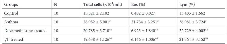
The BALF counting showed us the number of total cells, eosinophils and lymphocytes. As is shown in Table 1, the cell number of the asthma
subgroup was significantly higher than the control subgroup. And that of the dexamethasone-treated subgroup and γT-treated subgroup were decreased in comparison to the asthma subgroup. Obviously there was little difference between the dexameth-asone-treated subgroup and the γT-treated sub-group. In addition, the percentage of eosinophils and lymphocytes was noticeably lower following the addition of dexamethasone or γT. The rate of eos decreased by 15% in the dexamethasone-treated subgroup and γT-treated subgroup without a great difference, but showed a large reduction compared to the asthma subgroup. A similar phenomenon was also found in the Lym (%) measurement.
HE Staining
HE staining was used to represent the mor-phology of airway lumen in the four model sub-groups (Fig. 1). The staining results revealed the distinct characteristics between them. Firstly, the normal control subgroup contains the whole air-way epithelium with tiny amounts of inflamma-tory cells around without damage in the bronchi-al mucosbronchi-al epithelium and muscularis mucosae structure. In comparison with the staining results of the normal subgroup, the airway lumen of the asthma subgroup was found with evident incras-sation and blood vessels infiltrated a large major-ity of inflammatory cells, which could also be seen in the pulmonary mesenchyme and alveolar space. When referred to the dexamethasone-treated sub-group or γT-treated subsub-group, the above-men-tioned phenomenon was repeated to a certain de-gree, with not only the exudation of inflammatory cells but also the inhibited hyperplasia of goblet cells. Moreover, the asthma-catabatic function of γT was comparable to the dexamethasone,
there-Table 1. Comparison of cell count between the four groups (mean ± SD)
Groups N Total cells (×105/mL) Eos (%) Lym (%)
Control 10 15.321 ± 2.102 0.482 ± 0.027 13.405 ± 1.662 Asthma 10 28.952 ± 5.001* 21.734 ± 3.251* 36.981 ± 3.724* Dexamethasone-treated 10 20.785 ± 3.710*# 6.923 ± 1.840*# 22.729 ± 4.002*#
γT-treated 10 19.638 ± 1.126*# 6.146 ± 1.006*# 21.764 ± 3.152*#
* p < 0.01 vs. control; # p < 0.01 vs. asthma group.
fore γT might be applied instead of dexametha-sone for the treatment of asthma.
The Decreased Level of Eotaxin
Unlike the results of the control subgroup, the concentration of eotaxin in the asthma subgroup was obviously enhanced (Table 2). However, the eotaxin in BALF was remarkably reduced when treated with dexamethasone or γT compared to the asthma subgroup. Meanwhile, eotaxin in the serum decreased in comparison to the asthma sub-group. Furthermore, the effects of γT were even better than dexamethasone in eotaxin reduction which indicated the potential of γT in aspects of asthma relief.
The Decreased Level of IL-4
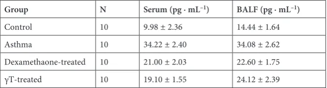
Next, we assess the IL-4 level in serum and BALF of the four distinct subgroups. The IL-4 level of the asthma mice was conspicuously higher than the in normal mice. The addition of dexametha-sone or γT decreased the IL-4 level, especially γT, which reduced the IL-4 in the serum of nearly half of the asthma subgroup better than dexametha-sone as is shown in Table 3. The function of γT to IL-4 in BALF was comparable to dexamethasone without large differences.
Discussion
Bronchial asthma is a chronic inflammatory disorder of the airways in which many cells, par-ticularly eosinophils, may play important roles
through the release of various mediators [16–18]. There is strong evidence that an imbalance be-tween the reducing and oxidizing systems favoring a more oxidative state is present in the airway in-flammation and a deficiency in the amount of an-tioxidants exists in the asthmatic airway [19, 20]. A plant source of vitamin E has been found to re-duce key mitochondrial dysfunctions, alleviate asthmatic features and serve as a marker of clin-ical status in asthma [5, 21]. In this study, using a mouse model of allergic asthma, we demonstrat-ed the potential function of γT, one of the vitamin E isoforms, in preventing airway inflammation with a distinct decline of eosinophilic granulocyte, followed by obvious remission in aspects of mor-phology.
In addition, eotaxin is the most specific and strongest factor which can affect the function of eosinophil [22]. The finding of an eotaxin decrease compared to the asthma subgroup in blood serum and BALF further showed the efficiency of γT in asthma relief. Furthermore, there were compara-ble or even better effects of γT than dexametha-sone, which is a good drug for asthma treatment but with some of side effects [23–25].
Eotaxin could regulate eosinophil traffick-ing into the airways along with other chemotac-tic factors, such as IL-4 which is a pleiotropic cy-tokine that elicits a wide spectrum of physiological and pathogenic events including cell prolifera-tion, differentiaprolifera-tion, apoptosis and inflammation [26–28]. The enhanced level of IL-4 in the asthma subgroup was attenuated with the addition of γT, with almost the same function as dexamethasone. It is most likely that a weakening of the level of IL-4 and eotaxin could reverse the unbalanced
Table 2. The detection of eotaxin in serum and BALF (mean ± SD)
Group N Serum (ng · L–1) BALF (ng · L–1)
Control 10 208.67 ± 19.97 427.13 ± 49.58 Asthma 10 297.30 ± 41.34 573.78 ± 52.82 Dexamethasone-treated 10 289.73 ± 28.79 492.13 ± 40.36 γT-treated 10 255.82 ± 25.84 445.48 ± 37.18
Table 3. The IL-4 level in serum and BALF (mean ± SD)
Group N Serum (pg · mL–1) BALF (pg · mL–1)
condition of immune response and contribute to the prevention or treatment of asthma. However, this hypothesis still needs to be verified in the near future.
In conclusion, the desirable capability of γT in reducing eotaxin and IL-4 in asthma mice serum or BALF has been discovered. A strategy in asthma ther-apy might be found utilizing this functional vitamin.
Acknowledgements. We thank Shaanxi College of Traditional Chinese Medicine for assisting us with the related laboratory equipment and venues.
References
[1] Burioka N: Chronotherapy of bronchial asthma. Nihon Rinsho 2013, 71, 2146–2152.
[2] Allan K, Devereux G: Diet and asthma: Nutrition implications from prevention to treatment. J Am Diet Assoc 2011, 111, 258–268.
[3] Dworski R, Han W, Blackwell TS, Hoskins A, Freeman ML: Vitamin E prevents NRF2 suppression by allergens in asthmatic alveolar macrophages in vivo. Free Radic Biol Med 2011, 51, 516–521.
[4] Han YY, Blatter J, Brehm JM, Forno E, Litonjua AA, Celedon JC: Diet and asthma: Vitamins and methyl donors. Lancet Respir Med 2013, 1, 813–822.
[5] Mabalirajan U, Aich J, Leishangthem GD, Sharma SK, Dinda AK, Ghosh B: Effects of vitamin E on mito-chondrial dysfunction and asthma features in an experimental allergic murine model. J Appl Physiol 2009, 107, 1285–1292.
[6] Ghaffari J, Farid Hossiani R, Khalilian A, Nahanmoghadam N, Salehifar E, Rafatpanah H: Vitamin e supple-mentation, lung functions and clinical manifestations in children with moderate asthma: A randomized double blind placebo-controlled trial. Iran J Allergy Asthma Immunol 2014, 13, 98–103.
[7] Liang W, Zhou Z, Ji Z, Wang Y, Xue W, Zhang X: Association study of bronchial asthma with polymorphisms of IL-4 and IL-4R receptor genes. Zhonghua Yi Xue Yi Chuan Xue Za Zhi 2014, 31, 97–100.
[8] Hernandez ML, Wagner JG, Kala A, Mills K, Wells HB, Alexis NE, Lay JC, Jiang Q, Zhang H, Zhou H, Peden DB: Vitamin E, gamma-tocopherol, reduces airway neutrophil recruitment after inhaled endotoxin challenge in rats and in healthy volunteers. Free Radic Biol Med 2013, 60, 56–62.
[9] Jiang Q: Natural forms of vitamin E: Metabolism, antioxidant, and anti-inflammatory activities and their role in disease prevention and therapy. Free Radic Biol Med 2014, 72, 76–90.
[10] Jian YR, Chang SY, Lin PY, Yang YH, Chuang YH: Inactivated influenza virus vaccine is efficient and reduces IL-4 and IL-6 in allergic asthma mice. Influenza Other Respir Viruses 2013, 7, 1210–1217.
[11] Kim CK, Callaway Z, Kim DW, Kita H: Eosinophil degranulation is more important than eosinophilia in identi-fying asthma in chronic cough. J Asthma 2011, 48, 994–1000.
[12] Paplinska M, Hermanowicz-Salamon J, Nejman-Gryz P, Bialek-Gosk K, Rubinsztajn R, Arcimowicz M, Placha G, Gora J, Chazan R, Grubek-Jaworska H: Expression of eotaxins in the material from nasal brushing in asthma, allergic rhinitis and COPD patients. Cytokine 2012, 60, 393–399.
[13] Wang ZD, Lian D, Shen JL, Sun R, Xu W, Xin Z, Lei L, Jin LH, Jin SD: Association between the interleukin-4, interleukin-13 polymorphisms and asthma: A meta-analysis. Mol Biol Rep 2013, 40, 1365–1376.
[14] Zimmermann N, Hogan SP, Mishra A, Brandt EB, Bodette TR, Pope SM, Finkelman FD, Rothenberg ME:
Murine eotaxin-2: A constitutive eosinophil chemokine induced by allergen challenge and IL-4 overexpression. J Immunol 2000, 165, 5839–5846.
[15] Moore PE, Church TL, Chism DD, Panettieri RA, Shore SA: IL-13 and IL-4 cause eotaxin release in human airway smooth muscle cells: A role for ERK 2002, 282, 847–853.
[16] De Vooght V, Smulders S, Haenen S, Belmans J, Opdenakker G, Verbeken E, Nemery B, Hoet PH, Vanoirbeek JA:
Neutrophil and eosinophil granulocytes as key players in a mouse model of chemical-induced asthma. Toxicol Sci 2013, 131, 406–418.
[17] Muniz-Junqueira MI, Barbosa-Marques SM, Junqueira LF Jr: Morphological changes in eosinophils are reliable markers of the severity of an acute asthma exacerbation in children. Allergy 2013, 68, 911–920.
[18] Tamaoki J: Bronchial asthma: Progress in diagnosis and treatments. Topics: IV. Subtype/particular type/comor-bidities; 2. Allergic rhinitis and obesity. Nihon Naika Gakkai Zasshi 2013, 102, 1412–1418.
[19] Sahiner UM, Birben E, Erzurum S, Sackesen C, Kalayci O: Oxidative stress in asthma. World Allergy Organ J 2011, 4, 151–158.
[20] Kirkham P, Rahman I: Oxidative stress in asthma and COPD: Antioxidants as a therapeutic strategy. Pharmacol Ther 2006, 111, 476–494.
[21] Wood LG, Garg ML, Blake RJ, Simpson JL, Gibson PG: Oxidized vitamin E and glutathione as markers of clini-cal status in asthma. Clin Nutr 2008, 27, 579–586.
[22] Beal DR, Stepien DM, Natarajan S, Kim J, Remick DG: Reduction of eotaxin production and eosinophil recruit-ment by pulmonary autologous macrophage transfer in a cockroach allergen-induced asthma model. Am J Physiol Lung Cell Mol Physiol 2013, 305, 866–877.
[23] Gong J, Ren D, Luo Y, Lai W, Li J, Wang W: Effects of dexamethasone on expressions of IL-21 and its receptor in lungs of experimental asthma mice. Xi Bao Yu Fen Zi Mian Yi Xue Za Zhi 2013, 29, 561–564.
[25] Meyer JS, Riese J, Biondi E: Is dexamethasone an effective alternative to oral prednisone in the treatment of pedi-atric asthma exacerbations? Hosp Pediatr 2014, 4, 172–180.
[26] Shimamura T, Husain SR, Puri RK: The IL-4 and IL-13 pseudomonas exotoxins: New hope for brain tumor therapy. Neurosurg Focus 2006, 20, E11.
[27] Weber F, Asher A, Bucholz R, Berger M, Prados M, Chang S, Bruce J, Hall W, Rainov NG, Westphal M, Warnick RE, Rand RW, Floeth F, Rommel F, Pan H, Hingorani VN, Puri RK: Safety, tolerability, and tumor response of IL4-pseudomonas exotoxin (NBI-3001) in patients with recurrent malignant glioma. J Neurooncol 2003, 64, 125–137.
[28] Rainov NG, Heidecke V: Long term survival in a patient with recurrent malignant glioma treated with intratu-moral infusion of an IL4-targeted toxin (NBI-3001). J Neurooncol 2004, 66, 197–201.
Address for correspondence:
Yan-Mei Wu
Department of Respiratory and Blood Oncology Xi’An XD Group Hospital
FengDeng North Street No. 97, 710077
Lianhu District, ShaanXi, Xi’an China
E-mail: [email protected]
Conflict of interest: None declared