© 2017 Hassan Mohamed Masoud et al. This is an open access article distributed under the terms of the Creative Commons Attribution License -NonCommercial-ShareAlikeUnported License (http://creativecommons.org/licenses/by-nc-sa/3.0/).
Journal of Applied Pharmaceutical Science Vol. 7 (05), pp. 123-128, May, 2017 Available online at http://www.japsonline.com
DOI: 10.7324/JAPS.2017.70522 ISSN 2231-3354
Purification and properties of an alanine aminopeptidase from camel
liver
Hassan Mohamed Masoud
*, Doaa Abdel-Khalek Darwish, Mohamed Salah-Eldin Helmy,
Mohamed Mosaad Abdel-Monsef
Molecular Biology Department, National Research Centre, Dokki, Giza, Egypt.
ARTICLE INFO ABSTRACT
Article history:
Received on: 03/10/2016 Accepted on: 20/01/2017 Available online: 30/05/2017
Alanine aminopeptidase is purified from camel liver to homogeneity and designated CLAAP. The purification procedure involved anion exchange chromatography on DEAE-cellulose column and gel filtration chromatography on Sephacryl S-300 column. The specific activity of CLAAP is increased to 9.9 folds over the crude extract with 25.3% yields. The purified CLAAP is homotrimmer protein with 180 kDa consists of three identical subunits of 60 kDa each. CLAAP displayed its optimum activity at pH 8.0 and the Km value is 0.083 mM alanine β-naphthylamide. The divalent cations CuCl2, MnCl2 and ZnCl2 inhibited CLAAP activity while CoCl2 and MgCl2 increased its activity. CLAAP was inhibited competitively with bestatin that has one binding site on the enzyme and Ki value of 14 µM. This report represents AAP purified from the camel liver as a safe source. Therefore, a task for the future will be the application of this purified CLAAP enzyme in the industry of meat and dairy products for flavor development.
Key words:
Alanine aminopeptidase; Camel liver; Purification; Characterization.
INTRODUCTION
Aminopeptidases are exopeptidases proteins that remove the N-terminal amino acid of their protein substrates (Niven, 1995; Erbeznik and Hersh, 1997). These enzymes are widespread in prokaryotes, mammals and plants and their substrate specificities depending on their physiological roles
(Barrett et al., 1998; Umetsu et al., 2003; Matsui et al., 2006;
Bogra et al., 2009; Mane et al., 2010; Renwrantz and Lam,
2010). These proteins have substantial tasks in numerous physiological processes as it regulates certain pathways in metabolism, controls the cell cycle and selectively degrades certain proteins (Brownless and Williams, 1993). They can be classified into two main types, one hydrolyzes the amino acids with hydrophobic side chains from N-terminus of the protein,
and the other removes other amino acid residues (Drag
et al., 2005). Alanine aminopeptidases are present in almost all
* Corresponding Author
Hassan Mohamed M. Masoud, Molecular Biology Department, National Research Centre, El-Tahrir st., Dokki, Giza, Egypt. Email: hssnasoud @ yahoo.com
mammalian tissues including intestine, kidney, liver and pancreas
(Mane et al., 2010). They are present in many human organs
including; seminal plasma (Huang et al., 1997), liver (Yamamoto
et al., 2000), placenta (Mizutani et al., 1993), pancreas
(Sidorowicz et al., 1980), kidney (Mantle et al., 1990), gallbladder
bile (Offner et al., 1994) and skeletal muscle (Mantle et al., 1983).
They are present in other mammalian tissues as bovine skeletal muscle (Ye and Ng, 2011) and porcine kidney (Itoh and Nagamatsu, 1995). Mammalian aminopeptidases take part in
metabolism of neurotransmitters and hormones (Hiroi et al., 1992;
Umetsu et al., 2003), used as kidneys damage biomarker and used
in diagnosis of certain kidney troubles as they are present at elevated urine percentages if there are kidney disorders (Flynn, 1990). For meat and dairy products industry, aminopeptidases share the flavor development as a direct action by removing bitter peptides which have high ratio of hydrophobic amino acids that are considered as perfect substrates for its action, or indirectly, where aminopeptidases may remove the aromatic amino acids that are main substrates of aromatic molecules present in cheese (Fernandez-Espla and Rul, 1999; Magboul and McSweeney, 1999;
This report represents purification and characterization of alanine aminopeptidase from camel liver as a safe source for the various industrial applications of aminopeptidases.
MATERIALS AND METHODS
Liver tissues
Six fresh camel liver samples were obtained from different individuals in a local slaughter-house and stored at -40 ˚C.
Chemicals
L-Alanine β-naphthylamide, Dithiothreitol (DTT), Fast
Garnet GBC salt, p-Hydroxymercuribenzoic acid (pHMBA), 1,10
Phenanthroline, Bovine serum albumin (BSA), Diethylaminoethyl cellulose (DEAE-Cellulose) and Sephacryl S-300 were purchased from Sigma-Aldrich Chemicals Co. The other chemicals were of analytical grade.
Assay of alanine aminopeptidase activity
The alanine aminopeptidase activity assay reaction mixture contained in 1.5 ml of 0.1 M Tris-HCl buffer pH 8, 0.4 mM alanine-β-naphthylamide and the enzyme solution. Incubate for 1 hour at 37˚C; stop the reaction with 0.5 ml Fast Garnet GBC solution (1 mg / ml) in 1 M acetate buffer pH 4.2 containing 10% Tween 20 and read absorbance at 525 nm against control lacking enzyme. The concentration of the liberated β-naphthylamine was calculated from a standard curve of β-naphthylamine treated similarly. One unit of alanine aminopeptidase activity is the amount of enzyme which catalyzes the liberation of 1 nmol of
β-naphthylamine per hour at 37 ˚C (Kawata et al., 1980).
Staining ofalanine aminopeptidase activity on PAGE
After electrophoresis, the gel was incubated in 50 ml 0.1
M sodium phosphate buffer pH 5.8 containing 1 mM CoCl2,
0.06% alanine β-naphthylamide HCl and 0.06% Fast Garnet GBC salt until the develop of the red bands. The stained gel was
washed with water and then fixed in 7% acetic acid (Chien et al.,
2002).
Purification of camel liver alanine aminopeptidase Preparation of crude extract
All of the procedures were performed at 4˚C unless
stated otherwise. 10 grams of camel liver were homogenized in
0.02 M sodium phosphate buffer pH 7.4 using Omni mixer homogenizer. Cell debris and insoluble materials were removed by
centrifugation at 5000 x g for 20 min and the supernatant was
saved and designated as crude extract.
DEAE-cellulose column Chromatography
The crude extract was chromatographed on DEAE-cellulose column previously equilibrated with 0.02 M sodium Phosphate buffer pH 7.4. The proteins were eluted with stepwise NaCl gradient ranging from 0 to 1 M followed by 0.5% Triton X-
100 prepared in the equilibration buffer. Fractions of 5 ml were collected at a flow rate of 60 ml/hour. Fractions containing alanine aminopeptidase activity were pooled and concentrated by lyophilization.
Sephacryl S-300 column Chromatography:
The concentrated solution containing the alanine aminopeptidase activity was applied onto a Sephacryl S-300 column (142 cm x 1.75 cm i.d.). The column was equilibrated and developed with 0.02 M sodium Phosphate buffer pH 7.4 at a flow rate of 30 ml / hour and 2 ml fractions were collected.
Electrophoretic analysis
Native gel electrophoresis was carried out with 7% PAGE (Smith, 1969). The subunit molecular weight of the purified enzyme was determined by 12% SDS-PAGE (Weber and Osborn, 1969; Laemmli, 1970). Coomassie brilliant blue R-250 was used to stain the proteins.
Protein determination
Protein content was determined by the dye binding assay method using BSA as a standard protein (Bradford, 1976).
RESULTS
Purification of camel liver alanine aminopeptidase
The purification of the major AAP from camel liver crude extract was monitored by ability of AAP to hydrolyze the substrate alanine-β-naphthylamide and the results of the purification is outlined in Table (1).
Table 1: Purification scheme of camel liver alanine aminopeptidase (CLAAP).
Purification step
To
ta
l
p
r
o
te
in
(m
g
)
A
c
ti
v
ity
(u
n
it)
S
p
e
c
ifi
c
a
c
ti
v
ity
R
e
c
o
v
e
ry
(%) Fo
ld
p
u
r
ifi
c
a
ti
o
n
Camel liver crude extract 228 99400 436.0 100.0 1.0 DEAE-cellulose fraction
(0.5 % Triton)
26.7 55736 2087.5 56.1 4.9
Sephacryl S-300 fraction 5.8 25120 4331.0 25.3 9.9
Analyses of CLAAP on polyacrylamide gel electrophoresis
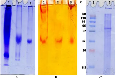
The CLAAP enzyme purification cycles including liver extract, anion exchange fraction and gel filtration fraction were seen on 7 % native PAGE (Fig. 2a). The purified enzyme was seen as a single protein band corresponded with the enzyme activity band (Fig. 2b) demonstrating the tentative purity of the preparation. CLAAP enzyme was seen on SDS-PAGE as a single band of 60 kDa (Fig. 2c) which indicates the subunit molecular weight of the purified enzyme.
A B C
Fig. 2: (a) Electrophoretic analysis of protein pattern of CLAAP on 7 % native PAGE: (1) crude extract, (2) DEAE-cellulose fraction, (3) Sephacryl S-300 purified fraction. (b) Electrophoretic analysis of alanine aminopeptidase isoenzyme pattern of CLAAP on 7 % native PAGE: (1) crude extract, (2) DEAE-cellulose fraction, (3) Sephacryl S-300 purified fraction (c) Molecular weight determination of CLAAP subunit by 12% SDS-PAGE: (1) Molecular weight marker proteins and (2) Purified CLAAP.
Substrate specificity, Optimum pH and Km value determination of CLAAP
The substrate specificity of the purified camel liver alanine aminopeptidase (CLAAP) was screened toward various substrates (Table 2). CLAAP cleaved preferentially
alanine-β-naphthylamide HCI (100 % relative activity) followed by
leucine-β-naphthylamide (87.8 %) and displayed low activity
.
toward glycine-β-naphthylamide (61.9 %). The optimum pH of
the purified CLAAP enzyme was tested with potassium phosphate buffer, pH (7.0 – 9.0). The pH profile of CLAAP displayed its optimum activity at pH 8.0 (Fig. 3a).
The Lineweaver-Burk plot calculates the Km value of the purified enzyme from (1/v) and (1/[S]) to be 0.083 mM alanine β-naphthylamide (Fig. 3b).
A B
Fig. 3: (a) Effect of pH on the purified CLAAP using 0.05 M potassium phosphate buffer, pH (7.0 – 9.0). (b) Lineweaver-Burk plot relating the reciprocal of the reaction velocity of the purified CLAAP to alanine β-naphthylamide concentration in mM.
Table 2: Substrate specificity of camel liver alanine aminopeptidase (CLAAP):
Substrate
C
o
n
c
e
n
tr
a
ti
o
n CLAAP
R
a
te
o
f
h
y
d
r
o
ly
si
s
R
e
la
ti
v
e
a
c
ti
v
ity
(%)
DL-Alanine-β-naphthylamide HCI 0.4 mM 0.352 100.0 Glycine-β-naphthylamide HCI 0.4 mM 0.218 61.9 L-Leucine-β-naphthylamide HCI 0.4 mM 0.309 87.8 The hydrolysis rate is the change in absorbance at 525 nm for β- naphthylamide derivatives per 30 min.
A B
Effect of divalent cations and various inhibitors on CLAAP activity
The purified camel liver alanine aminopeptidase CLAAP was pre-coupled with two different concentrations of all divalent cations at 37˚C then test the enzyme activity. These measures were
compared with a control test that has no cations. CuCl2, MnCl2 and
ZnCl2 inhibited the activity of CLAAP while CoCl2 and MgCl2
were found to be activators of the enzyme activity (Table 3). Furthermore, we preincubated the CLAAP with different inhibitors for 5 min at 37˚C then calculated the percent of inhibition in comparison with control with no inhibitors. Bestatin was the potent CLAAP enzyme activity inhibitor (Table 4).
Table 3: Effect of divalent cations on the purified camel liver alanine aminopeptidase CLAAP.
Reagent Concentration (mM) Residual Activity (%)
Control --- 100.0
CoCl2
1.0 105.4
2.0 122.7
MnCl2 1.0 50.7
2.0 13.6
FeCl2
1.0 94.7
2.0 82.6
ZnCl2
1.0 76.0
2.0 54.5
CuCl2 1.0 37.1
2.0 10.6
NiCl2
1.0 96.2
2.0 89.1
MgCl2 1.0 2.0 115.9 98.8
CaCl2 1.0 87.3
2.0 70.5
Table 4: Effect of inhibitors on the purified camel liver alanine aminopeptidase (CLAAP).
Reagent Concentration Inhibition
(%)
Control --- 0.0
Bestatin 100 µM 97.8
Cysteine 5 mM 14.2
DL-Dithiothreitol (DTT) 5 mM 4.7
Ethylenediamine tetraacetic acid (EDTA) 5 mM 15.3
N-Ethylmalimide 5 mM 5.5
p-Hydroxymercuribenzic acid (pHMBA) 1 mM 58.0
Glutathione (GSH) 5 mM 70.3
Iodoacetic acid 10 mM 19.3
β-Mercaptoethanol 5 mM 3.6
1,10 Phenanthroline 10 mM 80.1
Phenylmethylsulfonylfluoride (PMSF) 5 mM 62.0
Puromycin 1 mM 28.6
N-Tosyl-lysine chloromethyl ketone hydrochloride (TLCK)
5 mM 58.7
N-Tosylamide-L-phenylalanine chloromethyl ketone (TPCK)
5 mM 2.5
* These values represent % of the control and the means of triplicate experiments.
Kinetics of CLAAP inhibition by Bestatin
The purified enzyme CLAAP activity was inhibited strongly with bestatin. The enzyme was subjected to inhibition with increasing concentrations of bestatin making a titration curve
(Fig. 4a) demonstrating that 50% inhibition (I50) of CLAAP
activity was reached with 30 µM bestatin. Maximum enzyme
inhibition of 97.8 % was reached by 100 µM bestatin. When the Hill plot for CLAAP inhibition by bestatin was constructed a linear relationship was obtained and the slope of the Hill plot was found 1.11 (Fig. 4b). The CLAAP activity inhibition by bestatin was a competitive type (Fig. 4c) with a Ki value of 14 µM (Fig. 4d).
A B
C D
Fig. 4: (a) Inhibition of CLAAP by varying concentrations of Bestatin. (b) Hill plot for inhibition of CLAAP by Bestatin. (c) Lineweaver-Burk plots showing the type of inhibition of CLAAP by Bestatin. (d) Determination of the inhibition constant (Ki) value for the inhibition of the CLAAP by Bestatin.
DISCUSSION
Alanine aminopeptidases are widespread in mammalian tissues and body fluids. They have an important role in liberation of the neutral amino acids of proteins as Alanine, Leucine,
Methionine and Tyrosine (Mane et al., 2010). In this study, the
camel liver AAP is purified by a simple and reproducible method consists only of two column chromatographic steps which were found to be convenient to set a homogenous form of the enzyme. Separation of the camel liver crude extract on DEAE-cellulose column revealed the existence of one major form of camel liver alanine aminopeptidase termed CLAAP and another minor form (Fig. 1a). CLAAP eluted from the Sephacryl S-300 column as one peak of AAP activity with a native molecular weight of 180 kDa (Fig. 1b) while its subunit molecular weight is estimated at 60 kDa by SDS-PAGE indicating that the enzyme is homotrimmeric protein consists of three identical subunits (Fig. 2c). The molecular mass of CLAAP is similar to that of 180 kDa alanine
than that of 160 kDa chicken intestine (Jamadar et al., 2003), 153
kDa human seminal plasma (Huang et al., 1997) and 60 kDa
bovine skeletal muscle (Ye and Ng, 2011). The specific activity of the prepared CLAAP isoenzyme is increased to 4331 U/mg protein with 25.3% recovery and 9.9 folds purification (Table 1). Very low recovery of 3.43% was reported for chicken intestine alanine
aminopeptidase (Mane et al., 2010). CLAAP homogeneity is
proved by native polyacrylamide gel with a single band for the purified protein which coincided with the band of enzyme activity (Fig. 2a and 2b).
The CLAAP preferentially cleaved alanine- β
-naphthylamide followed by leucine- β -naphthylamide and
glycine-β-naphthylamide (Table 2) in agreement with chicken
intestine aminopeptidase (Jamadar et al., 2003), bovine skeletal
muscle alanine aminopeptidase (Ye and Ng, 2011) and the two
alanyl aminopeptidases of Trypanosoma congolense (Pillay et al.,
2013). The CLAAP displayed its optimum activity at pH 8 (Fig. 3). Various alanine aminopeptidases displayed the same optimum pH; alanine aminopeptidase from bovine skeletal muscle (Ye and
Ng, 2011), the two alanyl aminopeptidases of Trypanosoma
congolense (Pillay et al., 2013) and human pancreas alanine
aminopeptidase (Sidorowicz et al., 1980). The CLAAP displayed
Km value of 0.083 mM alanine β-naphthylamide (Fig. 3). This
value is closely related to 0.1 mM that of chicken intestine
aminopeptidase (Mane et al., 2010). The low Km value explains
the CLAAP high affinity toward alanine β-naphthylamide. The
CLAAP activity was increased in the presence of CoCl2 and
MgCl2, while CuCl2, MnCl2 and ZnCl2 inhibited the CLAAP
activity drastically (Table 3). Similar to CLAAP, the bovine
skeletal muscle alanine aminopeptidase is inhibited by Zn2+ and
Mn2+ and in contrast with it; it is strongly inhibited by Co2+ and
Mg2+ ions (Ye and Ng, 2011).
In this study, the effect of various inhibitors on the purified camel liver AAP revealed that, CLAAP was resistant to DTT, N-Ethylmalimide, β-Mercaptoethanol and TPCK. It was inhibited with bestatin, glutathione, 1,10 phenanthroline, PMSF, puromycin, soya bean trypsin inhibitor and TLCK. The purified CLAAP is suggested to have a cysteine protease activity due to its
sensitivity to the cysteinyl protease inhibitor p
-chloromercuribenzoic acid. It also suggested having a serine protease activity due to the sensitivity to PMSF and soya bean trypsin inhibitor suggesting the presence of serine residue in the enzyme active site. The CLAAP inhibition with 1, 10 phenanthroline indicates that CLAAP is a metalloenzyme in
similarity with porcine liver (Imamura et al., 1983) and rabbit
kidney aminopeptidases (Oliveira et al., 1999). Bestatin is reported
as a specific inhibitor of alanyl aminopeptidases (Tieku and
Hooper, 1992). Here, bestatin inhibited CLAAP vigorously since 100 µM bestatin inhibited the enzyme completely (Table 4) confirming that it is an alanine aminopeptidase. A straight line was obtained when drawing the Hill plot for the bestatin inhibition of the purified CLAAP. The Hill plot has a slope of 1.11 indicating that there is one binding site for bestatin on CLAAP molecule
(Fig. 4b). Bestatin inhibited CLAAP competitively since the bestatin existence did not alter the Vmax value and increased the
Km value (Fig. 4c). To determine the Ki value, we plotted slopes
of the reciprocal plots lines against the bestatin concentrations. The CLAAP bestatin inhibition Ki value was found to be 14 µM
directly from the plot X axis intercept (Fig. 4d). The low Ki value
proves that the bestatin is a potent inhibitor of alanine aminopeptidases. In conclusion, this report presents a simple, convenient and reproducible method for purification of alanine aminopeptidase from the camel liver as a safe, locally available and rich source. Large scale production of this well characterized enzyme will allow its using in different applications as in meat and dairy products industry in removing pitter peptides and in examination of the protein primary structure.
ACKNOWLEDGEMENTS
Financial support and sponsorship: The authors would gratefully acknowledge financial support and fund provided by the National Research Centre, Egypt under the contract No. P101101.
Conflict of Interests: There are no conflicts of interest.
REFERENCES
Barrett AJ., Rawlings ND., Woessner JF. 1998. Handbook of Proteolytic Enzymes. Academic Press, New York.
Bogra P., Singh J., Singh H. Purification and characterization of aminopeptidase B from goat brain. Proc. Biochem. 2009; 44: 776-780.
Bradford MM. A rapid and sensitive method for the quantitation of microgram quantities of protein utilizing the principle of protein-dye binding. Anal. Biochem 1976; 72: 248-254.
Brownless J., Williams CH. Peptidases, peptides and the mammalian blood–brain barrier. J. Neurochem. 1993; 60: 780-793.
Chien HCR., Lin LL., Chao SH., Chen CC., Wang WC., Shaw CY., Tsai YC., Hu HY., Hsu WH. Purification, characterization, and genetic analysis of a leucine aminopeptidase from Aspergillus sojae. Biochim. Biophys. Act. 2002; 1576: 119-126.
Drag M., Grembecka J., Pawełczak M., Kafarski P. α-Aminoalkylphosphonates as a tool in experimental optimization of P1 side chain shape of potential inhibitors in S1 pocket of leucine and neutral aminopeptidases. Eur. J. Med. Chem. 2005; 40: 764-771.
Erbeznik H., Hersh LB. A novel mammalian high-molecular weight aminopeptidase. Arch. Biochem. Biophys. 1997; 344: 228-234.
Fernandez-Espla MD., Rul F. Peptidase from Streptococcus thermophilus. A new member of the aminopeptidase T family of thermophilic bacteria. Eur. J. Biochem. 1999; 263: 502 - 510.
Flynn FV. Assessment of renal function: selected developments. Clin. Biochem. 1990; 23: 49-54.
Hiroi Y., Endo Y., Natori Y. Purification and properties of an aminopeptidase from rat-liver cytosol. Arch. Biochem. Biophys. 1992; 294: 440-445.
Huang K., Takahara S., Kinouchi T., Takeyama M., Ishida T., Ueyama H., Nishi K., Ohkubo I. Alanyl amino- peptidase from human seminal plasma: purification, characterization and immunohistochemical localization in the male genital tract. J. Biochem. 1997; 122: 779-787.
Imamura T., Kawata S., Ninomiya K., Makisumi S. Porcine liver aminopeptidase. Further characterization of its sulfhydryl groups. J. Biochem. 1983; 94: 267-273.
Jamadar VK., Jamadar SN., Dandekar SP., Harikumar P. Purification and characterization of aminopeptidase from chicken intestine. J. Food Sci. 2003; 68: 438-443.
Kawata S., Takayama S., Ninomiya K., Makisumi S. Purification and some properties of porcine liver aminopeptidase B. J. Biochem. 1980; 88: 1025-1032.
Laemmli UK. Cleavage of structural proteins during the assembly of the head of Bacteriophage T4. Nature 1970; 227: 680-685.
Magboul AAA., McSweeney PLH. Purifiation and
characterization of an aminopeptidase from Lactobacillus curvatus
DPC2024. Int. Dairy J. 1999; 9: 107-116.
Mane S., Damle M., Harikumar P., Jamdar S., Gade W. Purification and characterization of aminopeptidase N from chicken intestine with potential application in debittering Process. Biochemistry 2010; 45: 1011-1016.
Mantle D., Hardy MF., Lauffart B., Mcdermott JR., Smith AI., Pennington RJT. Purification and characterization of the major aminopeptidase from human skeletal muscle. Biochem. J. 1983; 211: 567-573.
Mantle D., Lauffart B., Mcdermott J., Gibson A. Characterization of aminopeptidases in human kidney soluble fraction. Clin. Chim. Acta 1990; 187: 105-113.
Martinez-Cuesta MC., Fernandez de Palencia P., Requena T., Pelaez C. Enzymatic ability of Lactobacillus casei subsp. casei IFPL731 for flavour development in cheese. Internl. Dairy J. 2001; 11: 577-585.
Matsui M., Fowler JH., Walling LL. Leucine aminopeptidases: diversity in structure and function. Biol. Chem. 2006; 387: 1535-1544.
Mizutani S., Goto K., Nomura S. Possible action of human placental aminopeptidase N in feto-placental unit. Res. Commun. Chem. Pathol. Pharmacol. 1993; 82: 65-80.
Niven GW. The characterization of two aminopeptidase activities from the cyanobacterium Anabaena flosaquae. Biochim. Biophys. Acta 1995; 1253: 193-198.
Offner GD., Gong D., and Afdhal NH. Identification of a 130-kilodalton human biliary concanavalin A binding protein as aminopeptidase N. Gastroenterology. 1994; 106: 755-762.
Oliveira SM., Freitas Jr J.O., Alves KB. Rabbit kidney aminopeptidases: purification and some properties. Immunopharmacol. 1999; 45: 215-221.
Pillay D., Boulangé AV., Coustou V., Baltz T., Coetzer THT. Recombinant expression and biochemical characterization of two alanyl aminopeptidases of Trypanosoma congolense. Exp. Parasit. 2013, 135: 675-684.
Renwrantz L., Lam A. Soluble and surface-bound aminopeptidase in eosinophilic blood cells from Mytilus edulis. J. Invert. Pathol. 2010; 103: 68-70.
Sidorowicz W., Iackson GC., Behal FJ. Multiple molecular forms of human pancreas alanine aminopeptidase. Clin. Chim. Acta 1980; 104: 169-179.
Smith I. Acrylamide gel disc electrophoresis. In: Smith, I., (Eds.), Electrophoretic techniques, Academic press, New York, 1969; pp. 365-515.
Tieku S., Hooper NM. Inhibition of aminopeptidases N, A and W. A re-evaluation of the actions of bestatin and inhibitors of angiotensin converting enzyme. Biochem. Pharmacol. 1992; 44: 1725–1730.
Umetsu H., Arai M., Ota T., Kudo R., Sugiura H., Ishiyama H., Sasak K. Purification and properties of an aminopeptidase from the mid -gut gland of scallop (Patinopecten yessoensis). Comp. Biochem. Physiol. 2003; 136: 935-942.
Weber K, Osborn M. The reliability of molecular weight determinations by dodecyl sulfate-polyacrylamide gel electroph. J. Biol. Chem. 1969; 244: 4406-4412.
Yamamoto Y., Li YH., Ushiyama I., Nishimura A., Ohkubo I., Nishi K. Puromycin-sensitive alanyl aminopeptidase from human liver cytosol: purification and characterization. Forensic Sci. Int. 2000; 113: 143-146.
Ye XJ., Ng TB. Purification and characterisation of an alanine aminopeptidase from bovine skeletal muscle. Food Chemistry 2001; 124: 634-639.
How to cite this article: