1. Introduction
strocytes are major cell types in brain with numerous important functions. In contrast to previous believes, they are not only supporting cells but also recent findings have shown their biological importance in physio- and pathophysiological
condi-Fluoxetin Upregulates Connexin 43 Expression in Astrocyte
Hossein Mostafavi1,2,5,6, Mojtaba Khaksarian1,5,7, Mohammad Taghi Joghataei1,2, Gholamreza Hassanzadeh3, Masoud Soleimani4,
Sanaz Eftekhari1,2, Mansooreh Soleimani2, Kazem Mousavizadeh2,8, Mahmoud Reza Hadjighassem1,2,9*
1. Department of Neuroscience, School of Advanced Medical Technologies, Tehran University of Medical Sciences, Tehran, Iran. 2. Division of Neuroscience, Cellular and Molecular Research Center, Iran University of Medical Sciences, Tehran, Iran. 3. Department of Anatomy, School of Medicine, Tehran University of Medical Sciences, Tehran, Iran.
4. Department of Hematology, Tarbiat Modares University, Tehran, Iran.
5. Stem Cell Technology Research Center, Molecular Biology and Genetic Engineering Department, Tehran, Iran. 6. Department of Physiology and Pharmacology, Zanjan University of Medical Sciences, Zanjan, Iran. 7- Department of Physiology, Lorestan University of Medical Sciences, Khorramabad, Iran. 8. Department of Basic Medical Sciences, Iran University of Medical Sciences, Tehran, Iran.
9. Neuroscience Institute, Brain and Spinal cord Injury Research Center, Tehran University of Medical Sciences, Tehran, Iran.
* Corresponding Author:
Mahmoud Reza Hadjighassem, MD., PhD.
Cellular and Molecular Research Center, Brain and Spinal cord injury research center, School of Advanced Technologies in Medicine, Tehran University of Medical Sciences.
Tel: +98-21-82944572 / Fax: +98-21-88622689
Introduction: Recent studies have shown that astrocytes play major roles in normal and disease condition of the central nervous system including multiple sclerosis (MS). Molecular target therapy studies in MS have revealed that connexin-43 (Cx43) and Aquaporin-4 (AQP4) contents of astrocytes undergo expression alteration. Fluoxetine had some effects in MS patients
unrelated to its known antidepressant effects. Some of fluoxetine effects were attributed to its
capability of cAMP signaling pathway stimulation. This study aimed to investigate possible
acute effects of fluoxetine on Cx43 and AQP4 expression in astrocyte.
Methods: Astrocytoma cells were treated for 24 hours with fluoxetine (10 and 20 μg/ml) with
or without adenyl cyclase (AC) and protein kinase A (PKA) inhibition. Cx43 expression at both mRNA and protein levels and AQP4 expression at mRNA level were evaluated.
Results: Acquired results showed that fluoxetine with and without AC and PKA inhibition
resulted in Cx43 up-regulation both in mRNA and protein levels, whereas AQP4 expression have not changed.
Discussion: In conclusion, data showed that fluoxetine alone and in the absence of serotonin
acutely up-regulated Cx43 expression in astrocytes that can be assumed in molecular target
therapy of MS patients. It seems that cAMP involvement in fluoxetine effects need more
researches.
A B S T R A C T
Article info:Received: 22 April 2013
First Revision: 21 May 2013
Accepted: 15 June 2013
Key Words:
Astrocyte, Fluoxetine, cAMP, Connexin-43, Aquaporin-4.
et al. 2010). These communication networks provide by some astrocyte elements including Cx43 and AQP4 (Ne-dergaard, Ransom et al. 2003).
Studies have shown that expression of Cx43 and AQP4 altered in multiple sclerosis animal models and patients (Brand-Schieber, Beelitz et al. 2005; Li, Zhang et al. 2009).
Deletion of Cx43, the primary protein forming gap junction channels in astrocytes, has been shown that led to dysmyelinating Phenotype (Lutz, Zhao et al. 2009). In an Animal Model of Multiple Sclerosis, it was found that Cx43 is down-regulated in inflamed white matter of centeral nervous system (Brand-Schieber, Beelitz et al. 2005).
AQP4 is the most important member of the aquapo-rin family in astrocytes and has key roles in maintaining water and ion homeostasis. It was suggested that AQP4 may have other functions that is unrelated to its water transport role (Verkman 2009).
Possible involvement of AQP4 in CNS inflamma -tory diseases has been suggested based on evidences that have demonstrated auto-antibodies existed against AQP4 in patients with neuromyelitis optica (NMO) (Graber1, Levy et al. 2008). Moreover, up-regulation of AQP4 in CNS of animal model of MS provided more support for the possible involvement of AQP4 in CNS inflammatory diseases (Miyamoto, Nagaosa et al. 2009).
In addition, correction of Cx43 and AQP4 expression alterations had relationship with re-myelination. In a guinea pig model of experimental autoimmune en-cephalomyelitis (EAE), expression of Cx43 was almost absent in all demyelinated lesions, on the other hand, treated animals showed up-regulation of Cx43 in all re-myelinating lesions (Roscoe, Messersmith et al. 2007). With regards to AQP4 expression and EAE, it was shown that AQP4 knockout mice had more attenuated EAE severity (Li, Zhang et al. 2009).
Some previous researches have revealed that enhanc-ing intracellular cAMP signallenhanc-ing pathways in astro-cytes in patients with MS may reduces the severity of the disease (Keyser, Zeinstra et al. 2003; Mostert, Admiraal-Behloul et al. 2008). Further more, cAMP signaling pathway relationship with Cx43 and AQP4 expressions have been shown (TenBroek, Lampe et al. 2001; Kortenoeven, Trimpert et al. 2012). Fluoxetine, as
a selective serotonin reuptake inhibitor (SSRI), widely used in depression (Rossi, Barraco et al. 2004), has been shown to elevate cAMP levels in astrocytes (Mostert, Admiraal-Behloul et al. 2008) and reduced the devel-opment of focal inflammatory lesions (Keyser, Laureys et al. 2010). Kong et al reported that fluoxetine needs AQP4 to induce antidepressive effect (Kong, Sha et al. 2009).
Motivated by the potential effects of fluoxetine on biological pathways in astrocytes, we studied possible effects of fluoxetine on expression of Cx43 and AQP4 in astrocytic cell lines through cAMP and its newly de-scribed downstream Epac (exchange protein directly ac-tivated by cAMP) (Kawasaki, Springett et al. 1998). As a model system, we used the human astrocytoma cell lines 1321N1 and U87MG, which have been shown to be use -ful cell lines to investigate signal transduction (Harden, Cotton et al. 1980 ).
2. Methods
2.1. MaterialsAll drugs in this study including Fluoxetine hydro-chloride, (F132), adenyl cyclase inhibitor, SQ 22,536 (S 153), PKA specific inhibitor (H-89) (B1427) were pur -chased from Sigma-Aldrich (USA). Qiazol and reverse transcriptase were purchased from Qiagen and vivantis respectively.
2.2. Cell Culture
The human astrocytoma cell lines including 1321N1 and U87MG were obtained from the Stem Cell Tech -nology Research Center, (Tehran, Iran) and were grown in DMEM containing 10% FBS, incubated at 5% CO2 and 37˚C temperature. Cells were plated in six-well plates for RNA extraction analysis. Treatment was done when the cell confluency reach to 70-80% following re -placement of medium with DMEM containing 1% FBS.
2.3. Real-time Reverse Transcription-Polymerase Chain Reaction Assay
Cx43 forward: 5′- GAT GAG GAA GGA AGA GAA G -3′,
Cx43 reverse: 5′- CGC TAG CTT GCT TGT TGT AA -3′,
AQP4 forward: 5′- CTG ATG TCA CTG GCT CAA TAG-3′,
AQP4 reverse :5′- ACC CAA TAT ATC CAA TGG-3′, Epac forward: 5′- CGA TTC ACT GAC TCC CTT AC -3′,
Epac reverse: 5′- CTT CCA AAT GTG TGA TAG ATT AG -3′,
HPRT1 forward: 5′- CCT GGC GTC GTG ATT AGT G -3′,
HPRT1 reverse: 5′- TCA GTC CTG TCC ATA ATT AGT CC -3′.
Gene expression levels were quantified by Rotor Gene 6000 (Corbett, Concorde, NSW, Australia). The relative expression ratio of genes was calculated using the rela-tive expression software tool (REST).
2.3. Western Blot Analysis
Immunoblotting assay were performed using corre-spond antibodies for detection of Cx43 and EPAC. Total
cell lysates were collected by using ice-cold PBS and homogenization in cold lysis buffer (RIPA buffer). ly-sate clarified by centrifugation at 12,000 rpm for 10 min, supernatant transferred to a new tube. Protein content was measured using the BCA protein kit (Pierce). Equal amount of protein (25μg) in each sample was loaded to 12.5% standard SDS-PAGE. By using a wet system (Bio-Rad) the proteins were transferred to PVDF mem-branes. After blocking, membranes were incubated over night with primary antibodies (1/1000), washed three times with TBST 20 (0.5%) incubated with secondary antibody (1/5000) for 1 hour at room temperature. Af-ter three washes, detection was performed by applying Pierce ECL Plus Western Blotting Substrate (#32134), emanating chemiluminescence from the membrane for manual x-ray film development. Protein levels were nor -malized to β-actin.
3. Results
3.1. EPAC expression in Cell Lines
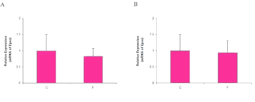
To evaluate possible effect of fluoxetine on expres -sion of EPAC in U87MG and 1321N1 cell lines, we performed Real- time PCR analysis on cDNA obtained from these cells using specific primers for detection of EPAC. We found that fluoxetine did not change the ex -pression of EPAC mRNA in both cell lines (Fig. 1).
Figure 1. Relative expression of Epac in 1321N1 (Fig. 1A) and U87MG (Fig. 1B) cell lines in un-treated (C) and treated cells with 10 μg/ml fluoxetine (F) HPRT1 was used as internal control.
* P<0.05; ** P<0.01; PTZ, Pentylenetetrazol; Ctrl, Control; SB, SB334867; ORX, Orexin-A; Lido., lidocaine
3.2. Effect of fluoxetine on Cx43 expression
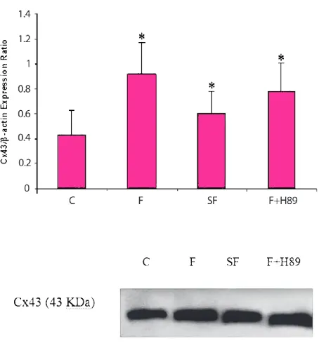
Treatment of cells with 10 and 20 μg/ml of fluoxetine for 24 hours resulted in up-regulation of Cx43 both in
pretreatments declined the fluoxetine effects on Cx43 expression, but not returned it to basal level. Suggest-ing that up-regulation of Cx43 seen in the presence of fluoxetine may have gone partially via cAMP-Epac and without PKA pathway.
3.3. Effect of Fluoxetine on AQP4 Expression
We reported in our previous study (not published yet) that AQP4 was not expressed in 1321N1 cells using RT-Figure 2. Fluoxetine effects on Cx43 mRNA expression with and without cAMP-PKA signaling pathway. 1321N1 cells treated with F (Fluoxetine), SF (AC inhibitor+ Fluoxetine) and F+H89 (Fluoxetine + PKA inhibitor) for 24 hours and changes in transcript amount expression were determined by real-time qPCR. It revealed an up-regulation of Cx43 in all of groups (*p < 0.01 compared to untreated controls).
Figure 3. Expression of Cx43 protein in the 1321N1 cells. Cx43 protein levels were normalized to that of β-actin pro-tein. Data are expressed as means ± S.E.M. Each immunob-lotting was performed in duplicate to increase the reliability of the measurements. Lower Panels show representative im-munoblots of Cx43 (43 kDa). C (un-treated), F (
Figure 4. Fluoxetine effects on AQP4 mRNA expression with and without cAMP-PKA signaling pathway. U87MG cells treated with F (Fluoxetine), SF (AC inhibitor+ Fluoxetine) and F+H89 (Fluoxetine + PKA inhibitor) for 24 hours and changes in transcript amount expression were determined by real-time qPCR. There were no significant differences in treated groups in compared to untreated controls.
PCR and real time PCR. So we used U87MG glioma cells to study fluoxetine possible effect on AQP4 ex -pression in this cell line. In U87MG cells, although 24 hours treatment with fluoxetine (10 and 20 μg/ml) de -creased AQP4 expression, but this effect was not signifi -cant. Pretreatment of cells with AC inhibitor and PKA inhibitor had no effect on AQP4 expression level (Fig. 4). These data suggested that expression of AQP4 in this cell line have other regulatory pathway.
4. Discussion
demonstrated that fluoxetine could reduce formation of new enhancing lesions and had neuroprotective effects against axonal degeneration (Mostert, Admiraal-Behloul et al. 2008). Since fluoxetine has been shown to have relationship with the cAMP signaling pathway and have regulatory effects on Cx43 and AQP4 (Darrow, Fast et al. 1996; Kortenoeven, Trimpert et al. 2012), this study aimed to investigate possible fluoxetine effects on Cx43 and AQP4 expression through cAMP-Epac pathway in astrocyte. We showed that 24 hours treatment of astro-cytoma cells with fluoxetine resulted in up-regulation of Cx43 both in mRNA and protein levels. Investigation of the signaling pathway demonstrated that inhibition of cAMP production by AC inhibitor decline the fluoxetine effects on Cx43 expression, but not returns it to basal level. This can be attributed to partial effect of cAMP in total effect of fluoxetine on Cx43 expression. We have seen the same effect of PKA inhibitor on Cx43 level.
Our results are in line with previous study that showed Cx43 protein expression was significantly increased fol -lowing treatment with fluoxetine (Fatemi, Folsom et al. 2008). In this study, we want to investigate acute effect of fluoxetine in the absence of serotonin. Acute effect of fluoxetine on MAP kinase pathway activation in cul -tured astrocytes has been showed (Mercier, Lennon et al. 2004). Our data showed that pre-inhibition of AC and PKA decreased fluoxetine effects on Cx43 expres -sion but this change was not significant. Although previ -ous studies have reported that antidepressant treatment resulted in activation of AC and increased cAMP activity (Donati, Thukral et al. 2001), but our results have not confirmed AC and cAMP involvement in fluoxetine ef -fects. We think that dosage and duration of treatment have role in this discrepancy observed in our study.
With regards to AQP4 expression, fluoxetine had no significant effect. we used U87MG cell, which have higher glioma grade (Stiborová, Rupertová et al. 2011). So ineffectiveness of fluoxetine in alteration of AQP4 expression may attribute to higher glioma grade of U87MG.
In conclusion, we showed that 24 hours treatment with fluoxetine that can be assumed as acute treatment in -creased Cx43 expression. Our data suggested that some fluoxetine effects can be attributed to its acute effects on Cx43 expression. So in MS patients that commonly have depression and based on researches have down-regulat-ed level of Cx43, fluoxetine as an antidepressant with capability of crossing the blood-brain-barrier can have dual benefit effects.
Acknowledgment
This study was supported by grant #12290 from Depu-ty of Research, Tehran UniversiDepu-ty of Medical Sciences. We also want to thank Stem Cell Technology Research Center, Tehran, Iran, for their support in supplying ma-terials.
References
Brand-Schieber, E., M. Beelitz, et al. (2005). "Connexin43, the Major Gap Junction Protein of Astrocytes, Is Down-Regulat-ed in InflamDown-Regulat-ed White Matter in an Animal Model of Multiple Sclerosis." Journal of Neuroscience Research 80: 798-808
Darrow, B., V. Fast, et al. (1996). "Functional and structural as-sessment of intercellular communication - Increased conduc-tion velocity and enhanced connexin expression in dibutyryl cAMP-treated cultured cardiac myocytes." CIRCULATION RESEARCH 79(2 ): 174-183.
Donati, R. J., C. Thukral, et al. (2001). "Chronic Treatment of C6 Glioma Cells with Antidepressant Drugs Results in a Redis-tribution of Gs‐." MOLECULAR PHARMACOLOGY 59(6): 1426- 1432.
Fatemi, S. H., T. D. Folsom, et al. (2008). "Chronic psychotropic drug treatment causes differential expression of connexin 43 and GFAP in frontal cortex of rats." Schizophrenia Research 104: 127-134.
Graber1, D. J., M. Levy, et al. (2008). "Neuromyelitis optica pathogenesis and aquaporin 4." Journal of Neuroinflamma-tion 5(22).
Harden, T., C. Cotton, et al. (1980 ). "Catecholamine-induced alteration in sedimentation behavior of membrane bound beta-adrenergic receptors." Science 210(4468): 441-3.
He, F. and Y. E. Sun (2007). "Glial cells more than support cells?" The International Journal of Biochemistry & Cell Biol-ogy 39: 661- 665.
Kawasaki, H., G. M. Springett, et al. (1998). "A Family of cAMP-Binding Proteins That Directly Activate Rap1." Science 282: 2275- 2279.
Keyser, J. D., G. Laureys, et al. (2010). "Astrocytes as potential targets to suppress inflammatory demyelinating lesions in multiple sclerosis." Neurochemistry International 57(4): 446-50.
Keyser, J. D., E. Zeinstra, et al. (2003). "Are Astrocytes Central Players in the Pathophysiology of Multiple Sclerosis?" Arch Neurol 60: 132- 136.
Kong, H., L.-l. Sha, et al. (2009). "Requirement of AQP4 for An-tidepressive Efficiency of Fluoxetine: Implication in Adult Hippocampal Neurogenesis." Neuropsychopharmacology 34: 1263-1276.
independent of protein kinase A and CREB but may involve Epac." Am J Physiol Renal Physiol 302(11): F1395-401.
Li, L., H. Zhang, et al. (2009). "Greatly attenuated experimen-tal autoimmune encephalomyelitis in aquaporin-4 knockout mice." BMC Neuroscience 10(94).
Lutz, S. E., Y. Zhao, et al. (2009). "Deletion of Astrocyte Con-nexins 43 and 30 Leads to a Dysmyelinating Phenotype and Hippocampal CA1 Vacuolation." The Journal of Neurosci-ence 29(24): 7743-7752.
Mercier, G., A. M. Lennon, et al. (2004). "MAP Kinase Activa-tion by Fluoxetine and Its RelaActiva-tion to Gene Expression in Cultured Rat Astrocytes." Journal of Molecular Neuroscience 24: 207- 216.
Miyamoto, K., N. Nagaosa, et al. (2009). "Upregulation of water channel aquaporin-4 in experimental autoimmune encepha-lomyeritis." J Neurol Sci 276: 103-107.
Mostert, J. P., F. Admiraal-Behloul, et al. (2008). "Effects of fluoxetine on disease activity in relapsing multiple sclero-sis: a double-blind, placebo-controlled, exploratory study." J Neurol Neurosurg Psychiatry 79: 1027-1031.
Nedergaard, M., B. Ransom, et al. (2003). "New roles for astro-cytes: Redefining the functional architecture of the brain." TRENDS in Neurosciences 26(10): 523- 530.
Roscoe, W. A., E. Messersmith, et al. (2007). "Connexin 43 Gap Junction Proteins Are Up-Regulated in Remyelinating Spinal Cord." Journal of Neuroscience Research 85: 945–9 53.
Rossi, A., A. Barraco, et al. (2004). "Fluoxetine: a review on evi-dence based medicine." Annals of General Hospital Psychia-try 3(2).
Stiborová, M., M. Rupertová, et al. (2011). "Cytochrome P450- and peroxidase-mediated oxidation of anticancer alkaloid el-lipticine dictates its anti-tumor efficiency." Biochim Biophys Acta 1814(1): 175-85.
TenBroek, E., P. Lampe, et al. (2001). "Ser364 of connexin43 and the upregulation of gap junction assembly by cAMP." JOUR-NAL OF CELL BIOLOGY 155 (7 ): 1307-1318.