Biologically Modified Titanium Substrates for Improved Surface Bioactivity
Mahsa Gheysour1, Shahab Faghihi1*
Abstract
Introduction
As a very promising biomaterial for the fabrication of den-tal implants, Titanium (Ti) and its alloys have attracted increasing scientific interest for the decades. The success of Ti-based implants is particularly due to their desirable mechanical strength, corrosion resistance and biocompati-bility [1, 2]. However the main problems associated with Ti-based dental implants are a poor and delayed osseointe-gration [3]and implant-related infections [4]. In fact, for a long-term stability this class of biomaterials still cannot satisfy all the criteria. Generally, it is well known that the response of a biomaterial to human tissue highly depends on its surface properties and biocompatibility [4]. In other words, the surface characteristics of biomaterial implants play a crucial role on the osseointegration process since the surface is the only part that is in contact with surround-ing biological environment [3, 5]. Thus, to enhance surface properties and accelerate osseointegration of implants var-ious surface modification approaches have been developed so far. These approaches can be classified into three major categories including the mechanical, chemical and physi-cal methods. These methods are used either for morpho-logical modifications such as increasing surface roughness, enhancing wettability, improving corrosion resistance and switching topography from the micro to the nanoscale, or for acquiring different coatings on the implant surface. To achieve a mixed synergic effect, a combination of morpho-logical changes and coatings could be applied. The
prima-ry aim of surface coating is to achieve a better osseointe-gration as well as to reduce bacterial infection of Ti sub-strates. It is known that after bone-implant interface, the host tissues begin to create an intermediate layer of colla-genous fibrous tissue in between the implant surface and the bone tissue as a biological response to a foreign body [6-8]. The formation of this soft fibrous tissue is one of the main reasons of failure of implant function [9]. Thus, sur-face coating main purpose is to avoid the soft tissue forma-tion and increase the funcforma-tional surface area of implants by creating a firmly integrated and interlocked transition between tissue and the implant surface [5, 9].
Up to now, various new materials have been employed to enhance surface properties of dental implants. The most common such materials are carbon and carbon-based mate-rials, bisphosphonates, bone stimulating factors, bioactive ceramics and polymers, hydroxyapatite, and calcium phos-phate[10]. Among these, carbon-based materials and bioactive polymers in particular have demonstrated prom-ising application potentials for use in biomedical implant coatings area. As one of the most important bioactive polymers, chitosan is a chitin derivatives with unique cha-racteristics such as biocompatibility, biodegradability, and antibacterial properties [11]. It has been reported that chitosan also can play a positive role in bone repair and regeneration by triggering osteoprogenitor cells differen-tiation. Bumgardner et al., [12], investigated the potential role of chitosan coating on morphological changes of Ti Post-surgery infections and not effective integration represent a serious issue in the
Titanium (Ti) based implants function for a long term stability. To reduce such issue various surface functionalization method including surface coating has been explored. Here we successfully coated Ti substrates with Graphene Oxide (GO), Chitosan (Cs), and nanocomposite of GO and Cs (GO/Cs) via spin coated method to evaluate the osteogenic properties of each coatings. Uncoated Ti substrates were used as control. Scanning electron microscopy was used to investigate the coating morphology. Surface roughness measurements were achieved from atomic force microscopy. To measure surface wettability, contact angels method was performed. Ti substrates coated with Cs (TiCs) and Cs/GO (TiCs/GO) showed the highest surface wettability compared to Ti substrates coated with GO (TiGO) and the control. The highest surface roughness was also observed in TiCs/GO. To test cellular attachment and proliferation the samples were exposed to human osteoblast-like MG63 cells after 2 hours, 4 hours, 6 hours, 1 day, 3 days, and 1 week. MTT [3-4,5 dimethyl-thiazol-2yl (2,5diphenyl-2H-tetrazoliumbromide)] assay was performed to measure the percentage of cellular attachment and proliferation for each coatings. Cell adhe-sion and cell proliferation was most improved in TiCs followed by TiCs/GO. Corro-sion resistance of the coatings was investigated using potentiodynamic polarization test in simulated body fluid. The result indicated that the nanocomposite coating could provide effective protection of Ti substrates from corrosion.
Keywords: Titanium, Chitosan, Graphene Oxide, Nanocomposite, Surface Modifi-cation
1. Stem cell and regenerative medicine group, National Institute of Genetic Engineering and Biotechnology, Tehran, Iran
* Corresponding Author Shahab Faghihi
Stem cell and regenerative medicine group, Na-tional Institute of Genetic Engineering and Bio-technology, Tehran, Iran
E-mail: [email protected]
Mahsa Gheysour, et al. Biologically Modified Titanium Substrates
178 Journal of Applied Biotechnology Reports, Volume 4, Issue 3, Summer 2017
substrates, and observed that surface wettability decreased compared to uncoated samples, but protein adsorption and cell attachment on coated Ti substrates increased. Pavitra
R et al., [13], investigated the biocompatibility and
antimi-crobial effect of chitosan coated Ti substrates and concluded that the coating increases biocompatibility and reduces microbial invasion.
One of the most considered carbon-based materials which has been proposed as promising candidate for use in biomedical fields is graphene [3]. Graphene oxide (GO) and reduced graphene oxide (rGO) are two derivatives of graphene. GO is prepared by oxidation of graphite. By taking advantage of the presence of several functional groups in its structure, GO has the potential to combine with several materials. This ability offers great opportunity to make bio-composites with desirable properties for achieving a more synergic effect [3]. Besides, various excellent properties such as desirable biocompatibility, proper mechanical strength, and high conductivity have made GO a desirable candidate for use in dental implants coating application. Moreover, some studies also reported that GO could show antibacterial properties. Li et al., (2017) [3] fabricated reduced graphene oxide coatings on Ti6Al4V alloy substrates to evaluate the osteogenic prop-erties of coated and uncoated substrates, and reported that the coating has a positive influence on biocompatibility and oseoinductive performance of Ti6Al4V alloy sub-strates. To accelerate bone regeneration Ho et al., [14] modified commercially pure Ti surfaces with nanoscale reduced graphene oxide coating, and according to the re-sults, pre-osteoblast cells cultured on the coated samples showed higher cell viability and cell attachment than those on pure samples. Zhao [4] investigated the potential appli-cation of GO in bone repair, and observed that GO-coated samples show excellent biocompatibility with MC3T3-E1 cells, the differentiation of the cells was also enhanced Although, these studies hold great promise for demonstrat-ing the positive effects of surface coatdemonstrat-ing on enhancdemonstrat-ing surface characteristics of Ti substrates, but in terms of long term replacement there is not a general solution to meet the requirements for an ideal implant surface. For example, although combination of polymer-coated or modified implant surfaces with therapeutic agents showed great potentials to improve implant-tissue integration and reduce foreign body infection [15, 16], but the main problem remains with their poor wear resistance and some degrada-tion products such as lactic acid which has a negative influence on cell proliferation by generating acidic condi-tions [17]. Thus, there is an urgent need to improve both mechanical properties and coatings adhesion to the implant surfaces simultaneously [18]. So, development in surface modification techniques and proper surface coating materials candidates would require for more advances in this field.
In this study, first we coated Ti substrates with chitosan, GO, and the combination of chitosan and GO to achieve a more synergistic biological function using spin coating method. After surface characterization of coated substrates with atomic force microscopy (AFM) and contact-angle measurements, cell substrate interactions were investigated
using human osteoblast-like MG63 cells through evalua-tion of cell attachment, proliferaevalua-tion, morphology, corro-sion resistance, and antibacterial activity.
Materials and Methods
Materials
The titanium substrates (sheets of Ti–6Al–4V) were com-mercially available from McMaster-Carr Company (Los Angeles, CA, USA). Chitosan, C2H4O2, H2SO4, HNO3, K2MnO4, H2O2 and HCl were purchased from Merck. Dul-becco’s modified eagle medium (DMEM) and tripsin were purchased from Gibco BRL, France. Graphene Oxide (GO), MTT [3-4,5-dimethylthiazol-2yl (2,5diphenyl-2H-tetrazoliumbromide], and fetal bovine serum (FBS), PBS and penicillin/streptomycin (PS) were purchased from Sigma-Aldrich (USA).
Synthesis of Graphene Oxide
The modified Hummer's method was used to prepare the GO sheets. In a typical experiment, natural graphite powd-er (1 g) was suspended in 120 ml of sulfuric acid (98%). The mixture was cooled in ice bath and under moderate stirring (200 RPM) NaNO3 (500 mg) was added. KMnO4 (6 g) was added over 60 min. The temperature of the mixture was then allowed to warm to 35°C with constant stirring. After 48 h the brownish green solution becomes too viscose to stir. 400 ml double distilled water (DDW) was slowly added to the reaction, keeping the temperature at 70°C for one hour. Finally, 30% H2O2 (10 ml) was add-ed until the color of mixture changadd-ed to brilliant yellow. The mixture was rested for 2 days to allow precipitation of graphene oxide nanosheets. The supernatant was decanted away and the residuals was then rewashed again with 0.5 M aqueous HCl for 10 times to remove metal ions and then washed with DDW to remove the acid residue. To obtain nano-sized mono layer graphene oxide sheet, a probe-typed ultrasonic treatment (200W, 2H) was used to disperse GO suspension. The resultant brown solution was dried under freeze drying to produce a fine nanographene oxide powder.
Preparation of nanocomposite solution
Chitosan solution (1%) was produced by adding Cs powd-er to the acetic aside solution (1%). The resulting mixture was stirred at medium speed for 24 hours. Then, GO powder was mixed with distilled water (1% weight ratio) and placed on a steady magnet for 1 hour. The mixture was sonicated for 1 hour by a probing ultrasonic device to sep-arate the GO nanosheets and to obtain a homogenous solu-tion. The Cs and GO solutions was then mixed with an equal volume and sonicated for 1 hour. In all the above steps, the ultrasonic device was set at 40 Hz and 0.5 rpm.
Titanium sample preparation
Titanium (Ti) discs 13 mm in diameter and 2 mm in thick-ness, were mechanically polished with P1500 silicon car-bide paper and Grinder/Polisher machine (MP-28) to achieve smooth surfaces before coating. Subsequently, after placing the Ti discs in an ultrasound bath for 1 hour, they were cleaned ultrasonically in ethanol, acetone, iso-propanol and distilled water for 20 min separately to re-move the surface contaminants. The cleaned Ti discs were then autoclaved at 121°C for 30 min and dried at 70°C in a
conventional oven for 12 hours. Spin coating method was performed to create Ti discs surface coating. The titanium discs were spin-coated with composite solution for 60s at 2500 rpm at the laboratory temperature by a spin coater device (2M.T.D.92). The samples were kept in the sub-strate holder of the spin coater and subjected to coating treatment 5 times in a row. Then, by placing the samples in a conventional oven at 50°C for 24 hours, the heating was applied to connect the coating to the discs surface. The samples were then washed twice with distilled water and BPS for further experiments. All samples were dried at room temperature then exposed to UV-light for 20 minutes to sterilize surfaces prior to MG-63 cell culturing experi-ments.
Surface characterization of Ti substrates
The coating morphology was observed by scanning elec-tron microscope (SEM, AMRY 1-1910FE). The sessile-drop contact angle method (OCA 15 plus; Data physics) was used to determine surface wettability of the uncoated and coated Ti samples at room temperature. A small deio-nized water droplet (0.4 μl) was placed on the sample sur-face, then after placing the drop at 37°C, the angle between the droplet and the surface was measured during five seconds. Samples in triplicate were tested for each compo-site coating. Surface roughness of the samples was ana-lyzed by atomic force microscope (AFM, Veeco Instru-ments Inc., Woodbury, NY, USA). AFM image provides quantitative three-dimensional topographical structure with the calculation of dimensional roughness parameters with sharp probing tip at room temperature. NanoScope Analysis software was used to process the images.
Cell culture
Human osteoblast-like MG63 cells were used for this study, the cells were cultured in Dulbeccos modied essen-tial medium (DMEM) (Gibco BRL, France) containing 10% of fetal bovine serum (FBS), 100 U/ml penicillin and 100 mg/ml streptomycin (PS) (Gibco). Cell suspension was plated in a cell culture dish and incubated at 37°C in a humidified atmosphere of 5% CO2 and 95% air and the culture medium was refreshed every 2 day. Cultured cells were detached by trypsinization, suspended in new culture medium and used for the designed experiments.
I- Cell morphology on Ti substrates
The samples were placed with the coated side downwards into a 24-well plate and cells were seeded on to the tita-nium discs with a final density of 15,000 cells per well. After three days, media was removed and washed with PBS, then, all samples were fixed in 2.5% glutaraldehyde (w/v) for 24 h at 4°C. After three rinses with PBS, the samples were then dehydrated in a sequential series of ethanol solutions (30, 40, 50, 60 70, 80, 90, 100%) for 15 min each and air-dried at room temperature for 24h. The samples were then sputter-coated with gold. The surface of the specimens and the cell morphology on different sam-ples was observed by scanning electron microscopy (SEM; EM3200).
II- Cell attachment and viability
An MTT test was used to determine the number of viable adherent cells and the rate of cell viability. MG63 cells were seeded (10000 cells for cell viability, 50000 cells per
ml for adhesion tests) on of each titanium sample in 24-well tissue culture polystyrene plates. After each time point, the samples were transferred to new cell culture plates to analyze only those cells on the samples surface. Non-adherent cells were removed by washing the surface gently with PBS (pH 7.4). One ml of fresh medium and 0.5 mg/ml MTT was added to each well and then the plates were incubated for another 3 h to form formazan. After 24 h incubation the formazan crystals were dissolved in solu-bilizing solution and transferred to a 96-well plate. Absor-bance of each solution was measured at a wavelength of 570 nm, with subtraction of the 650 nm background, using a UV–Vis spectrophotometer. The viable adherent cells and cell proliferation rate in samples surface was com-pared with the control and repeated three more times.
Electrochemical characterization
The corrosion resistance behavior of the samples was eva-luated by potentiodynamic polarization studies using con-ventional three electrode cell (reference electrode: satu-rated calomel, counter electrode: graphite rod, working electrode: test material). The polarization experiments were carried out using a potentiostat (model PG State 30, Auto Lab the Netherland B.V) controlled by personal computer and the GPES Version 6.0 soft-ware. To achieve a steady circuit potential (OCP), all samples were im-mersed into simulated body fluid (SBF) and stabilized for 60 min. The potential was applied on the working elec-trode at a scan rate of 1 mV/s from cathodic to anodic di-rection. The corrosion parameters were investigated using Tafel extrapolation method. To achieve the test reproduci-bility, the experiments were carried out in triplicate.
Statistical analyses
Differences in contact angle, protein absorption and cell attachment between the Ti and chitosan coated-Ti surfaces were analyzed using ANOVA and were statistically signif-icant P< 0:05.
Results and Discussion
Surface roughness and wettability
Mahsa Gheysour, et al. Biologically Modified Titanium Substrates
178 Journal of Applied Biotechnology Reports, Volume 4, Issue 3, Summer 2017
14.1 ± 2.08, 4.82 ± 2.55 nm and 27.45 ± 1.56 nm for TiCS/GO, TiCS, TiGO and Ti, respectively (Table 1). It can be seen that the highest roughness was observed on the sur-face of TiCS/GO sample. This result may relate to the folding and bending characteristics of GO multi-layer sheets on Cs polymeric chain which may cause surface crumbling. This finding is in agreement with our AFM and SEM results [21].
Figure 1. Water contact angle measurements of samples (a) Ti GO, (b) Ti Cs, (c), control (d) Ti Cs/GO .
Table 1. Roughness and wettability values of different samples and control * P<0.05 compared to control, Ti Cs/GO, Ti Cs, and control, ** P<0.05 compared Ti Cs, Ti GO, and control.
Substrate Contact angle (deg) Ra (μm)
TiCs 60.5 ± 0.33 14.1 ± 2.08
TiGO 109.4 ± 1.27* 4.82 ± 2.55
TiCs/GO 82.65 ± 0.46 33.58 ± 2.09**
Control 95 ± 0.81 27.45 ± 1.56
Surface morphology of the coatings
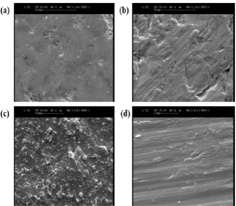
The surface characteristics of each samples was examined using scanning electron microscopy (SEM). As shown in the Figure 3, TiGO consist of a multilayer sheets structure and the individual GO layer sheet stacked one above the other and exhibited a wrinkled structure in some parts. It is possible to recognize the edge of each layer from the image. Comparing with TiCS, the TiCS/GO demonstrated a more coarse morphology. This may contribute to the pres-ence of GO in the composite sample. The Figure 3 clearly shows the folding and bending behavior of GO sheets on TiCS/GO. The TiCS sample on the other hand showed a con-tinuous structure with high uniformity. The presence of a large amount of oxygen-functional groups in GO structure enabled it to make physical and electrostatic interactions with Cs, resulting in a well dispersed GO sheets in the Cs matrix [11, 22].
Cell attachment and proliferation
MTT assay involves a reduction reaction which reduces MTT reagent to a blue formazan product when incubated with viable cells and the absorbance of formazan indirectly reflects the level of cell viability in culture. Figure 5 shows the number of viable MG63 cells that adhere to the sample surface. After 2 hours of culture, no significant difference
is seen in between the cell adhesion rates all samples. However after 4 hours, the cell adhesion to the TiCS and TiCS/GO samples were increased in comparison with the other samples. After 6 hours, the cell adhesion to the TiCS and TiCS/GO surfaces was significantly higher than that of TiGO and Ti samples. At all time points, the cell adhesion in Cs was higher than all the other samples (P<0.05) (Fig. 4).
Figure 2. AFM images and root mean square roughness values (rms) of (a) Ti GO, (b) Ti Cs, (c) Ti Cs/GO, (d) control.
Figure. 3 Scanning electron microscope (SEM) images of each coatings and control. (a) Ti Cs, (b) Ti GO, (c) Ti Cs/GO, and (d) con-trol.
This is believed to be related to the attraction between the net positive charge of CS and the net negative charge of MG63 cells [23]. Moreover, previous studies have suggested that cell attachment could be further improved on the rougher surfaces.
Figure. 4 MG63 osteoblast cells adhesion on titanium surfaces after 1, 2 and 4 h. Data is presented as mean ± SD; n = 3. * P<0.05 compared toTi Cs/GO, Ti GO and control, **
pared toTi Cs, Ti GO and control.
The reported increased adhesion to rougher been explained with an increase of the surface ble for adhesion. As we mentioned above, sample showed the highest surface roughness tics among the other samples. So, these findings plain why the cells prefer to attach more TiCS/GO samples. Cell proliferation of the samples evaluated via MTT assay, after direct cultivation cells on the samples for 1, 3 and 7 days.
uncoated samples were used as control. shown in Figure 5. After 1 day of culture, significant difference between MTT values cells seeded on control and the coated samples. days, TiCS and TiCS/GO samples had MTT higher than that of the Ti. After 7 days of culture, and TiCS/GO surfaces displayed significantly values than the control (P<0.05).
Figure 5. Proliferation of MG63 cells after 1, 3
titanium surfaces was measured with MTT assay. Each bar represents the mean of cell proliferation ± SD (n=3). * compared to Ti Cs/GO, Ti GO and control, ** P<0.05 compared to Ti Cs, Ti GO, and control.
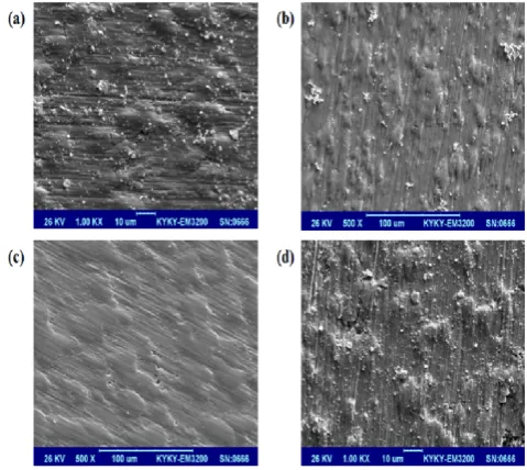
SEM observations (Fig. 6) confirmed the results. It is well known that cell attachment depending on expression and adsorption
MG63 osteoblast cells adhesion on titanium surfaces after 1, 2 and 4 h. Data is presented as mean ± SD; n = 3. * and control, ** P<0.05
com-rougher surfaces has surface area availa-above, the composite roughness
characteris-findings may ex-more on TiCS and
samples was also cultivation of the Cells grown on The results are culture, there was no values detected in the samples. After 3 MTT values slightly culture, the TiCS significantly higher MTT
roliferation of MG63 cells after 1, 3 and 7 days on was measured with MTT assay. Each bar represents the mean of cell proliferation ± SD (n=3). * P<0.05 <0.05 compared to
the proliferation attachment is highly of extracellular
matrix proteins such as fibronectin, Previous reports have corroborated hydrophilicity can enhance protein angle results confirmed that
demonstrated the higher hydrophilic other sample. Moreover, due
glycosaminoglycan, an exracellular has the potential to promote expression in human osteoblast
Cell morphology
Scanning electron microscopic the morphology of MG63 cells days on different Ti surfaces shown in Figure3. The observations tion between cells in TiCS/GO with control and TiGO samples, inent filopodia and lamellipodia in TiCS and TiCS/GO samples. Our SEM images clearly showed completely covered the TiCS cell contacts of the filopodias samples than that of TiGO and morphology on these samples surface roughness properties. can modulate the local regulatory osteoblast-like MG-63 cells findings is in agreement with supported that surface roughness, influence cell behavior and enhance ration, and differentiation on
Figure. 6 Scanning electron microscope cells cultured on titanium samples Ti GO, (b) Ti Cs, (c) Ti Cs/GO, (d) control.
Corrosion property of the samples
To evaluate the corrosion under aggressive electrochemical zation experiment was performed. potentiodynamic polarization corrosion potential value extracted from the polarization
fibronectin, vinculin, and actin. corroborated that increased surface
protein adsorption. Our contact that the TiCS and TiCS/GO samples hydrophilic properties than the ue to similar structure of CS to exracellular matrix molecule, CS promote extracellular matrix proteins
osteoblast cells [12, 22].
microscopic (SEM) was used to study cells after culturing for three surfaces (Fig. 6). The results are observations demonstrate
interac-CS/GO and Tiinterac-CS/GO. In comparison samples, a large population of prom-lamellipodia extensions were observed
showed that the MG63 cells
CS and TiCS/GO samples. More
filopodias were also observed in these and control samples. The better samples can be ascribed to their properties. Surface roughness, in fact,
regulatory factors produced by at the molecular level. These with previous studies which roughness, and wettability can enhance cell adhesion,
titanium surfaces.
microscope (SEM) images of MG63 samples after 2 days of incubation, (a)
control.
samples
vulnerability of the samples electrochemical environment, the
Mahsa Gheysour
178
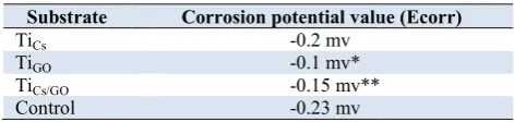
listed in Table 2. As it can be seen the Ecorr (-0.1 mv) and TiCS/GO (-0.15 mv) samples were ly higher than TiCS (-0.2 mv) and pure Ti ( ples. No significant difference was also observed Ecorr value of TiCS and pure Ti samples. This sion resistance in TiGO and TiCS/GO samples buted to the closer stacking of GO multilayer spin coating and drying. This ability offers for GO sheets to act as a barrier in prohibiting lyte from reaching the metal surface [11].
Figure 7. Potentiodynamic polarization plots for and coated samples.
Table 2. Corrosion potential value of different control * P<0.05 compared to TiCs and control, compared TiCs and control.
Substrate Corrosion potential value (Ecorr) TiCs -0.2 mv
TiGO -0.1 mv* TiCs/GO -0.15 mv** Control -0.23 mv
Conclusion
In this study, we successfully fabricated Cs, GO and Cs GO nanocomposite coatings on Ti substrates through spin coating method to evaluate the osteogenic properties of each coatings and uncoated Ti substrates. Our results ind cated that the addition of GO into Cs had a positive effect on the attachment and morphology of MG63 cells grown on Ti substrates. The resulting Cs/GO coatings also showed higher surface wettability compared to pure Ti substrates. More importantly, the nancomposite coating could reduce the bacterial growth to the su
vide effective corrosion protection of the Ti substrates. Therefore, our study suggests that combination of Cs and GO may be a promising coating material to increase the osteogenic properties of Ti based implants.
Acknowledgments
We gratefully acknowledge the National Institute of Genetic Engineering and Biotechnology for the financial support of this work.
630
Mahsa Gheysour, et al. Biologically Modified Titanium Substrates
Journal of Applied Biotechnology Reports, Volume corr value of TiGO
were significant-(-0.23 mv) sam-observed between This notable corro-samples can be attri-multilayer sheets during
offers great potential prohibiting the
electro-for control sample
different samples and control, Ti, ** P<0.05
Corrosion potential value (Ecorr)
0.15 mv**
fabricated Cs, GO and Cs-GO nanocomposite coatings on Ti substrates through spin coating method to evaluate the osteogenic properties of each coatings and uncoated Ti substrates. Our results indi-cated that the addition of GO into Cs had a positive effect
n the attachment and morphology of MG63 cells grown on Ti substrates. The resulting Cs/GO coatings also showed higher surface wettability compared to pure Ti substrates. More importantly, the nancomposite coating could reduce the bacterial growth to the surface and pro-vide effective corrosion protection of the Ti substrates. Therefore, our study suggests that combination of Cs and GO may be a promising coating material to increase the osteogenic properties of Ti based implants.
ly acknowledge the National Institute of Genetic Engineering and Biotechnology for the financial
References
1. Long, M., Rack, H., Titanium alloys in total joint replacement—a materials science perspective
1998, Vol. 19, pp. 1621-1639.
2. Donachie, M.J., Titanium: a technical guide international.
3. Li, X., Lin, K., Wang, Z., Enhanced growth and osteogenic differentiation of MC3T3-E1 cells on Ti6Al4V alloys modified with reduced graphene oxide.RSC Adv
14437.
4. Zhao, C., Lu, X., Zanden, C., Liu, J., The promising application of graphene oxide as coating materials in orthopedic implants: preparation, characterization and cell behavior
Mater, 2015, Vol. 10, pp. 015019.
5. Abraham, C.M., Suppl 1: A Brief Historical Perspective on Dental Implants, Their Surface Coatings and Treatments Dent J, 2014, Vol. 8, pp. 50-55.
6. Williams, D.F., On the mechanisms of biocompatibility Biomaterials, 2008, vol. 29, pp. 2941
7. Ratner, B.D., Bryant, S.J., Biomaterials: where we have been and where we are going. Annu Rev Biomed
pp. 41-75.
8. Anderson, J.M., Biological responses to materials Mater Res, 2001, Vol. 31, pp. 81
9. Bosco, R., Van Den Beucken, J., Leeuwenburgh, S., Jansen, J., Surface engineering for bone implants: a trend from passive to active surfaces.Coatings, 2012, V
10. Xuereb, M., Camilleri, J., Attard, N.J., Systematic review of current dental implant coating materials and novel coating techniques.Int J Prosthodont, 2015, V
11. Shi, Y., Li, M., Liu, Q., Jia, Z., Xu, X., Cheng, Y., Zheng, Y., Electrophoretic deposition of graphene oxide reinforced chitosan–hydroxyapatite nanocomposite coatings on Ti substrate Journal of Materials Science: Mater Med
12. Bumgardner, J.D., Wiser, R., Gerard, P.D., Bergin, P., Chestnutt, B., Marini, M., Ramsey, V., Elder, S.H., Gilbert, J.A., Chitosan: potential use as a bioactive coating for orthopaedic and craniofacial/dental implants. J Biomater Sci
2003, Vol. 14, pp. 423-438.
13. Pavithra, R., Logesh Kumar, S., Vijayalakshmi, C., Padmapriya, P., Nithyakalyani, K., Surface modification of titanium with chitosan extraction from crab shell by spin c Int J Cric Theor App, 2016, Vol. 9(9)
14. Jung, H.S., Choi, Y.-j., Jeong, J., Lee, Y., Hwang, B., Jang, J., Shim, J.-H., Kim, Y.S., Choi, H.S., Oh, S.H., Nanoscale graphene coating on commercially pure titanium for accelerated bone regeneration.RSC Adv, 2016, V
15. Garvin, K.L., Miyano, J.A., Robinson, D., Giger, D., Novak, J., Radio, S., Polylactide/polyglycolide antibiotic implants in the treatment of osteomyelitis. A canine model
1994, Vol. 76, pp. 1500-1506.
16. Wei, J., Lu, J., Yan, Y., Li, H., Ma, J., Wu, X., Dai, C., Liu, C., Preparation and characterization of well ordered mesoporous diopside nanobiomaterial.J Nanosci N
pp. 10746-10749.
17. Ikada, Y., Surface modification of polymers for medical a plications.Biomaterials, 1994, V
18. Zablotsky, M.H., Hydroxyapatite coatings in implant dent stry.Implant dentistry, 1992, vol. 1, pp. 253
19. Zuldesmi, M., Waki, A., Kuroda, K., Okido, M., Hydrothe mal treatment of titanium alloys for the enhancement of oste conductivity.Mater Sci Eng: C, 2015, V
20. Gittens, R.A., Scheideler, L., Rupp, F., Hyzy, S.L., Geis Gerstorfer, J., Schwartz, Z., Boyan, B.D., A review on the wett bility of dental implant surfaces II: biological and clinical a pects.Acta Biomater, 2014, Vol. 10, pp. 2907
, Volume 4, Issue 3, Summer 2017 Long, M., Rack, H., Titanium alloys in total joint a materials science perspective. Biomaterials, Titanium: a technical guide. 2000: ASM Li, X., Lin, K., Wang, Z., Enhanced growth and osteogenic E1 cells on Ti6Al4V alloys modified RSC Adv, 2017, Vol. 7, pp. 14430-Zhao, C., Lu, X., Zanden, C., Liu, J., The promising application of graphene oxide as coating materials in orthopedic implants: preparation, characterization and cell behavior.Biomed
ol. 10, pp. 015019.
Abraham, C.M., Suppl 1: A Brief Historical Perspective on Dental Implants, Their Surface Coatings and Treatments. Open Williams, D.F., On the mechanisms of biocompatibility.
l. 29, pp. 2941-2953.
Ratner, B.D., Bryant, S.J., Biomaterials: where we have been Annu Rev Biomed Eng., 2004, Vol. 6, Anderson, J.M., Biological responses to materials. Ann Rev
ol. 31, pp. 81-110.
Bosco, R., Van Den Beucken, J., Leeuwenburgh, S., Jansen, J., Surface engineering for bone implants: a trend from passive to
, 2012, Vol. 2, pp. 95-119.
Xuereb, M., Camilleri, J., Attard, N.J., Systematic review of dental implant coating materials and novel coating
, 2015, Vol. 28, pp.
Shi, Y., Li, M., Liu, Q., Jia, Z., Xu, X., Cheng, Y., Zheng, Y., Electrophoretic deposition of graphene oxide reinforced omposite coatings on Ti substrate. Materials Science: Mater Med, 2016, Vol. 27, pp. 48. Bumgardner, J.D., Wiser, R., Gerard, P.D., Bergin, P., Chestnutt, B., Marini, M., Ramsey, V., Elder, S.H., Gilbert, J.A., bioactive coating for orthopaedic and J Biomater Sci, Polymer Edition, Pavithra, R., Logesh Kumar, S., Vijayalakshmi, C., P., Nithyakalyani, K., Surface modification of chitosan extraction from crab shell by spin coating.
, 2016, Vol. 9(9), pp. 4027-4032.
j., Jeong, J., Lee, Y., Hwang, B., Jang, H., Kim, Y.S., Choi, H.S., Oh, S.H., Nanoscale commercially pure titanium for accelerated
, 2016, Vol. 6, pp. 26719-26724. Garvin, K.L., Miyano, J.A., Robinson, D., Giger, D., Novak, J., Radio, S., Polylactide/polyglycolide antibiotic implants in the itis. A canine model.J Bone Joint Surg, Wei, J., Lu, J., Yan, Y., Li, H., Ma, J., Wu, X., Dai, C., Liu, C., Preparation and characterization of well ordered mesoporous J Nanosci Nanotechnol, 2011, Vol. 11, Ikada, Y., Surface modification of polymers for medical
ap-, 1994ap-, Vol. 15ap-, pp. 725-736.
Zablotsky, M.H., Hydroxyapatite coatings in implant denti-, 1992denti-, vol. 1denti-, pp. 253-257.
Zuldesmi, M., Waki, A., Kuroda, K., Okido, M., Hydrother-mal treatment of titanium alloys for the enhancement of
osteo-, 2015osteo-, Vol. 49osteo-, pp. 430-435. Gittens, R.A., Scheideler, L., Rupp, F., Hyzy, S.L.,
Geis-er, J., Schwartz, Z., Boyan, B.D., A review on the wetta-bility of dental implant surfaces II: biological and clinical
21. Marimuthu, M., Veerapandian, M., Ramasundaram, S., Hong, S.W., Sudhagar, P., Nagarajan, S., Raman, V., Ito, E., Kim, S., Yun, K., Sodium functionalized graphene oxide coated titanium plates for improved corrosion resistance and cell viability. Appl Surf Sci, 2014, Vol. 293, pp. 124-131.
22. Lahiji, A., Sohrabi, A., Hungerford, D.S., Frondoza, C.G., Chitosan supports the expression of extracellular matrix proteins in human osteoblasts and chondrocytes. J Biomed Mater Res, 2000, Vol. 51, pp. 586-595.