RESEARCH ARTICLE
Application of solid phase
microextraction–gas chromatography–mass
spectrometry method for the detection
of active moulds on historical objects
Tomasz Sawoszczuk
*and Justyna Syguła‑Cholewińska
Abstract
The main purpose of these studies was to assess the possibility of applying the technique of solid phase microextrac‑ tion (SPME)–gas chromatography (GC)–mass spectrometry (MS) to detect the activity of moulds on historical objects, based on the analysis of microbial volatile organic compounds (MVOCs). The studies were performed for selected species of moulds, which were inoculated onto model samples of silk, cellulose, parchment and wool that had been prepared on microbiological medium, in vials for headspace sampling. After a few days of incubation, the MVOCs in the vials were sampled by using SPME fibre, and then they were analysed in the GC–MS system. The acquired chroma‑ tograms were qualitatively and quantitatively assessed, and it was ascertained that among the identified compounds are markers of mould activity which can be used to detect the vital mould growing on actual historic items. This usefulness of the method was additionally confirmed by analysis of MVOCs emitted by keratinolytically active mould inoculated on a sample of historical wool prepared in a Petri dish without a medium.
Keywords: Biodeterioration, Historic objects, MVOCs, GC/MS
© The Author(s) 2017. This article is distributed under the terms of the Creative Commons Attribution 4.0 International License (http://creativecommons.org/licenses/by/4.0/), which permits unrestricted use, distribution, and reproduction in any medium, provided you give appropriate credit to the original author(s) and the source, provide a link to the Creative Commons license, and indicate if changes were made. The Creative Commons Public Domain Dedication waiver (http://creativecommons.org/ publicdomain/zero/1.0/) applies to the data made available in this article, unless otherwise stated.
Background
Moulds occurring in the museums, libraries, archives,
can be derived from various sources [1–3]. However,
they grow only at certain values of temperature and relative humidity of air and at high water activity in the object [4–6]. Therefore, the protection of historic objects against biodeterioration relies primarily on keeping the parameters of microclimate at an appropriate level at the place of storage or exhibition of the objects [6]. However, it has been confirmed that some species can develop on materials even at quite low water activity, especially in the places with a low rate of air exchange [7, 8]. Also, malfunctioning of the heating, air-conditioning or ventilation system may lead to sudden fluctuations in temperature and humidity, potentially enabling the devel-opment of microorganisms [5, 9]. A likewise serious risk
is posed when the central heating system or the water pipes failure, or when the roof is leaking [10]. All of the circumstances described above may enhance the risk of microbial growth on or within the object [11].
Nevertheless, even if there are any visible changes on the object that suggest the presence of moulds, first of all, it should be investigated if these moulds are active. Based on this information the decision to disinfect the object should be made. There is no need for disinfection when the moulds are inactive. This is especially impor-tant in the case of the naturally aged, brittle, historical objects made of a protein material (silk, wool, parch-ment) because the organic materials are very sensitive to the destructive effect of most of the available disinfection methods [12–18]. The investigation of moulds activity should be done with a test method that can detect the fungi growing on the surface of object as well as inside its structure (a hidden growth of moulds inside the histori-cal objects with complex structure). A method that meets these expectations is the measurement of secondary
Open Access
*Correspondence: [email protected]
volatile metabolites, which are emitted by active moulds at every stage of their development [19–22]. The so-called microbial volatile organic compounds (MVOCs) easily diffuse through porous barriers like textiles [23, 24] so they can be used to detect moulds even if they are growing inside the structure of a historical object [25, 26].
Although the moulds emit circa 150 MVOCs, in the lit-erature it has been proposed to detect the active myce-lia of moulds based on the identification of the following compounds: 1-octen-3-ol, 3-octanol, and 1-octen-3-one, 2-octanone, 3-octanone (classified into the so-called
eight-carbon compounds, C8 complex); heptanone,
hex-anone as well as terpenes and sesquiterpenes [22, 25, 27–32] However, the majority of articles related to stud-ies of these compounds concern the analysis of MVOCs emitted by moulds growing on building and construction materials or in microbiologically contaminated rooms. These analyses are mainly related to the assessment of the health risk level for people staying in spaces with the active fungi growth, in the context of the so-called sick building syndrome (SBS) [20, 24–27, 29, 32]. Only few authors analysed the MVOCs emitted by moulds grow-ing on historic objects, e.g. on historic paper [21] or his-torical cinematographic film [22]. An interesting paper on the topic of the analyses of MVOCs with the regard to cultural heritage objects has been presented by Joblin et al. [23].
The main purpose of this study was to assess the pos-sibility of detecting the activity of moulds on the his-torical objects based on the measurements of MVOCs carried out with the SPME–GC–MS method which has been previously developed and optimized for this meas-urement [19]. This study will also reveal the information about the applicability of the new method. The investiga-tions were accomplished for selected species of moulds, which were inoculated onto various types of model sam-ples and one real historical sample. After a specific time of incubation, the MVOCs emitted by the fungi were analysed in the GC–MS set. For the acquired chroma-tograms, a qualitative and quantitative analysis was performed in order to ascertain whether the identified compounds included indicators of moulds activity which might be used to detect fungi growing on actual histori-cal objects.
Methods
Chemical reagents and microbiological media
All the chemical reagents used as reference substances and for the preparation of microbiological media were of analytical purity (Avantor Performance Materials, Poland). One type of microbiological media was bought as ready made-to-order preparations (BTL, Poland). The
second one was prepared in a laboratory. The ordered medium contained neither organic carbon nor nitrogen nor sulphur that could be available for fungi, as it was assumed on the basis of the list of ingredients provided by the manufacturer. However, the absence of these elements was confirmed indirectly by inoculating the selected species of moulds on this medium (without an organic material sample). No growth was observed after 2 weeks of incubation. The goal of the experiment was to ensure that the main nutrient for moulds was the model organic materials, which was placed on the surface of the medium. Firstly, this was a way to confirm that the chosen mould really has the cellulolytic or proteolytic activity (when it can grow on cellulose, silk, parchment or wool substrate, respectively). Furthermore, the appli-cation of media without organic nutrients makes it very probable that the volatiles emitted by moulds originated mostly from the enzymatic decomposition of the organic material and not from the metabolism of sugar which is usually in the media. Media were prepared in the shape of a slope (using 5 ml of medium) inside 20 ml sterile vials designed for headspace (HS) sampling. One type of medium used for cultivation of moulds was a modified version of Weary and Canby’s medium (W&C) [33, 34]. The second type of medium was a modified version of Czapek–Dox medium (Cz&D–BTL, Poland).
The W&C medium was prepared without keratin, whereas the Cz&D medium did not contain sucrose. The other components in the media, i.e. set of salts and agar, were the same as in their unmodified version.
Model materials
The model samples that were chosen to resemble historical objects were the following organic materials
• Whatman cellulose filter paper (Sigma-Aldrich), grammage 100 g m−2, thickness 200 µm, ash content
0.008%,
• NOVO Test Paper 2 (KLUG-CONSERVATION, Immenstadt, Germany), grammage 70 g m−2, 100%
bleached wood pulp, 12–15% kaolin, sized with resin and with the addition of aluminium sulphate. pH 4.5 (adjusted with alum); without the optical brightening agents,
• Natural degummed silk, grammage 8 g m−2,
unbleached, undyed, Habutai type,
• Natural wool fabric, 100% wool, unbleached, undyed, plain weave, grammage 250 g per linear meter,
• Handmade parchment, the limed calfskin, with-out any additives or additional treatment, thickness 1.5 mm.
the surface of a sterile microbiological media inside the vials.
All samples of organic materials were disinfected prior to the experiment. The procedure of samples disinfection followed the one described in [19]. The applied disinfec-tion methods caused a degradadisinfec-tion of model samples which results in a change in their structure and colour [12–18].
The efficiency of sterilization was confirmed after ran-domly chosen samples of model materials were incubated on Potato Dextrose Agar (PDA, BTL, Poland) at 28 °C. No growth of moulds was observed on the materials even with incubation extended to 14 days.
Fungal species
The growth of 16 species of mould on the sterile model samples, prepared on the W&C and Cz&D media, was initially evaluated to select the species which can dete-riorate the model materials. The fungi were purchased as identified and certified species at the Belgian Coor-dinated Collection of Microorganisms (IHEM Culture Collection, Brussels, Belgium). For further investigations only one species was chosen for each type of organic material based on the observation of moulds growth intensity on the samples. This has always been a mould which growth was the most vigorous on a given mate-rial. The fungal species chosen for this study have already been isolated from historic objects [2, 3, 7, 8, 11]. The
list of studied species contained: Chaetomium
globo-sum IHEM:6552, Aspergillus fumigatus IHEM:1363,
Aspergillus niger IHEM:2312, Cladosporium herbarum
IHEM:5294, Trichoderma viride IHEM:3170. For each
species, an inoculum was prepared in the same way as it was described in [19]. 0.1 ml of the spore suspension was pipetted onto the model samples of materials, which were placed on the surface of the medium inside the vials. The next steps followed the procedure demonstrated in [19].
Historical sample
In order to confirm the results of measurements obtained for the samples of model material, analyses were also car-ried out on MVOCs emitted by moulds growing on an actual historic artefact. The sample was a destroyed frag-ment of the seventeenth century woollen textile, which was removed during conservation, with dimensions 3 × 2 cm. The sample was disinfected in a chamber with ethylene oxide, and then conditioned until the gas had evaporated completely, as confirmed by GC–MS analysis. The conditioned sample was cut into two pieces, each of which was placed in sterile Petri dishes and then mois-tened with a small amount of saline solution, so that the samples were moist but not covered with water. In this case, no microbiological medium was used. Next, one
sample was inoculated with T. viride IHEM:3170 while
the other remained sterile as the reference sample. The samples were incubated for 6 days at 23 °C.
In summary, the following set of samples was prepared A. niger—silk—W&C medium
A. fumigatus—calfskin parchment—Cz&D medium C. herbarum—NOVO test paper—Cz&D medium T. viride—Whatman paper—Cz&D medium C. globosum—wool—W&C medium
T. viride—historical wool
The choice between W&C and Cz&D media for each material—mould set was made based on the observation of the intensity of fungal growth, which was influenced by the composition of salts in the medium. In each case the medium with the model sample which stronger intensi-fied the growth of moulds was selected. By assumption, the more intensive growth of moulds the higher MVOCs emission could have been measured. Contrary, the stud-ies on historical sample were carried out without any medium to determine whether it is possible to measure the MVOCs emission when the only substrate is the wet sample of organic material which did not contain microelements.
Headspace–solid phase microextraction
SPME (solid phase microextraction) was chosen as the method of MVOCs sampling. The so-called sandwich fibre consisting of three different sorbents (DVB/CAR/
PDMS) was chosen for MVOCs sorption [35]. Prior to
repeated four times for ever set of mould—model mate-rial—microbial medium.
The same measurement procedure was used to analyse the so-called blank samples. Those were vials with media and samples of the model materials on which no fungal spores were inoculated. The chromatographs acquired for these samples specify the volatile organic compounds emitted from the media, the seal, and the cap materials, which is the so-called background emission. These chro-matograms were subtracted in the data analysis process from the chromatograms obtained for samples inocu-lated with moulds, to make sure that the further inter-pretation of the results would only be carried out for the MVOCs emitted by fungi growing on the samples.
The so-called sandwich fibre was also used for the
col-lection of MVOCs emitted by T. viride growing on the
sample of wool. The fibre was placed in a partly open Petri dish and the needle was pulled out to be exposed just above the inoculated or non-inoculated sample of wool, respectively. The MVOCs were collected onto the fibre for 12 h in this case. After finishing the sorption, the fibre was placed in the injector of the chromatograph, and the anal-ysis was started. The measurements were repeated three times for the sample of historical wool with and without mould. It should be noted that in this case the analysis of MVOCs was carried out for a mould growing on actual historic wool sample without medium, sorption of the microbial volatiles was carried out in a shorter time than for the samples in vials, and additionally, the sorption was performed in the open system. Results of this experi-ment were to provide information whether it is possible to carry out measurements of MVOCs emitted by moulds developing on actual historic or artwork objects (without microbial medium), in situ, i.e. in the museum, library or archive, in the open system, in which concentration of MVOCs in the air decreases with the increase of the dis-tance from the surface of the mould, due to diffusion.
Investigations of MVOCs emitted by fungi growing on the model samples and the historic sample comprised both the qualitative and the quantitative analysis. A cali-bration was carried out with the aim of performing the quantitative analysis of MVOCs which are the indicators of the active growth of moulds. The calibration proce-dure followed the one found in the article [19]. Alcohols: 1-octen-3-ol, 3-octanol and ketones: 1-octen-3-one, 3-octanone, 2-hexanone, 2-heptanone were used as ref-erence compounds. The detection and quantification lim-its were determined for each reference compound based on standard deviation of the response and the slope of calibration curve. The highest quantification limit was found for hexanone which was the least abundant com-pound in chromatograms acquired during the calibration procedure. The quantification limit for hexanone was
3.0 µg m−3. The quantification limits determined for all
other compounds were between 3.0 and 0.7 µg m−3. The
detection and quantification limits were also determined for a group of terpenes and sesquiterpenes based on the calibration procedure carried out for the reference odour compounds mix ISO 17943 (Sigma-Aldrich). The limit of quantification calculated for the least abundant com-pound which was present in the reference odour mix, geosmin, was 0.5 µg m−3.
GC–MS analysis
The analysis of the MVOCs was performed in the gas chromatograph (GC, Trace 1310)–quadrupole mass spec-trometer (MS, ISQ) system, manufactured by Thermo Scientific Inc. (USA). The injector was equipped with a liner intended for desorbing SPME fibres. During desorp-tion of MVOCs from the fibre, the injector was set to the splitless mode. The parameters of the capillary column and a temperature program of MVOCs analysis were the same as the ones used for the investigations which are described in [19]. The work parameters of the MS detec-tor were almost the same as the ones reported in [19]. The only exemption was the range of mass-to-charge ratio which was scanned in MS. In this case, the range was 33–650 m/z. The MS worked in a total ion current mode (TIC). Reference libraries (NIST/EPA/NIH Mass Spectral Library) from NIST MS search program version 2.0 were used to identify the MVOCs. When required, a reference compound was used as a standard for confirming the presence of particular volatile compounds.
Results and discussion
Selection of moulds
The moulds investigated in these studies have already been described in literature as cellulolytic and
proteo-lytic species. A. niger IHEM:2312 was recognized as a
fungus capable to decay the silk [36, 37], A. fumigatus IHEM:1363 shows collagenolytic properties [38, 39]. C. herbarum IHEM:5294 and T. viride IHEM:3170 have cel-lulolytic properties [3, 40]. C. globosum IHEM:6552 and T. viride IHEM:3170 are known not only as cellulolytic moulds but they possess a keratinolytic properties [25, 41, 42]. All of the mentioned species have been identi-fied in the indoor air of museums, archives and libraries [1–3, 8, 40]. These species have also been isolated from the surfaces of historic textile and archival documents [3, 39–42].
Qualitative and quantitative analysis of the chromatograms
Qualitative analysis
model samples was performed in the AMDIS program (Automated Mass Spectral Deconvolution and Identifi-cation System, version 2.70, 13 May 2011). Mass spectra registered under the chromatograms were subjected to deconvolution, and then an identification of volatile com-pounds was carried out based on the reference librar-ies (NIST/EPA/NIH Mass Spectral Library) which are available in the NIST MS search program. The obtained results demonstrate that each of the studied mould spe-cies cultured on different model materials produces more than 140 volatile organic compounds (Fig. 1). These com-pounds belong to different groups of organic comcom-pounds [22, 25, 32, 43]: hydrocarbons (heptane, isoprene), aromatic hydrocarbons (benzene, styrene), alcohols (1-octen-3-ol, 3-octanol, 3-methyl-1-butanol), aldehydes, ketones (1-octanone, 1-octen-3-on, 3-octanone), organic acids, ethers, esters (butyl acetate), terpenes (limonene), sesquiterpenes (humulene, nerolidol, bisabolene), organic compounds containing sulphur and nitrogen.
Qualitative analysis was focused only on the com-pounds that could have been identified with probability
higher than 90%. In some cases, when the probability was lower than 90%, identification was confirmed by car-rying out the analysis for the reference substances. This was essential in the case of compounds that had already been described in literature as the indicators of the active growth of moulds. The results of analysis confirmed that the individual profiles of MVOCs emitted by each mould were different (Fig. 1). Moreover, it was possible to dis-cover in each chromatogram the volatiles that were char-acteristic only for a given mould. However, few of the identified volatiles were emitted by all species regardless of the type of medium and the type of organic sample on which they grew. These were mostly the six-, seven- and eight-carbon ketones as well as the eight-carbon alcohols.
The analysis of the microbial volatiles was focused on the compounds with eight carbons in their chains, the so-called C8-complex MVOCs [31], particularly the alcohols: 1-octen-3-ol, and 3-octanol; and ketones: 1-octen-3-one, 2-octanone and 3-octanone because they are indicators of the moulds activity, as it was mentioned in the intro-duction [22, 25, 30, 31]. These compounds are released in
high concentrations and are adequate for detecting the metabolic activity of many species [22, 30, 43]. Addition-ally, it has been demonstrated in the literature that some enzymatic pathways are finished at other volatile prod-ucts like heptanone and hexanone beside or instead of the C8 compounds [31]. Other authors have also shown that terpenes and sesquiterpenes are useful bioindicators of the mould activity [28, 44]. Thus, it can be determined that all of the aforementioned groups of compounds are bioindicators that can be used for the detection of the vegetative mycelium because they distinguish between active and inactive fungi.
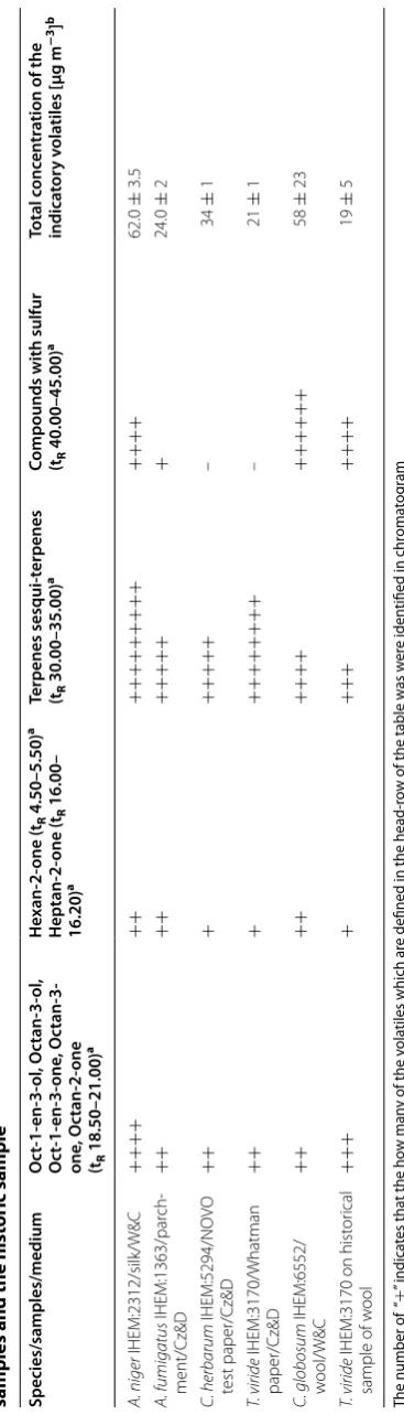
Taking into account the arguments presented above, it was assumed that the most important part of the quali-tative analysis of the chromatograms was to determine whether the moulds developing on the model materi-als imitating historic objects, and on the actual historic sample, emit volatiles belonging to all of the three above-mentioned groups. Additionally, it was significant to find whether the chromatograms acquired for MVOCs emit-ted by moulds growing on protein materials contained volatiles having a sulphur atom. The presence of sulphur containing compounds confirms that fungi actually did metabolize the specific protein material because fibroin, keratin and collagen contain cysteine with sulphur atom. The results of the qualitative analysis have been pre-sented in Table 1.
Based on the data presented in Table 1 it can be con-cluded that all of the species of mould emitted volatile
compounds belonging to the C8 complex, regardless of
the kind of material and microbial media on which the fungi were incubated. Furthermore, in all of the cases the studied fungi also emitted six- and seven-carbon ketones. All species of moulds emitted terpenes and sesquiterpe-nes. The emission of volatiles with the sulphur atom was detected only for moulds growing on materials which contained the proteins with cysteine in the polypeptide chain. These were the esters of sulfuric acids with long-chain alcohols. In the case of two species which grew on the cellulosic samples no emission of sulphur contain-ing compounds was detected. In this case, the media and both types of paper did not contained organic com-pounds with sulphur atom.
By analysing the data presented in Table 1 it can be confirmed that the largest number of compounds consid-ered as bioindicators of the metabolic activity of moulds
was emitted by A. niger IHEM:2312 growing on silk. A
slightly smaller number of the indicatory MVOCs was emitted by the other four species growing on various organic materials.
The chromatograms acquired for MVOCs emitted by T. viride IHEM:3170 growing on the historical sample of wool were subjected to a qualitative and quantitative
analysis, using the same methodology as for the experi-ment in the vials. It could be confirmed that T. viride IHEM:3170 growing on a sample of seventeenth century wool emitted 2-octanone, 1-octen-3-on, 3-octanone, heptanone and three volatile compounds belonging to the group of terpenes and sesquiterpenes. One of them was geosmin.
Quantitative analysis
The MVOCs emitted by moulds growing on various media were also quantitative analysed. This analysis was carried out for compounds that are already defined in the literature as indicators of the active growth of moulds. These data were calculated after integrating the areas under the peaks found for indicators in the chromato-grams (Genesis Peak Integration, Xcalibur). The area under the peaks was then converted into the correspond-ing quantity of indicatory MVOCs expressed in [µg m−3] on the basis of the established calibration curves (see “Headspace–solid phase microextraction”). The results have been presented in Table 1.
A comparison of the quantities of indicatory MVOCs emitted by the particular species of mould growing on different samples demonstrated that the highest level of
emission was measured for A. niger IHEM:2312
grow-ing on silk and for C. globosum IHEM:6552 growing on
wool. Smaller amount of bioindicators was emitted by
C. herbarum IHEM:5294 incubated on the samples of
acidic paper and the lowest emission was measured for
A. fumigatus IHEM:1363 incubated on parchment and
Table 1 Q ualita tiv e and quan tita tiv e analy sis of chr oma to gr ams ac quir ed f or MV OCs emitt ed b y selec ted sp ecies of moulds gr owing on the mo del ma terial
samples and the hist
oric sample
The number of
“
+
” indica
tes tha
t the ho
w man
y of the v
ola
tiles which ar
e defined in the head
‑r
ow of the table w
as w
er
e iden
tified in chr
oma
tog
ram
a R
et
en
tion time in chr
oma
tog
rams [min]
b
This is the sum of mean c
onc en tr ations det er mined f
or each c
ompound list
ed in the head
‑r
ow of the sec
ond
, thir
d and f
our
th c
olumn; the standar
d devia
tion w
as calcula
ted f
or each sum based on the v
ar ianc es Species/samples/medium Oc t-1- en-3-ol , Oc tan-3-ol , Oc t-1- en-3-one , Oc tan-3-one , Oc tan-2-one (tR 18.50–21.00) a He xan-2-one (tR 4.50–5.50) a Heptan-2-one (tR 16.00– 16.20) a Terpenes sesqui-t erpenes (tR 30.00–35.00) a
Compounds with sulfur (t 40.00–45.00)R
a Total c onc en tr ation of the indica tor y v ola tiles [µg m − 3] b A. niger IHEM:2312/silk/W&C ++++ ++ +++++++++ ++++ 62.0 ± 3.5 A. fumigatus IHEM:1363/par ch ‑ ment/Cz&D ++ ++ +++++ + 24.0 ± 2 C. herbarum IHEM:5294/NO VO test paper/Cz&D ++ + +++++ – 34 ± 1 T. viride IHEM:3170/Whatman paper/Cz&D ++ + ++++++++ – 21 ± 1 C. globosum IHEM:6552/ w ool/W&C ++ ++ ++++ ++++++ 58 ± 23 T. viride
IHEM:3170 on hist
or
ical
sample of w
on parchment is also not typical for this fungus. In this case probably the alkaline pH of limed parchment sam-ple was inhibiting the metabolism of mould. The results of calculations shown that for all of the studied sets of mould—sample—medium the emission of compounds which are indicators of the active growth of mould was over their quantification limits established during the cal-ibration procedure.
Trichoderma viride IHEM:3170 inoculated on the his-toric sample of wool emitted three times smaller
quan-tity of the indicatory MVOCs compared with A. niger
IHEM:2312 cultivated on silk or C. globosum IHEM:6552
on wool. The low level of MVOCs concentration measured for T. viride IHEM:3170 growing on historical wool, when no medium was used, was influenced by three factors: the sampling was carried out in an open system; the time of sampling was shortened to 12 h; and the only source of nutrients for the mould was the sample of wool without a medium containing microelements. In spite of these con-ditions of sampling, the emission of compounds which are indicators of the active growth of mould was over their quantification limits established during the calibra-tion procedure. The level of the mean emission measured for the volatiles which are indicators of the active growth of fungus was 19 µg m−3. Moreover, the measured level of emission was equal to levels determined for T. viride IHEM:3170 incubated on the Whatman paper samples.
Conclusions
The level of the indicatory MVOC emission, measured both in closed and open system, was low; however, it was above the quantification limit determined for every indi-cator of the active growth of moulds. The obtained data show that all the species of mould emitted volatile com-pounds belonging to the C8 complex, regardless of the kind of material and microbiological medium on which they were cultivated. In all of the analysed cases, fungi also emitted ketones with six- and seven-carbons as well as ter-penes and sesquiterter-penes. Among the MVOCs emitted by moulds growing on protein materials, volatile compounds containing sulphur were identified. Those were mainly sul-furic acid esters. This fact confirms the assumption that fungi decomposed the organic materials on which they were cultivated because each of the protein materials used as the substrate contained cysteine, an amino acid having a sulphur atom. The obtained results support the hypoth-esis that the metabolic activity of moulds growing on the organic materials imitating historical objects can be con-firmed based on the detection of the aforementioned three groups of the indicatory volatiles.
Moreover, the results of MVOCs measurements
car-ried out in an open system for T. viride IHEM:3170
growing on the historical sample of wool without a
medium confirmed that the mould emitted all of the compounds reported in the literature as the indict-ors of active moulds growth. Moreover, the concen-trations of MVOCs collected on the SPME fibre when sampling was carried out in a relatively short time, in an open system, are over the quantification limits deter-mined during the calibration procedure. Thus, the fact that measured concentrations of indicatory MVOCs are high supports the hypothesis that these bioindicators can be used to detect moulds actively growing on his-torical objects.
Author contributions
TS was responsible for: planning all experiments, carrying out the measure‑ ments, analyzing chromatograms, interpreting the results, writing the article, improvement of the text. JSCh has made substantial contributions to concep‑ tion of experiments, and was responsible for: preparing the microbial broths and the species of fungi, inoculating the fungi on samples, carrying out their incubation. All authors read and approved the final manuscript.
Acknowledgements
The authors gratefully acknowledge the financial support from the National Science Centre in Poland which has allowed implementing the project entitled Investigations of biodeterioration of historical objects based on analysis of volatile organic compounds emitted by moulds, decision reference DEC–2012/05/B/HS2/04094.
Competing interests
The authors declare that they have no competing interests.
Publisher’s Note
Springer Nature remains neutral with regard to jurisdictional claims in pub‑ lished maps and institutional affiliations.
Received: 15 December 2016 Accepted: 5 April 2017
References
1. Valentín N, García R, De Luis O, Maekawa S. Microbial control in archives, libraries and museums by ventilation systems. Restaurator. 1998;19:85–107. 2. Florian ML. Fungal‑problem monitoring for heritage collections: the need
for baseline reference levels for fungal structures and beta‑glucans. In: Koestler RJ, Koestler VH, Charola AE, Nieto‑Fernandez FE, editors. Art, biol‑ ogy, and conservation. New York: Biodeterioration of Works of Art; 2002. 3. Zyska B. Fungi isolated from library materials: a review of literature. Int
Biodeterior Biodegrad. 1997;40:43–51.
4. Adan OCG. On the fungal defacement of interior finishes. Eindhoven: Technische Universiteit Eindhoven; 1994.
5. Sedlbauer K. Prediction of mould fungus formation on the surface of and inside building components. Stuttgart: Fraunhofer Institute for Building Physics; 2001.
6. Martens M. Climate risk assessment in museums: degradation risks determined from temperature and relative humidity data. Eindhoven: Technishe Universiteit Eindhoven; 2012.
7. Micheluz A, Manente S, Tigini V, Prigione V, Pinzari F, Ravagnan G, Varese GC. The extreme environment of a library: xerophilic fungi inhabiting indoor niches. Int Biodeterior Biodegrad. 2015;99:1–7.
8. Borrego S, Lavin P, Perdomo I, Gomez de Saravia S, Guiamet P. Determination of indoor air quality in archives and biodeteriora‑ tion of the documentary heritage. ISRN Microbiol. 2012;2012:680598.
doi:10.5402/2012/680598.
Environmental monitoring in four European museums. Atmos Environ. 2001;35:127–40.
10. http://cstmuseum.techno‑science.ca/en/the‑museum‑is‑closed.php. 11. Strzelczyk AB. Observations on aesthetic and structural changes induced
in Polish historic objects by microorganisms. Int Biodeterior Biodegrad. 2004;53:151–6.
12. Porck H, Teygeler R. An overview of recent developments in research on the conservation of selected analog library and archival materials. In: Treatment. Washington: Council on Library and Information Resources; 2000.p.14–5.
13. Grosvenor AJ, Morton JD, Dyer JM. determination and validation of mark‑ ers for heat‑induced damage in wool proteins. AJAC. 2012;3:431–6. 14. Chamberlin Walde E, Barr M, Edgar R. The degradation of silk, wild silk and
wool by steam. Text Res J. 1936;6:235–40.
15. Watt IC. Properties of wool fibers heated to temperatures above 100 °C. Text Res J. 1975;45:728–35.
16. Hashimoto T, Taniguchi Y, Kameda T, Tamada Y, Kurosu H. Changes in the properties and protein structure of silk fibroin molecules in autoclaved fabrics. Polym Degrad Stab. 2015;112:20–6.
17. Dolgin B, Bulatov V, Schechter I. Non‑destructive assessment of parchment deterioration by optical methods. Anal Bioanal Chem. 2007;388:1885–96.
18. Manfredi M, Bearman G, France F, Shor P, Marengo E. Quantitative multi‑ spectral imaging for the detection of parchment ageing caused by light: a comparison with ATR–FTIR, GC–MS and TGA analyses. Int J Conserv Sci. 2016;6:3–14.
19. Sawoszczuk T, Syguła‑Cholewińska J, Del Hoyo‑Meléndez JM. Opti‑ mization of headspace solid phase microextraction for the analysis of microbial volatile organic compounds emitted by fungi: application to historical objects. J Chromatogr. 2015;1409:30–45.
20. Polizzi V, Delmulle B, Adams A, Moretti A, Susca A, Picco AM, Rosseel Y, Kindt R, Van Bocxlaer J, De Kimpe N, Van Peteghem C, De Saeger S. JEM spotlight: fungi, mycotoxins and microbial volatile organic compounds in mouldy interiors from water‑damaged buildings. J Environ Monit. 2009;11:1849–58.
21. Canhoto O, Pinzari F, Fanelli C, Mangan N. Application of electronic nose technology for the detection of fungal contamination in library paper. Int Biodeterior Biodegrad. 2004;54:303–9.
22. Bingley GD, Verran J, Munro LJ, Craig E, Banks CE. Identification of micro‑ bial volatile organic compounds (MVOCs) emitted from fungal isolates found on cinematographic film. Anal Methods. 2012;4:1265–71. 23. Joblin Y, Moularat S, Anton R, Bousta F, Orial G, Robine E, Picon O, Bour‑
ouina T. Detection of moulds by volatile organic compounds: application to heritage conservation. Int Biodeterior Biodegrad. 2010;64:210–7. 24. Lavine BK, Mirjankar N, LeBouf R, Rossner A. Prediction of mold contami‑
nation from microbial volatile organic compound profiles using solid phase microextraction and gas chromatography/mass spectrometry. Microchem J. 2012;103:37–41.
25. Fiedler K, Schütz E, Geh S. Detection of microbial volatile organic com‑ pounds (MVOCs) produced by moulds on various materials. Int J Hyg Environ Health. 2001;204:111–21.
26. Moularat S, Robine E, Ramalho O, Oturan MA. Detection of fungal devel‑ opment in closed environment through the identification of specific VOC: demonstration of a specific VOC fingerprint for fungal develop‑ ment. Sci Total Environ. 2008;407:139–46.
27. Wilkins K, Nielsen EM, Wolkoff P. Patterns in volatile organic compounds in dust from moldy buildings. Indoor Air. 1997;7:128–34.
28. Polizzi V, Adams A, Malysheva S, De Saeger S, Van Peteghem C, Moretti A, Picco AM, De Kimpe N. Identification of volatile markers for indoor fungal
growth and chemotaxonomic classification of Aspergillus species. Fungal Biol. 2012;116:941–53.
29. Polizzi V, Adams A, Picco AM, Adriaens E, Lenoir J, Van Peteghem C, De Saeger S, De Kimpe N. Influence of environmental conditions on produc‑ tion of volatiles by Trichoderma atroviride in relation with the sick building syndrome. Build Environ. 2011;46:945–54.
30. Wilkins K, Larsen K. Variation of volatile organic compound patterns of mold species from damp buildings. Chemosphere. 1995;31:3225–36. 31. Wilkins K, Larsen K, Simkus M. Volatile metabolites from mold
growth on building materials and synthetic media. Chemosphere. 2000;41(3):437–46.
32. Schuchardt S, Kruse H. Quantitative volatile metabolite profiling of common indoor fungi: relevancy for indoor air analysis. J Basic Microbiol. 2009;49:350–62.
33. Weary MA, Canby CM. Keratinophilic activity of Trichophyton schoenleini, T. rubrum and T. mentagrophytes. J Investig Dermatol. 1967;48:240–8. 34. Safranek WW, Goos RD. Degradation of wool by saprotrophic fungi. Can J
Microbiol. 1981;28:137–40.
35. Betancourt DA, Krebs K, Moore SA, Martin SM. Microbial volatile organic compound emissions from Stachybotrys chartarum growing on gypsum wallboard and ceiling tile. BMC Microbiol. 2013;13:283.
36. Seves A, Romano M, Maifrenic T, Soraa S, Ciferria O. The microbial degradation of silk: a laboratory investigation. Int Biodeterior Biodegrad. 1998;42:203–11.
37. Sato M. The effects of molds on fibers and their products. VIII. Scanning electron microscopic study on the destruction of silk yarns damaged by molds. Kyoto‑furitsu Daigaku Gakujutsu Hokoku Rigaku Seikatsu Kagaku. 1976;27:59–64.
38. Polacheck I. Damage to an ancient parchment document by Aspergillus. Mycopathologia. 1989;106:89–93.
39. Pinzari F, Colaizzi P, Maggi O, Persiani AM, Schütz R, Rabin I. Fungal bioleaching of mineral components in a twentieth‑century illuminated parchment. Anal Bioanal Chem. 2012;402:1541–50.
40. Flannigan B. Microorganisms in indoor air. In: Flannigan B, Samson RA, Miller JD, editors. Microorganisms in home and indoor work environ‑ ments. Boca Raton: Taylor & Francis Group; 2011. p. 17–31.
41. Abdel‑Kareem O, Alfaisal R. Treatment, conservation and restoration of the Bedouin dyed textiles in museum of Jordan heritage. Mediterr Arch Archaeometr J. 2010;10:25–36.
42. Cybulska M, Jedraszek‑Bomba A, Kuberski S, Wrzosek H. Methods of chemical and physicochemical analysis in the identification of archaeo‑ logical and historical textiles. Fibres Text East Eur. 2008;16:67–73. 43. Kuske M, Romain AC, Nicolas J. Microbial volatile organic compounds as
indicators of fungi. Can an electronic nose detect fungi in indoor environ‑ ments? Build Environ. 2005;40:824–31.
44. Demyttenaere JCR, Moriña RM, De Kimpe N, Sandra P. Use of headspace solid‑phase microextraction and headspace sorptive extraction for the detection of the volatile metabolites produced by toxigenic Fusarium species. J Chromatogr. 2014;1027:147–54.
45. Korpi A, Järnberg J, Pasanen AL. Microbial volatile organic compounds. Crit Rev Toxicol. 2009;39:139–93.
46. Šimonovičová A, Kraková L, Pangallo D, Majorošová M, Piecková E, Bodoriková S, Dörnhoferová M. Fungi on mummified human remains and in the indoor air in the Kuffner family crypt in Sládkovičovo (Slovakia). Int Biodeterior Biodegrad. 2015;99:157–64.