R E S E A R C H
Open Access
A gait abnormality measure based on root mean
square of trunk acceleration
Masaki Sekine
1*, Toshiyo Tamura
1, Masaki Yoshida
1, Yuki Suda
2, Yuichi Kimura
3, Hiroaki Miyoshi
4, Yoshifumi Kijima
5,
Yuji Higashi
5and Toshiro Fujimoto
5Abstract
Background:Root mean square (RMS) of trunk acceleration is seen frequently in gait analysis research. However, many studies have reported that the RMS value was related to walking speed. Therefore, the relationship between the RMS value and walking speed should be considered when the RMS value is used to assess gait abnormality. We hypothesized that the RMS values in three sensing axes exhibit common proportions for healthy people if they walk at their own preferred speed and that the RMS proportions in abnormal gait deviate from the common proportions. In this study, we proposed the RMS ratio (RMSR) as a gait abnormality measure and verified its ability to discriminate abnormal gait.
Methods:Forty-seven healthy male subjects (24–49 years) were recruited to examine the relationship between walking speed and the RMSR. To verify its ability to discriminate abnormal gait, twenty age-matched male hemiplegic patients (30–48 years) participated as typical subjects with gait abnormality. A tri-axial accelerometer was attached to their lower back, and they walked along a corridor at their own preferred speed. We defined the RMSR as the ratio between RMS in each direction and the RMS vector magnitude.
Results:In the healthy subjects, the RMS in all directions related to preferred walking speed. In contrast, RMSR in the mediolateral (ML) direction did not correlate with preferred walking speed (rs =−0.10,p = 0.54) and represented the similar value among the healthy subjects. Moreover, the RMSR in the ML direction for the hemiplegic patients was significantly higher than that for the healthy subjects (p < 0.01).
Conclusions:These results suggest that the RMSR in the ML direction exhibits a common value when healthy subjects walk at their own preferred speed, even if their preferred walking speed were different. For subjects with gait
abnormality, the RMSR in the ML direction deviates from the common value of healthy subjects. The RMSR in the ML direction may potentially be a quantitative measure of gait abnormality.
Keywords:Gait abnormality measure, Trunk acceleration, Root mean square (RMS) ratio
Background
Recently, accelerometry has been widely used to study human body movement as an alternative approach to conventional motion analysis techniques, such as opto-electronic and force plate motion analyses. The benefits of accelerometers are as follows: low cost compared with commonly used motion analysis equipment; measure-ment not being limited to a laboratory environmeasure-ment; and accelerometers being small in size and lightweight,
which avoids interference with a subject’s movement [1]. Typical applications of accelerometry are gait analysis, balance evaluation and screening of falling risk [2-17]. By using the characteristics of acceleration patterns, several researchers have proposed algorithms to obtain spatiotemporal parameters such as walking cycle, step duration, stance and swing durations [2,3]. Gait abnor-mality and falling risk have also been assessed by apply-ing frequency analysis includapply-ing harmonic analysis [2,4-8], time-frequency analysis [9,10], non-linear ana-lysis such as entropy anaana-lysis [11,12] and fractal anaana-lysis [13,14] to body acceleration.
* Correspondence:[email protected]
1
Faculty of Biomedical Engineering, Osaka Electro-Communication University, 18-8 Hatsucho, Neyagawa, Osaka 572-8530, Japan
Full list of author information is available at the end of the article
Root mean square (RMS) of acceleration is also seen frequently in gait analysis research [3-8,11,12,15-17]. This parameter constitutes a statistical measure of the magnitude of acceleration. Computation of the RMS is extremely simple and requires no preconditions like an optimal threshold and accurate peak detection to obtain characteristics of the signal pattern. Thus, the physical meaning of an RMS value is clear, and the value is easy to use in clinical practice. Moe-Nilssen showed that the RMS value along the mediolateral direction in a slightly balance-impaired subject increased compared with that in normal subjects [4]. On the other hand, Mizuike et al. demonstrated that RMS values of all three directions in stroke patients were significantly lower than those of normal subjects [16]. It is expected that one of the main factors of the discrepancy in these findings for abnormal gait comes from the relationship between RMS and walking speed. Moe-Nilssen studied the difference of RMS between normal and abnormal walking at the same walking speed [4]. Mizuike et al. evaluated the RMS of healthy subjects and stroke patients at their preferred walking speed [16]. In these studies, the conditions of walking speed differed. Many studies reported that the RMS had a high correlation with walking speed [3-6,11,15]. Therefore, Menz et al. and Latt et al. sug-gested that the usefulness of RMS is limited in experi-ments involving modulation of walking speed as subjects will have higher RMS when they walk faster [5,6]. In other words, it is necessary to consider the walking speed when RMS is used to assess gait abnormality.
It is generally known that some gait parameters exhibit common values or ratios in normal gait at an individual’s preferred walking speed (natural walking speed). For ex-ample, a walking cycle lasts around 1 s; stance and swing durations are consistently approximately 60% and 40% of a walking cycle in normal gait [18]. It is known that these parameters change with abnormal gait. For stance, Stolze et al. reported that a significantly in-creased walking cycle with a prolonged stance and double limb support duration was found in cerebellar ataxic gait [19]. We hypothesized (1) that the RMS values in three sensing axes also exhibit common pro-portions for healthy people if they walk at their own pre-ferred speed and (2) that the RMS proportions in abnormal gait deviate from the common proportions. In this study, we propose the RMS ratio (RMSR described in the Analysis section) as a gait abnormality measure and verify its ability to discriminate abnormal gait.
Methods
Subjects
To examine the relationship between individual’s pre-ferred walking speed and the RMSR, forty-seven healthy male subjects (age: 21–49 years; height: 172.5 ± 4.6 cm
(mean ± S.D.); weight: 64.7 ± 8.6 kg) were recruited. None had a history of neurological, musculoskeletal or gait disorder, or any painful condition likely to affect their balance or mobility. Auvinet et al. reported that gait vari-ables such as walking speed and characteristics of vertical acceleration started to change after 50 years of age for men [2]. Therefore, our condition of“healthy subjects” excludes people with age-related functional decline.
To verify the ability of RMSR to discriminate abnor-mal gait, twenty age-matched abnor-male post-stroke hemiple-gic patients (age: 28–48 years; height: 170.8 ± 5.9 cm; weight: 65.9 ± 8.2 kg; Brunnstrom stage III: 3, IV: 9, V: 5 and VI: 3) participated as typical subjects with gait abnormality. In addition, an elderly male hemiplegic (age: 76 years; height: 163 cm; weight: 60 kg; Brunnstrom stage V) participated to confirm the changes of RMSR along with recovery in an acute period. Mazzà et al. de-scribed that a significant gender difference was found in the RMS at the pelvis [17]. To exclude an effect of gender, only male subjects and male patients were adopted in the experiment.
This study was approved by the ethics committees of Fujimoto General Hospital and Chiba University, and all the subjects gave written informed consent before examination.
Data collection
A wireless sensor unit was used, which contained a tri-axial accelerometer (MMA7260Q, Freescale Semicon-ductor, USA), an infrared (IR) remote control receiver (NJL21H380A, New Japan Radio, Japan), a microcontrol-ler and a Bluetooth module. This sensor unit was devel-oped in our laboratory and its electronic design and characteristics permit a measuring range of ± 4 g, sensitiv-ity of 0.002 g and a range of response frequency from 0 Hz to 30 Hz for the accelerometer. The accelerometer and IR receiver outputs were digitized at a sampling rate of 100 Hz by the microcontroller and sent to a computer via the Bluetooth module for further off-line analysis. The unit size and weight are 52 × 54 × 18 mm and 55 g includ-ing a battery, respectively.
sensor unit. The timing was also used to extract the acceleration signals associated with the walking speed.
Procedure
The wireless sensor unit was attached to the lower back, around the L3-L4 vertebrae, using an elastic belt. This position was chosen owing to its proximity to the center of mass of the human body during standing. The sensing axes were oriented along the anatomical anteroposterior (AP), mediolateral (ML) and vertical (V) directions.
Forty-one of the healthy subjects and the patients were instructed to walk along a 14-m walking path at their own preferred speed. The healthy subjects wore their usual shoes, excluding low-heeled footwear such as san-dals or slippers. If the hemiplegic patients usually used a cane and/or a short leg brace, they also used them in the measurement.
The measurement was carried out once per each sub-ject after he walked along the walking path once to de-termine the optimal height of the photoelectric sensors for the individual subject. Only the elderly post-stroke hemiplegic patient carried out the measurement six times. Each measurement was performed after 13, 17, 21, 25, 31 and 40 days of the onset.
The other six healthy subjects were asked to walk at 0.83, 1.11, 1.39, 1.67 and 1.94 m/s on a treadmill for 2 min each.
Analysis
The recorded acceleration signal consists of both a dy-namic component reflecting changes in velocity during walking and a static acceleration caused by gravity. The sensing axes of the accelerometer may not be aligned with the axes of the horizontal-vertical coordinate sys-tem even if the sensor unit is attached to the human body carefully. This discordance of axes introduces small calculation errors. To remove the static component and transform the measured dynamic accelerations into the horizontal-vertical coordinate system, the algorithm pro-posed by Moe-Nilssen [20] was applied to the recorded acceleration signal. This algorithm is based on a simple trigonometric computation and transforms by using the acceleration signal reflecting the tilt angle of the sensor under static conditions.
After corrections of the signal, RMS and RMSR were calculated using the following equations:
RMST¼
ffiffiffiffiffiffiffiffiffiffiffiffiffiffiffiffiffiffiffiffiffiffiffiffiffiffiffiffiffiffiffiffiffiffiffiffiffiffiffiffiffiffiffiffiffiffiffiffiffiffiffiffiffiffiffiffiffiffiffi RMSAP2þRMSML2þRMSv2 p
ð1Þ
RMSRx¼RMSx=RMST ð2Þ
where x represents the direction of acceleration. The RMS is a statistical measure of the magnitude of acceler-ation in each direction. In this study, the RMS coincides with the standard deviation since the acceleration signals
were transformed to give a mean equal to zero. The RMSR represents the ratio between RMS in each direc-tion and the RMS vector magnitude (RMST). In other
words, the RMSR is the RMS normalized by theRMST. Some researchers have reported that the RMS demon-strated a non-linear relationship to walking speed [3,4,15]. Therefore, Spearman’s rank correlation coefficient rswas used to evaluate the relationship among gait parameters and subject characteristics. This coefficient is a non-parametric measure of statistical dependence between two variables. For RMS and RMSR, Mann-Whitney’s U Test was used to test the difference between the healthy sub-jects and the hemiplegic subsub-jects.
Results
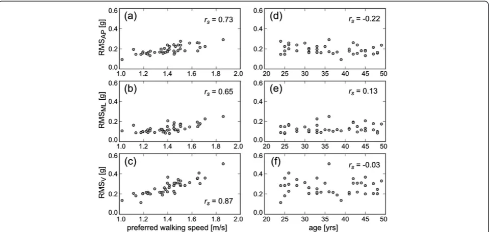
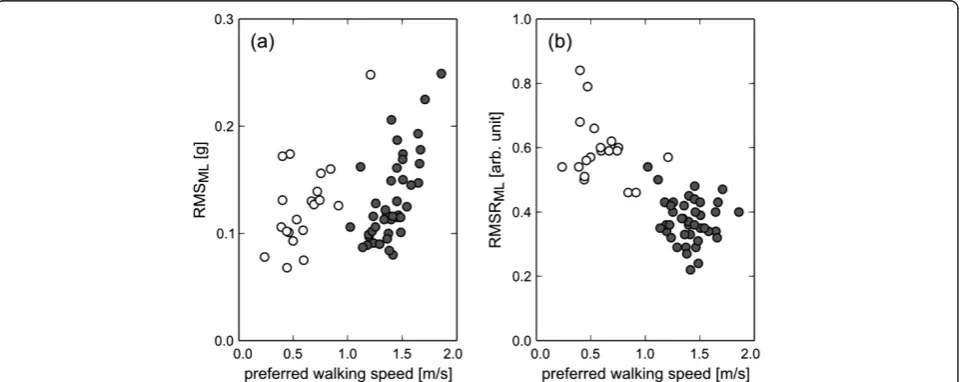
Figure 1 shows the relationship between preferred walking speed and the acceleration RMS in the healthy subjects. The range of preferred walking speed was 1.02 - 1.86 m/s. This result means that the fastest subject selected a preferred walking speed nearly twice that of the slowest subject. The acceleration RMS in all directions increased as the preferred walking speed became faster. The Spear-man's rank correlation coefficients between preferred walking speed and the RMS values in AP, ML and V direc-tions were 0.73 (p< 0.01), 0.65 (p< 0.01) and 0.87 (p< 0.01), respectively. In contrast, there were no strong relation-ships between age and the RMS values in all directions (AP: rs=−0.22, p= 0.16; ML: rs= 0.13, p= 0.42; V: rs=−0.03, p= 0.86). On the other hand, although the RMSR in AP and V directions were still influenced by pre-ferred walking speed (AP: rs=−0.40, p< 0.01; V:rs= 0.42, p< 0.01), the RMSR in the ML direction did not correlate with preferred walking speed (rs=−0.10,p= 0.54), as shown in Figure 2. In addition, there was no strong relationship between age and RMSR in the ML direction (rs= 0.22, p= 0.17). The mean ± SD of RMSR in the ML direction was 0.37 ± 0.07 in the healthy subjects.
When the six healthy subjects were instructed to walk at the five controlled speeds from 0.83 to 1.94 m/s, the RMS in the ML direction showed an exponential rela-tionship with walking speed (Figure 3). This is consistent with previous studies [3-6,11,15]. On the other hand, the RMSR in the ML direction showed a U-shaped relation-ship to walking speed. The minimum value of the RMSR was 0.45 at 1.39 m/s. After the experiment, all subjects answered that the preferred speed was 1.39 m/s among the five controlled speeds. These results indicate that the RMSR is minimal at the preferred walking speed, and in-creases with faster or slower walking.
The hemiplegic patients selected a significantly slower walking speed than the healthy subjects (p < 0.01); how-ever, there was no significant decrease in the RMS of the hemiplegic patients compared with that in healthy sub-jects (p= 0.23). These results indicate that RMS was influenced by gait abnormality. On the other hand, the
RMSR in the ML direction for the hemiplegic patients was 0.59 ± 0.09, which was significantly higher than that in the healthy subjects (p< 0.01).
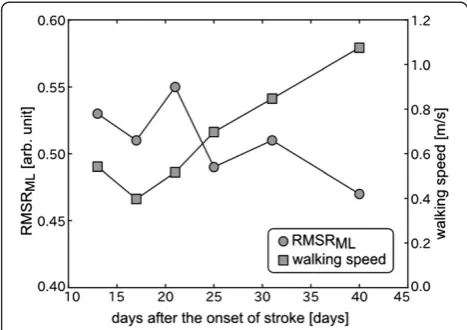
Figure 5 shows the changes in walking speed and the RMSR in the ML direction in an acute period of the elderly hemiplegic patient. The walking speed increased Figure 1The effect of preferred walking speed on RMS in AP (a), ML (b) and V directions (c), and the effect of age on RMS in AP (d), ML (e) and V directions (f).Each circle shows the RMS of each healthy subject at his preferred walking speed.rsrepresents Spearman’s rank
correlation coefficients.
Figure 2The effect of preferred walking speed on RMS ratio (RMSR) in AP (a), ML (b) and V directions (c), and the effect of age on RMSR in AP (d), ML (e) and V directions (f).Each circle shows the RMSR of each healthy subject at his preferred walking speed.rsrepresents
along with the recovery from motor disorder by physical therapy. The RMSR decreased while slightly fluctuating and reached its minimum value on the last measurement day.
Discussion
In this study, a new measure, RMSR, was proposed to evaluate gait abnormality. Our major finding is that acceleration RMSR in the ML direction represented the similar value in the healthy subjects when they walked at their own preferred speed, even if their preferred walk-ing speed were different, as shown in Figure 2(b). This finding suggests that the RMSR in the ML direction exhibits a common value in normal gait at an individual’s
preferred walking speed like walking cycle, stance and swing durations. In addition, although RMSR in AP and V directions were influenced by preferred walking speed, RMSR in the sagittal plane also adopts a common value, the-oretically. This is because RMSRML2+RMSRs2= 1, where
RMSRS¼
ffiffiffiffiffiffiffiffiffiffiffiffiffiffiffiffiffiffiffiffiffiffiffiffiffiffiffiffiffiffiffiffiffiffiffiffiffiffiffiffiffiffi RMSRAP2þRMSRV2
p
represents RMSR in the sagittal plane.
During walking, the gravitational potential energy of the center of body mass is exchanged for forward kinetic energy [21]. Thus, forward motion is mainly caused by AP and V movements in the sagittal plane. ML move-ment plays only a small role in external work [21]. The acceleration RMS in the ML direction increased in a slightly balance-impaired subject compared with that in Figure 3The effect of controlled walking speed on RMS (a) and RMSR (b) in the ML direction for the healthy subjects.
normal subjects, when they walked at the same speed [4]. Although the measurement site was slightly different from that in our study, Menz et al. reported that walking on an irregular surface resulted in significant increases in acceleration RMS at the pelvis when subjects walked on the irregular surface while maintaining their pre-ferred speed [5]. When the RMSR was calculated from the RMS values shown in their study, the RMSR in the ML direction increased under the irregular surface con-ditions. Therefore, we speculate that the RMSR in the ML direction are primarily associated with walking bal-ance. If the subjects are permitted to choose their own speed, they will select a speed that minimizes the energy cost per distance [22,23]. In addition, walking faster or slower requires more energy per step [22,23]. These fac-tors suggest that subjects optimize their body movement when they walk at their own preferred speed. These findings are similar to the relationship between RMSR and walking speed, as shown in Figure 3. The RMSR was minimized at the preferred walking speed, and in-creased at a faster or slower walking speed. As a result of such body movement optimization, it was considered that the RMSR in the sagittal plane and the ML direc-tion converged to the common values in normal gait.
To confirm that the RMSR in the ML direction can be used as a gait abnormality measure, the age-matched post-stroke hemiplegic patients were evaluated as typical subjects with gait abnormality. Compared with the healthy subjects, the RMSR in the ML direction signifi-cantly increased in the hemiplegic patients, although there was no significant difference of the RMS value in ML direction between the healthy subjects and the hemiplegic subjects. This result suggests that it is diffi-cult to assess gait abnormality by the directly use of the
RMS value in ML direction. It is known that almost all hemiplegic patients walk slower than healthy people [16]. In fact, the hemiplegic patients selected a slower walking speed than the healthy subjects in this study. Acceleration RMS was closely associated with preferred walking speed in the healthy subjects, as shown in Fig-ure 1(a)-(c). If the same relationship holds true for hemi-plegic patients, the RMS value should be small. Our results suggested that the RMS value is related to not only walking speed but also gait abnormality. Moreover, it was actually confirmed, as shown in Figure 5, that the RMSR in the ML direction decreased along with recov-ery in the acute period, although this was a single-case experiment. Changes in the RMSR and walking speed showed different patterns in terms of the time courses. As described above, the RMSR in the ML direction ex-hibited a common value when the normal subjects walked at their own preferred speed. Therefore, the in-creased RMSR in the ML direction represents gait abnor-mality. Hemiplegic stroke patients frequently present gait abnormalities because of decreased muscle strength, range of movement, abnormal muscle tone, motor coordination, sensory organization, cognition, and multisensory integra-tion [24]. The RMSR can not specify the cause of abnor-mal gait. However, the results indicate that it is possible to evaluate comprehensive gait abnormality and to provide objective evidence for treatment outcome during rehabili-tation by the RMSR in the ML direction.
Moe-Nilssen compared RMS values in the ML direc-tion between normal subjects and a subject with slight balance impairment at the same walking speed [4]. How-ever, it might be difficult to unite the walking speeds in clinical practice. For stroke patients, Mizuike et al. nor-malized RMS by the square of the walking speed and multiplied it by the average step length to exclude the effect of walking speed [16]. Although there was no de-scription of the relationship between the normalized RMS and walking speed, they suggested that the normal-ized RMS might reflect the degree of functional recovery after stroke better than raw RMS. By taking into account walking speed, the RMS value can be used for indicator of the smoothness or dynamics of the walking pattern [16]. If walking speed is included as a computation vari-able and is measured accurately, it requires other equip-ment such as a photoelectric sensor, as described in the Data collection section. The advantage of the RMSR is that this value can be calculated using only the tri-axial acceleration and provide information of gait abnormality considering the difference of individual walking speed. The RMSR takes its minimum value at the individual preferred walking speed for each subject, and increases at abnormal gait. By only involving the instruction to walk at a preferred walking speed, it is simple, but can assess abnormality of gait. The convenience of RMSR Figure 5Changes in walking speed and the RMS ratio (RMSR)
may make it useful as a screening marker of gait abnor-mality and a record of recovery from impairment for practical use.
Conclusions
In this study, we demonstrated that the acceleration RMSR in the ML direction exhibits a common value in healthy subjects under the condition that they walk at their own preferred speed. We also showed that this RMSR in the ML direction is increased in hemiplegic patients with gait abnormality under the same condi-tion. In addition, although the experiment during acute period was done for only one hemiplegic patient, the process of recovery from impairment could be quanti-fied with the RMSR in the ML direction. The RMSR can be computed only by measuring walking at a sub-ject’s own preferred speed using a tiny accelerometer, and may potentially be a quantitative measure of gait abnormality.
Competing interests
The authors declare that they have no competing interests.
Authors’contributions
MS performed the design of the study, measurements of healthy subjects, data analysis, and drafted the manuscript. TT and MY were involved in interpretation of results and critical revision of the manuscript for important intellectual content. YS participated in the measurements of healthy subjects, data analysis, and drafted the manuscript. YK was involved in interpretation of results and drafting the manuscript. HM participated in the measurements of healthy subjects and drafted the manuscript. YK and YH performed the measurements of hemiplegic patients and assisted with drafting the manuscript. TF coordinated the study and assisted with drafting the manuscript. All authors read and approved the final manuscript.
Acknowledgements
This work was partly supported by grants-in-aid from the Regional Innovation Strategy Support Program 2011, Ministry of Education, Culture, Sports, Science and Technology, Japan.
Author details
1
Faculty of Biomedical Engineering, Osaka Electro-Communication University, 18-8 Hatsucho, Neyagawa, Osaka 572-8530, Japan.2Graduate School of
Engineering, Chiba University, Chiba, Chiba, Japan.3Faculty of Biology Oriented Science & Technology, Kinki University, Kinokawa, Wakayama, Japan.
4
Sharp Corporation, Tenri, Nara, Japan.5Fujimoto General Hospital, Miyakonojyo, Miyazaki, Japan.
Received: 26 December 2012 Accepted: 17 December 2013 Published: 26 December 2013
References
1. Culhane KM, O'Connor M, Lyons D, Lyons GM:Accelerometers in rehabilitation medicine for older adults.Age Ageing2005,34:556–560. 2. Auvinet B, Berrut G, Touzard C, Moutel L, Collet N, Chaleil D, Barrey E:
Reference data for normal subjects obtained with an accelerometric device.Gait Posture2002,16:124–134.
3. Henriksen M, Lund H, Moe-Nilssen R, Bliddal H, Danneskiod-Samsøe B:
Test-retest reliability of trunk accelerometric gait analysis.Gait Posture 2004,19:288–297.
4. Moe-Nilssen R:A new method for evaluating motor control in gait under real-life environmental conditions. Part 2: Gait analysis.Clin Biomech 1998,13:328–335.
5. Menz HB, Lord SR, Fitzpatrick RC:Acceleration patterns of the head and pelvis when walking on level and irregular surfaces.Gait Posture2003,18:35–46.
6. Latt MD, Menz HB, Fung VS, Lord SR:Walking speed, cadence and step length are selected to optimize the stability of head and pelvis accelerations.Exp Brain Res2008,184:201–209.
7. Senden R, Savelberg HH, Grimm B, Heyligers IC, Meijer K: Accelerometry-based gait analysis, an additional objective approach to screen subjects at risk for falling.Gait Posture2012,36:296–300.
8. Menz HB, Lord SR, Fitzpatrick RC:Age-related differences in walking stability.Age Ageing2003,32:137–142.
9. Sekine M, Akay M, Tamura T, Higashi Y, Fujimoto T:Investigating body motion patterns in patients with Parkinson's disease using matching pursuit algorithm.Med Biol Eng Comput2004,42:30–36.
10. Akay M, Sekine M, Tamura T, Higashi Y, Fujimoto T:Unconstrained monitoring of body motion during walking.IEEE Eng Med Biol Mag2003,
22:104–109.
11. Kavanagh JJ:Lower trunk motion and speed-dependence during walking.
J Neuroeng Rehabil2009,6:9.
12. Lamoth CJ, van Deudekom FJ, van Campen JP, Appels BA, de Vries OJ, Pijnappels M:Gait stability and variability measures show effects of impaired cognition and dual tasking in frail people.J Neuroeng Rehabil 2011,8:2.
13. Sekine M, Akay M, Tamura T, Higashi Y, Fujimoto T:Fractal dynamics of body motion in patients with Parkinson’s disease.J Neural Eng2004,
1:8–15.
14. Akay M, Sekine M, Tamura T, Higashi Y, Fujimoto T:Fractal dynamics of body motion in post-stroke hemiplegic patients during walking.J Neural Eng2004,1:111–116.
15. Helbostad JL, Moe-Nilssen R:The effect of gait speed on lateral balance control during walking in healthy elderly.Gait Posture2003,18:27–36. 16. Mizuike C, Ohgi S, Morita S:Analysis of stroke patient walking dynamics
using a tri-axial accelerometer.Gait Posture2009,30:60–64.
17. Mazzà C, Iosa M, Picerno P, Cappozzo A:Gender differences in the control of the upper body accelerations during level walking.Gait Posture2009,
29:300–303.
18. Murray MP, Kory RC, Clarkson BH:Walking patterns in healthy old men.
J Gerontol1969,24:169–178.
19. Stolze H, Klebe S, Petersen G, Raethjen J, Wenzelburger R, Witt K, Deuschl G:
Typical features of cerebellar ataxic gait.J Neurol Neurosurg Psychiatry 2002,73:310–312.
20. Moe-Nilssen R:A new method for evaluating motor control in gait under real-life environmental conditions. Part 1: the instrument.Clin Biomech 1998,13:320–327.
21. Cavagna GA, Willems PA, Heglund NC:The role of gravity in human walking: pendular energy exchange, external work and optimal speed.
J Physiol2000,528:657–668.
22. Ralston HJ:Energy-speed relation and optimal speed during level walking.Int Z Angew Physiol1958,17:277–283.
23. Inman VT:Human locomotion.Can Med Assoc J1966,94:1047–1054. 24. de Oliveira CB, de Medeiros IR, Frota NA, Greters ME, Conforto AB:Balance
control in hemiparetic stroke patients: main tools for evaluation.
J Rehabil Res Dev2008,45:1215–1226.
doi:10.1186/1743-0003-10-118
Cite this article as:Sekineet al.:A gait abnormality measure based on root mean square of trunk acceleration.Journal of NeuroEngineering and Rehabilitation201310:118.
Submit your next manuscript to BioMed Central and take full advantage of:
• Convenient online submission
• Thorough peer review
• No space constraints or color figure charges
• Immediate publication on acceptance
• Inclusion in PubMed, CAS, Scopus and Google Scholar
• Research which is freely available for redistribution