Open Access
Review
Protein expression profiling arrays: tools for the multiplexed
high-throughput analysis of proteins
Jens R Sydor
2and Steffen Nock*
1Address: 1Promab Biotechnologies Inc., 1300 Bancroft Ave, San Leandro, CA 94577, USA and 2Infinity Pharmaceuticals, 780 Memorial Drive,
Cambridge, MA 02139, USA
Email: Jens R Sydor - [email protected]; Steffen Nock* - [email protected] * Corresponding author
Abstract
The completion of the human genome sequence has led to a rapid increase in genetic information. The invention of DNA microarrays, which allow for the parallel measurement of thousands of genes on the level of mRNA, has enabled scientists to take a more global view of biological systems. Protein microarrays have a big potential to increase the throughput of proteomic research. Microarrays of antibodies can simultaneously measure the concentration of a multitude of target proteins in a very short period of time. The ability of protein microarrays to increase the quantity of data points in small biological samples on the protein level will have a major impact on basic biological research as well as on the discovery of new drug targets and diagnostic markers. This review highlights the current status of protein expression profiling arrays, their development, applications and limitations.
Introduction
The analysis of the entire set of proteins of a biological sys-tem, commonly called proteomics, represents a research area that has emerged in the past decade as a largely tech-nology-driven field [1–4]. Techniques like mass spectrom-etry in combination with separation tools such as two-dimensional gel electrophoresis or multitwo-dimensional liq-uid chromatography, allow for the parallel analysis of abundances of dozens to hundreds of proteins [5–7]. These techniques, however, are very labor intensive and require a significant amount of biological material. In par-ticular, two-dimensional gel electrophoresis lacks the sen-sitivity to detect low abundance proteins.
These disadvantages of existing proteomics technologies have driven the development of novel miniaturized tools for the investigation of proteomes. An emerging technol-ogy in this field is the protein microarray [8–12].
Depend-ing on the configuration, these arrays can measure protein expression levels, protein interactions, protein-small molecule interactions as well as enzymatic activities. Protein expression profiling arrays are the most advanced in their development and therefore the major focus of this review.
The concept of protein expression profiling arrays was inspired by DNA microarrays, which enable the measure-ment of mRNA expression level of thousands of genes in a single experiment [13]. DNA microarrays have proven to be very powerful tools for the multiplexed comparative analysis of gene expression and led to important insights into gene expression patterns associated with disease states [14–20]. The possibility of performing similar anal-yses at the level of proteins – the functional products of almost all genes – is therefore very attractive.
Published: 10 June 2003
Proteome Science 2003, 1:3
Received: 31 May 2003 Accepted: 10 June 2003
This article is available from: http://www.Proteomesci.com/content/1/1/3
There are, however, several reasons why DNA microarray technology cannot readily be adapted towards the devel-opment of protein microarrays. First, design and synthesis of gene-specific capture probes is straightforward since it is based on simple base-pairing rules and standard solid phase phosphoramidite chemistry, respectively. In con-trast, the development of capture agents for protein arrays is far more complicated and requires significantly more time for development. Currently the preferred capture agents for protein expression profiling arrays are antibod-ies or antibody fragments, which have a very long and costly development time (see below). Furthermore pro-tein expression levels span a huge range (up to 8 orders of magnitude). To avoid multiple measurements of the same sample at different dilutions, protein capture agents with different affinities have to be developed to address such dramatic differences in expression level.
Second, an appropriate surface attachment strategy has to be implemented to immobilize the protein capture agents onto the array while retaining their binding activity. Fur-thermore, during the dispensing and immobilization process, the proteins must remain hydrated to assure the integrity of their three-dimensional structures, an issue not relevant to the production of DNA arrays.
Third, especially for high density protein expression pro-filing arrays, novel detection schemes with adequate sen-sitivity are required to monitor the specific binding of proteins by the immobilized capture reagents on the microarray.
This review will summarize these issues and how they are addressed. Despite these limitations, impressive advances have been made towards the development of protein expression profiling arrays and several publications have been appeared over the last years showing the use and power of this technology.
Assay formats
The simplest protein array format consists of a large number of protein capture reagents bound to defined spots on a planar support material. This array is then exposed to a complex protein sample. The binding of the specific analyte proteins to the individual spots can then be monitored using different approaches (Figure 1). In cases where the analytes have been pre-labeled with a flu-orescent dye, the binding can be monitored directly using a fluorescence scanner. A major limitation of this assay configuration lies in the often disappointing sensitivity, which prohibits the measurement of low abundance pro-teins. More often, however, the classical antibody sand-wich type format is used, in which two protein binding reagents simultaneously bind to the same antigen: one antibody is immobilized onto the surface, and the other
one is fluorescently labeled or conjugated to an enzyme that can produce a fluorescent, luminescent or colored product when supplied with the appropriate substrate. The disadvantage of this sandwich assay format lies in the fact that two highly specific protein capture agents must be developed. The assay itself, however, is more reliable because direct sample labeling, which is not very consist-ent between samples, is not required. Furthermore this assay setup has been successfully used to monitor protein expression levels at physiologically relevant concentra-tions [21–23].
Capture agents for protein profiling microarrays
Current estimations assume that the human genome encodes about 30,000 – 40,000 genes. Due to splice vari-ants on the mRNA level and a variety of post-translational modifications, the number of functionally distinct pro-teins is significantly higher, probably approaching one million. The development of protein microarrays for highly multiplexed protein profiling, similar to the multi-plexing capabilities of DNA microarrays, would therefore require a large number of capture agents. The develop-ment of these capture agents is currently the most chal-lenging bottleneck in protein microarray research.
Monoclonal antibodies or their antigen-binding frag-ments are currently the preferred choice for capture agents due to their high specificity, affinity and stability. They have been used in a variety of classical single analyte pro-tein profiling assays such as enzyme-linked immunosorb-ent assays (ELISA) since the sevimmunosorb-enties. The long development times and labor-intensive nature of the process is, however, a major disadvantage [24,25].
To overcome this problem, different 'display' methods are currently being used and industrialized for the high-throughput development of protein capture agents. Phage-display libraries of antibody fragments offer the potential for antibody production at proteomic scales. These libraries can be used to isolate high-affinity binding agents against protein targets in a significantly shorter time frame than it is possible with immunization-based methods [26–30]. Furthermore, such methods can be used to create binding agents against proteins that are toxic, highly conserved or murine in origin, which are problematic for tradition mouse monoclonal antibody generation.
the successful isolation of high affinity capture agents. Only very complex libraries with many billions of individ-ual clones are suited for the isolation of high qindivid-uality bind-ing reagents. Several companies have developed highly complex libraries over the last years including Dyax, Cam-bridge Antibody Technology and Morphosys. The con-struction of high complexity phage libraries does, however, require a great deal of labor and is limited by the transformation efficiency of bacteria.
Due to the fact that the size of capture agent libraries is one of the major limitations for the development of high-affinity binding agents, additional in vitro evolution
meth-ods have emerged that circumvent the size-limitation of phage libraries. Ribosome display and mRNA display are completely in vitro methods that rely on physically linking the library proteins to their encoding mRNA sequences. Such methods have successfully been used to select high-affinity binding reagents to target proteins [31].
These library-based methods provide capture agents based on antibody fragments or other protein scaffolds. One inherent limitation of protein-based capture agents is their lack of stability during array dispensing processes and subsequent array storage. Several groups have there-fore taken a completely different approach to develop
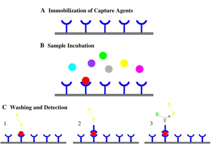
Figure 1
Basic principle of a protein expression profiling array and different detection schemes. (A) Capture agents with different ana-lyte specificity are immobilized on a surface. (B) Incubation with protein samples leads to specific capture of target proteins in a concentration dependent manner. (C) After washing, the specifically bound target protein can be visualized by a variety of detection schemes, such as (1) direct fluorescence labeling of the analyte with a fluorophore (F) (labeled prior to the experi-ment), (2) detection of the target protein by a fluorescently-labeled detection antibody or (3) an enzyme-conjugated detection antibody, allowing an ELISA-based detection with a fluorescent, chemiluminescent or colorimetric readout (S = substrate, P = product).
F
F
P S
E
A
Immobilization of Capture Agents
B
Sample Incubation
C
Washing and Detection
high affinity protein capture reagents for protein biochips. Aptamers are single stranded RNA or DNA molecules orig-inating from in vitro selection experiments (termed SELEX: systematic evolution of ligands by exponential enrich-ment) with high affinities to proteins [32,33]. Aptamers against a number of proteins have been successfully selected from libraries with a complexity of over 1014
mol-ecules (for review see [34]). A further development in aptamer technologies are so called photoaptamers. These molecules have an additional attribute that enhances their utility as protein capture reagents. They carry the photoac-tivatible crosslinking group 5'-bromodeoxyuridine, which, when activated by UV light, can cause covalent crosslinking with bound target proteins [35]. The photo-crosslinking event provides a second dimension of specif-icity similar to the binding of a secondary detection anti-body in a sandwich immunoassay. In a study by Golden et al. photoaptamers were used to crosslink low picomo-lar concentrations of fibroblast growth factor, in the pres-ence of serum, with very high specificity [36].
Regardless of the type of capture agent, high specificity and affinity are crucial. Due to the multiplexed nature of microarray assays, high specificity is absolutely required to avoid cross-reactivity. Extensive cross-reactivity scans between binding agents to be used together on a microar-ray must be performed as part of the screening process to identify suitable reagents. Another important criterion is the affinity of the capture agents. Protein microarrays are especially attractive for protein expression profiling of low abundance proteins that cannot be visualized by 2D-PAGE techniques. The capture agents should have affini-ties not more than 2 orders of magnitude higher than the biological concentrations of the target proteins for reliable detection. Since some important proteins are present at low picomolar concentrations in serum, for example, antibodies with sub-nanomolar affinities should be used.
In order to create these protein capture reagents, a target protein must be synthesized and purified. Several groups have started to develop methods for rapid parallel expres-sion and purification of proteins. The most widely used
system is based on expression in E. coli, but a large
number of human proteins expressed in this system are misfolded and insoluble. These misfolded proteins are often not useful for the development of antibodies that recognize the native form of a protein with high affinity. The refolding of proteins from inclusion bodies is difficult to perform in high throughput. For most human proteins, it will therefore be therefore necessary to access alternative expression systems that rely on mammalian or insect cells. Automation of some of these eukaryotic expression sys-tems is currently being explored [37].
Immobilization of capture agents for protein microarrays: where chemistry meets biology
The nature of the surface substrate and attachment strat-egy is one of the major factors for determining the quality of data obtained during protein microarray experiments. For optimal sensitivity and reproducibility, the activity of the immobilized capture agent has to be retained and non-specific binding of proteins to the surface must be minimized.
A wide variety of surface substrates and attachment chem-istries have been evaluated for the immobilization of cap-ture agents on protein microarrays. They fall into two basic categories. The simplest way to immobilize proteins on a solid support relies on non-covalent interactions. These immobilizations can be either based on hydropho-bic or van der Waals interactions, hydrogen bonding or electrostatic forces. Examples of electrostatic immobiliza-tion include the use of materials such as nitrocellulose and poly-lysine- or aminopropyl silane-coated glass slides [38]. Protein microarrays were also fabricated by means of physical adsorption onto plastic surfaces of 96-well plates [21]. A big advantage of these immobilization concepts is their ease of use. Usually no protein modification is needed prior to printing onto the surface. The disadvan-tage is that proteins often get denatured on these fairly undefined surfaces due multiple uncontrolled interac-tions between the protein and the surface material.
Physical adsorption of proteins onto surfaces can also lead to problems with protein desorption during the assay, which can lead to signal loss. It is therefore more desirable to attach the protein capture molecule cova-lently and in a controlled way onto the surface. An exam-ple of covalent attachment of proteins to the surface has been described by MacBeath and Schreiber [39]. However in this case the immobilization was random, which can lead to a decreased sensitivity compared to an oriented immobilization. In the ideal setup, a single covalent bond would mediate the attachment. Due to the very high affin-ity of streptavidin to biotin, the immobilization of bioti-nylated proteins onto streptavidin surfaces can be considered quasi covalent. Using this strategy, Peluso et al. [40] were able to demonstrate that an oriented single site attachment of an antibody fragment leads to an increase in sensitivity over random attachment in a micro-array assay.
substrates, for example, require different blocking strate-gies to gain optimal data quality. MacBeath and Schreiber used BSA-coated slides to reduce non-specific binding [39]. A more sophisticated approach was taken by Ruiz-Taylor et al. [41,42]. They engineered surfaces to avoid non-specific protein adsorption using poly (ethylene gly-col) derivates as coatings. Protein microarrays based on this method of attachment of capture molecules onto oth-erwise protein-resistant surfaces have shown to be of very high quality [40].
Detection strategies for multiplexed protein microarray applications
The preferred method for detecting binding events on a protein microarray relies on fluorescence. As described above, there are two ways to incorporate fluorophores into an assay: (1) direct fluorescent labeling of the protein sample, and (2) sandwich immunoassays with labeled detection antibodies. The use of a miniaturized sandwich assay also allows for incorporation of enzymes which then can be used for signal amplification. For example Huang [43] has shown the simultaneous detection of dif-ferent cytokines from conditioned media and patient sera using an array-based enzyme-linked immunosorbent assay in combination with enhanced chemiluminescence (ECL).
The choice of the detection strategy is partially determined by the application. A direct labeling of the protein sample can be applied to the analysis of cell lysates or purified protein samples. Miller et al. used a direct Cy3/Cy5 labe-ling strategy to perform differential profilabe-ling of prostate cancer biomarkers in serum samples [44]. They were able to identify five proteins that had significantly different expression levels between prostate cancer samples and normal controls. For quantitative studies with a limited number of specificities on a chip, however, a sandwich immunoassay format is preferred. Medium density arrays of antibodies against cytokines and other medically important proteins have been developed [21–23,45].
The fact that multiplexed protein microarray assays are performed on a flat surface adds certain restrictions to an ELISA-based assay setup. The resulting enzyme product must be either an insoluble precipitate or attached to a certain component of the microarray spot. Wiese et al. [23] performed a cytokine profiling microarray assay with an alkaline phosphatase-conjugated detection antibody, which generated a fluorescent precipitate. Another highly sensitive ELISA-based detection strategy has been described which uses rolling circle amplification (RCA) as a detection strategy [46,47]. This on-chip signal amplifica-tion strategy was used to perform very sensitive assays for highly multiplexed cytokine profiling and detection of allergen-specific IgE's in serum samples [48,49]. Further
improvement in sensitivity involves the application of flu-orescent labels in combination with waveguide technol-ogy. Rowe-Taitt et al. [50] have applied this technology to detect clinical analytes and biohazardous agents in com-plex samples at physiologically relevant concentrations. Thin film waveguides generated from a high-refractive
material such as Ta2O5 have been successfully used by
Duveneck et al. [51].
Protein microarrays: Beyond protein profiling
Although the major use of protein microarrays is currently in the field of expression profiling, several applications of functional arrays and protein-protein interaction arrays have been described. Zhu et al. [52] cloned and arrayed into nanowells 119 protein kinases from yeast. They then assayed their activity using 17 different substrates in the presence of radiolabeled ATP. Following the incorpora-tion of radioactive phosphate, they were able to identify the substrate preferences of most of these kinases. The same later undertook the heroic effort to clone and express nearly all of the 5,800 yeast open reading frames [53]. An array of these yeast proteins was created and probed for binding to calmodulin and certain lipids. They were able to identify 6 known calmodulin-binding pro-teins and several lipid-binding propro-teins. Once this approach is transferred to human proteins, a variety of novel protein-protein as well as protein-small molecule interactions will be discovered.
Another emerging microarray format consists of peptide arrays for the profiling of protein activity. An array of pep-tides on a gold surface was developed to monitor c-Src kinase activity. Phosphorylation of the immobilized pep-tide substrates was shown using radioactivity, fluores-cence or surface plasmon resonance as detection [54]. Another group has demonstrated the use of fluorescently labeled phospho-specific antibodies to detect the phos-phorylation event on immobilized peptide substrates [55].
Small-molecule microarrays have also been developed to detect the binding of proteins to an array of immobilized compounds [56,57]. These new microarray platforms will prove invaluable to basic biological research and have the potential to accelerate the pace of discovery of drug targets as well as lead compounds.
Conclusions
This technology platform, however, is still not at a stage where it could be compared with the commercial success of DNA microarrays. A few companies, such as Zyomyx, Ciphergen Pierce, Zeptosens, and BD Clontech have com-mercialized protein microarrays, but their applicability and competitive advantage over other, more macroscopic protein profiling platforms still needs to be proven.
Acknowledgement
We would like to thank David Wilson for the critical reading of the manuscript.
References
1. Patterson SD and Aebersold RH: Proteomics: the first decade and beyondNat Genet 2003, 33 Suppl:311-323.
2. Chambers G, Lawrie L, Cash P and Murray GI: Proteomics: a new approach to the study of diseaseJ Pathol 2000, 192:280-288. 3. Dutt MJ and Lee KH: Proteomic analysisCurr Opin Biotechnol 2000,
11:176-179.
4. Pandey A and Mann M: Proteomics to study genes and genomes
Nature 2000, 405:837-846.
5. Aebersold R and Mann M: Mass spectrometry-based proteomicsNature 2003, 422:198-207.
6. Washburn MP, Wolters D and Yates JR,III: Large-scale analysis of the yeast proteome by multidimensional protein identifica-tion technologyNat Biotechnol 2001, 19:242-247.
7. Wolters DA, Washburn MP and Yates JR,III: An automated multi-dimensional protein identification technology for shotgun proteomicsAnal Chem 2001, 73:5683-5690.
8. Zhu H and Snyder M: Protein arrays and microarraysCurr Opin Chem Biol 2001, 5:40-45.
9. Kodadek T: Protein microarrays: prospects and problems
Chem Biol 2001, 8:105-15.
10. Templin MF, Stoll D, Schrenk M, Traub PC, Vohringer CF and Joos TO: Protein microarray technology Trends Biotechnol 2002,
20:160-166.
11. Wilson DS and Nock S: Recent developments in protein micro-array technologyAngew Chem Int Ed Engl 2003, 42:494-500. 12. Wilson DS and Nock S: Functional Protein MicroarraysCurr Opin
Chem Biol 2002, 6:81-85.
13. Schena M, Shalon D, Davis RW and Brown PO: Quantitative mon-itoring of gene expression patterns with a complementary DNA microarrayScience 1995, 270:467-470.
14. Golub TR, Slonim DK, Tamayo P, Huard C, Gaasenbeek M, Mesirov JP, Coller H, Loh ML, Downing JR, Caligiuri MA, Bloomfield CD and Lander ES: Molecular Classification of Cancer: Class Discovery and Class Prediction by Gene Expression MonitoringScience
1999, 286:531-537.
15. Alizadeh AA, Eisen MB, Davis RE, Ma C, Lossos IS, Rosenwald A, Boldrick JC, Sabet H, Tran T, Yu X, Powell JI, Yang L, Marti GE, Moore T, Hudson J.,Jr., Lu L, Lewis DB, Tibshirani R, Sherlock G, Chan WC, Greiner TC, Weisenburger DD, Armitage JO, Warnke R, Staudt LM and .: Distinct types of diffuse large B-cell lymphoma identi-fied by gene expression profilingNature 2000, 403:503-511. 16. Perou CM, Sorlie T, Eisen MB, van de Rijn M., Jeffrey SS, Rees CA,
Pol-lack JR, Ross DT, Johnsen H, Akslen LA, Fluge O, Pergamenschikov A, Williams C, Zhu SX, Lonning PE, Borresen-Dale AL, Brown PO and Botstein D: Molecular portraits of human breast tumours
Nature 2000, 406:747-752.
17. Sorlie T, Perou CM, Tibshirani R, Aas T, Geisler S, Johnsen H, Hastie T, Eisen MB, van de Rijn M., Jeffrey SS, Thorsen T, Quist H, Matese JC, Brown PO, Botstein D, Eystein Lonning P. and Borresen-Dale AL:
Gene expression patterns of breast carcinomas distinguish tumor subclasses with clinical implicationsProc Natl Acad Sci
2001, 98:10869-10874.
18. Dhanasekaran SM, Barrette TR, Ghosh D, Shah R, Varambally S, Kura-chi K, Pienta KJ, Rubin MA and Chinnaiyan AM: Delineation of prognostic biomarkers in prostate cancer Nature 2001,
412:822-826.
19. Nielsen TO, West RB, Linn SC, Alter O, Knowling MA, O'Connell JX, Zhu S, Fero M, Sherlock G, Pollack JR, Brown PO, Botstein D and van
de Rijn M.: Molecular characterisation of soft tissue tumours: a gene expression studyLancet 2002, 359:1301-1307.
20. Pomeroy SL, Tamayo P, Gaasenbeek M, Sturla LM, Angelo M, McLaughlin ME, Kim JY, Goumnerova LC, Black PM, Lau C, Allen JC, Zagzag D, Olson JM, Curran T, Wetmore C, Biegel JA, Poggio T, Mukherjee S, Rifkin R, Califano A, Stolovitzky G, Louis DN, Mesirov JP, Lander ES and Golub TR: Prediction of central nervous system embryonal tumour outcome based on gene expressionNature 2002, 415:436-442.
21. Moody MD, Van Ardel SW, Orencole SF and Burns C: Array-based ELISAs for high-throughput analysis of human cytokines Bio-techniques 2001, 31:186-194.
22. Mendoza LG, McQuary P, Mongan A, Gangadharan R, Brignac S and Eggers M: High-throughput microarray-based enzyme-linked immunosorbent assay (ELISA)Biotechniques 1999, 27:778-788. 23. Wiese R, Belosludtsev Y, Powdrill T, Thompson P and Hogan M:
Simultaneous multianalyte ELISA performed on a microar-ray platformClin Chem 2001, 47:1451-1457.
24. Liddell JE and Cryer A: A practical guide to monoclonal antibodies Chich-ester, John Wiley & Sons; 1991.
25. Goding JW: Monoclonal Antibodies: Principles and Practice 3rdth edition.
San Diego, Academic Press; 1996.
26. Sheets MD, Amersdorfer P, Finnern R, Sargent P, Lindquist E, Schier R, Hemingsen G, Wong C, Gerhart JC, Marks JD and Lindqvist E: Effi-cient construction of a large nonimmune phage antibody library: the production of high-affinity human single-chain antibodies to protein antigens [published erratum appears in Proc Natl Acad Sci U S A 1999 Jan 19;96(2):795]Proc Natl Acad Sci U S A 1998, 95:6157-6162.
27. de Haard HJ, van Neer N, Reurs A, Hufton SE, Roovers RC, Hen-derikx P, de Bru, Arends JW and Hoogenboom HR: A Large Non-immunized Human Fab Fragment Phage Library That Per-mits Rapid Isolation and Kinetic Analysis of High Affinity AntibodiesJ Biol Chem 1999, 274:18218-18230.
28. Knappik A, Ge L, Honegger A, Pack P, Fischer M, Wellnhofer G, Hoess A, Wolle J, Pluckthun A and Virnekas B: Fully synthetic human combinatorial antibody libraries (HuCAL) based on modular consensus frameworks and CDRs randomized with trinucleotidesJ Mol Biol 2000, 296:57-86.
29. Vaughan TJ, Williams AJ, Pritchard K, Osbourn JK, Pope AR, Earn-shaw JC, McCafferty J, Hodits RA, Wilton J and Johnson KS: Human antibodies with sub-nanomolar affinities isolated from a large non-immunized phage display libraryNat Biotechnol 1996,
14:309-314.
30. Wittrup KD: Protein engineering by cell-surface displayCurr Opin Biotechnol 2001, 12:395-399.
31. Wilson DS, Keefe AD and Szostak JW: The use of mRNA display to select high-affinity protein-binding peptidesProc Natl Acad Sci U S A 2001, 98:3750-3755.
32. Brody EN and Gold L: Aptamers as therapeutic and diagnostic agentsJ Biotechnol 2000, 74:5-13.
33. Robertson MP and Ellington AD: In vitro selection of nucleopro-tein enzymesNat Biotechnol 2001, 19:650-655.
34. Jayasena SD: Aptamers: an emerging class of molecules that rival antibodies in diagnosticsClin Chem 1999, 45:1628-1650. 35. Petach H and Gold L: Dimensionality is the issue: use of
pho-toaptamers in protein microarraysCurr Opin Biotechnol 2002,
13:309-314.
36. Golden MC, Collins BD, Willis MC and Koch TH: Diagnostic potential of PhotoSELEX-evolved ssDNA aptamers J Biotechnol 2000, 81:167-178.
37. Gilbert M and Albala JS: Accelerating code to function: sizing up the protein production lineCurr Opin Chem Biol 2002, 6:102-105. 38. Haab BB, Dunham MJ and Brown PO: Protein microarrays for highly parallel detection and quantitation of specific proteins and antibodies in complex solutions Genome Biol 2001,
2:RESEARCH0004.
39. MacBeath G and Schreiber SL: Printing proteins as microarrays for high-throughput function determination Science 2000,
289:1760-1763.
Publish with BioMed Central and every scientist can read your work free of charge "BioMed Central will be the most significant development for disseminating the results of biomedical researc h in our lifetime."
Sir Paul Nurse, Cancer Research UK
Your research papers will be:
available free of charge to the entire biomedical community
peer reviewed and published immediately upon acceptance
cited in PubMed and archived on PubMed Central
yours — you keep the copyright
Submit your manuscript here:
http://www.biomedcentral.com/info/publishing_adv.asp
BioMedcentral 41. Ruiz-Taylor LA, Martin TL and Wagner P: X-ray photoelectron
spectroscopy and radiometry studies of biotin-derivatized poly(L-lysine)-grafted-poly(ethylene glycol) monolayers on metal oxidesVolume 17. Langmuir; 20017313-7322.
42. Ruiz-Taylor LA, Martin TL, Zaugg FG, Witte K, Indermuhle P, Nock S and Wagner P: Monolayers of derivatized poly(L-lysine)-grafted poly(ethylene glycol) on metal oxides as a class of biomolecular interfacesProc Natl Acad Sci U S A 2001, 98:852-857. 43. Huang RP: Simultaneous detection of multiple proteins with an array-based enzyme- linked immunosorbent assay (ELISA) and enhanced chemiluminescence (ECL)Clin Chem Lab Med 2001, 39:209-214.
44. Miller JC, Zhou H, Kwekel J, Cavallo R, Burke J, Butler EB, Teh BS and Haab BB: Antibody microarray profiling of human prostate cancer sera: Antibody screening and identification of poten-tial biomarkersProteomics 2003, 3:56-63.
45. Tam SW, Wiese R, Lee S, Gilmore J and Kumble KD: Simultaneous analysis of eight human Th1/Th2 cytokines using microarraysJ Immunol Methods 2002, 261:157-165.
46. Lizardi PM, Huang X, Zhu Z, Bray-Ward P, Thomas DC and Ward DC: Mutation detection and single-molecule counting using isothermal rolling-circle amplificationNat Genet 1998, 19: 225-232.
47. Schweitzer B, Wiltshire S, Lambert J, O'Malley S, Kukanskis K, Zhu Z, Kingsmore SF, Lizardi PM and Ward DC: Immunoassays with roll-ing circle DNA amplification: a versatile platform for ultra-sensitive antigen detection Proc Natl Acad Sci U S A 2000,
97:10113-10119.
48. Schweitzer B, Roberts S, Grimwade B, Shao W, Wang M, Fu Q, Shu Q, Laroche I, Zhou Z, Tchernev VT, Christiansen J, Velleca M and Kingsmore SF: Multiplexed protein profiling on microarrays by rolling-circle amplificationNat Biotechnol 2002, 20:359-365. 49. Wiltshire S, O'Malley S, Lambert J, Kukanskis K, Edgar D, Kingsmore
SF and Schweitzer B: Detection of multiple allergen-specific IgEs on microarrays by immunoassay with rolling circle amplificationClin Chem 2000, 46:1990-1993.
50. Rowe-Taitt CA, Hazzard JW, Hoffmann KE, Cras JJ, Golden JP and Ligler FS: Simultaneous detection of six biohazardous agents using a planar waveguide array biosensorBiosens Bioelectron
2000, 15:579-589.
51. Duveneck GL, Pawlak M and Neuschaefer D: Novel bioaffinity sen-sors for trace analysis based on luminescence excitation by planar waveguidesSens Actuators B 1997, B38:88-95.
52. Zhu H, Klemic JF, Chang S, Bertone P, Casamayor A, Klemic KG, Smith D, Gerstein M, Reed MA and Snyder M: Analysis of yeast protein kinases using protein chipsNat Genet 2000, 26:283-289. 53. Zhu H, Bilgin M, Bangham R, Hall D, Casamayor A, Bertone P, Lan N, Jansen R, Bidlingmaier S, Houfek T, Mitchell T, Miller P, Dean RA, Gerstein M and Snyder M: Global Analysis of Protein Activities Using Proteome ChipsScience 2001, 293:2101-2105.
54. Houseman BT, Huh JH, Kron SJ and Mrksich M: Peptide chips for the quantitative evaluation of protein kinase activity Nat Biotechnol 2002, 20:270-274.
55. Lesaicherre ML, Uttamchandani M, Chen GY and Yao SQ: Antibody-based fluorescence detection of kinase activity on a peptide arrayBioorg Med Chem Lett 2002, 12:2085-2088.
56. MacBeath G, Koehler AN and Schreiber SL: Printing small mole-cules as microarrays and detecting protein-ligand interac-tions en masseJ Am Chem Soc 1999, 121:7967-7968.
57. Kuruvilla FG, Shamji AF, Sternson SM, Hergenrother PJ and Schreiber SL: Dissecting glucose signalling with diversity-oriented syn-thesis and small-molecule microarraysNature 2002, 416: 653-657.
58. Ekins RP and Chu FW: Multianalyte microspot immunoassay--microanalytical "compact disk" of the futureClin Chem 1991,