Keel petal incision: a simple and efficient method
for genetic crossing in
Medicago truncatula
Veerappan
et al.
M E T H O D O L O G Y
Open Access
Keel petal incision: a simple and efficient method
for genetic crossing in
Medicago truncatula
Vijaykumar Veerappan
1, Khem Kadel
1, Naudin Alexis
1, Ashley Scott
1, Igor Kryvoruchko
2, Senjuti Sinharoy
2,
Mark Taylor
2, Michael Udvardi
2and Rebecca Dickstein
1*Abstract
Background:Genetic crossing is an essential tool in both forward and reverse genetic approaches to understand the biological functions of genes. ForMedicago truncatula(barrel medic) various crossing techniques have been used which differ in the methods used to dissect the female parent’s unopened flower bud to remove immature anthers for prevention of self-pollination. Previously described methods including front, side or back incision methods may damage the flower bud, impeding successful fertilization and/or seed development because they may allow pollen to dislodge and floral organs to desiccate after crossing, all of which diminish the success rates of crossing.
Results:We report the keel petal incision method for genetic crossing inM. truncatulaecotype R108 and demonstrate successful crosses with two otherM. truncatulaecotypes, A17 and A20. In the method presented here, an incision is made along the central line of the keel petal from the bottom 1/3rd of the female parent’s flower bud to its distal end. This allows easy removal of anthers from the flower bud and access for cross-pollination. After pollination, the stigma and the deposited pollen from the male donor are covered by the keel petal, wing petals and standard petal, forming a natural pouch. The pouch prevents dislodging of deposited pollen from the stigma and protects the internal floral organs from drying out, without using cling-film or water-containing chambers to maintain a humid environment. The keel petal incision method showed an approximate 80% success rate in theM. truncatulaR108 ecotype and also in other ecotypes including Jemalong A17 and A20.
Conclusions:Our keel petal incision protocol shows marked improvement over existing methods with respect to the ease of crossing and the percentage of successful crosses. Developed for theM. truncatulaR108 ecotype, the protocol has been demonstrated with A17 and A20 ecotypes and is expected to work with other ecotypes. Investigators of varying experience have achieved genetic crosses inM. truncatulausing this method.
Keywords:Legume, Genetic crossing, Barrel medic,Medicago truncatula, Artificial hybridization, Keel petal
Background
Medicago truncatula (barrel medic) is an important model legume species extensively used to study symbiotic interactions with soil rhizobia and arbuscular mycorrhizal fungi leading to symbiotic nitrogen fixation and mycorrhi-zation respectively [1-3]. It has also found use as a model for studies on secondary metabolism [4-6], plant patho-gens [7,8], leaf development [9-11] and other processes. M. truncatula, in the Galegoid clade of the Papilionoideae legume subfamily, is closely related to economically im-portant crops also in the Galegoid clade including alfalfa
(M. sativa), pea (Pisum sativum) and lentil (Lens culinaris) as well as crop legumes in the Phaseoloid clade including soybean (Glycine max) and common bean (Phaseolus vulgaris) [12]. In the past two decades, extensive genomic and genetic resources have been developed forM. trunca-tula, making it an elite legume model amenable for func-tional genomics as well as for genetic studies to identify key regulators in important processes. These resources include a global gene expression atlas [13] and a nearly complete genome sequence [14]. M. truncatula’s genome may contain up to 50,000 protein coding genes, most of unknown biological function [14]. To facilitate genetic studies in M. truncatula, several populations of mutants have been developed [15], including those treated with ethyl methane sulfonate (EMS) [16], fast neutron bombardment
* Correspondence:[email protected] 1
Department of Biological Sciences, University of North Texas, 1155 Union Circle #305220, Denton, Texas 76203, USA
Full list of author information is available at the end of the article
PLANT METHODS
© 2014 Veerappan et al.; licensee BioMed Central Ltd. This is an Open Access article distributed under the terms of the Creative Commons Attribution License (http://creativecommons.org/licenses/by/2.0), which permits unrestricted use, distribution, and reproduction in any medium, provided the original work is properly credited. The Creative Commons Public Domain Dedication waiver (http://creativecommons.org/publicdomain/zero/1.0/) applies to the data made available in this article, unless otherwise stated.
(FNB) [17-19] and a near saturation insertion mutant popu-lation carrying the tobaccoTnt1retrotransposon [20-23].
An efficient technique for genetic crossing in M. truncatula is essential in our efforts to characterize mu-tants and to help identify their defective genes. Genetic crossing is commonly used to cross-fertilize mutants with their wild-type parents and to outcross mutants to different ecotypes to map the mutation of interest. Mutant populations generated using EMS, FNB or Tnt1 transposition harbor multiple mutations in the same plant which may require backcrossing to the wild-type parent to remove extraneous mutations not linked to the mutant phenotypes under study and to investigate the mode of inheritance of phenotypes/traits. Back- and out-crossing provide valuable tools to identify causal mutations under-lying mutant phenotypes by co-segregation/linkage analysis. The M. truncatula genome contains many gene families with redundant functions due to gene duplications [12]. To study the functions of redundant genes, double mutants can be created by genetic crossing [24]. Genetic crossing is also essential for linkage mapping of natural variations and for genome-wide association studies [25,26].
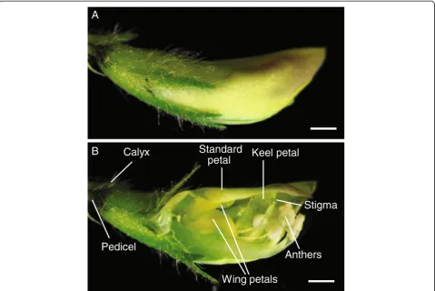
M. truncatulaflowers are hermaphroditic, with both male and female organs in the same flowers. A M. truncatula flower is comprised of a calyx, a large standard petal, two small wing petals on both sides of the flower and a fused keel petal which cover both male and female floral organs (Figure 1A, B). The stigma is surrounded by eight fused anthers (Figure 1B). M. truncatula is a self-pollinated species and fertilization occurs when the flower is still closed (cleistogamy). When the flower reaches maturity, its anthers grow close to the stigma surface and rupture to release the pollen grains (anthesis) on the stigma surface which allows the flower to self-fertilize. When performing a genetic cross, it is important to artificially open the closed flower bud to visualize and access the anthers and stigma before anthesis. In a cross, for the flower bud serving as a female, all the anthers are removed before they release pollen. To artificially fertilize the female flower, pollen grains from mature anthers of a male donor are deposited onto the stigma surface of the female flower to allow cross-pollination to occur.
Several detailed genetic crossing techniques have been described for M. truncatula, which differ by how the immature flower bud is opened to allow removal of anthers and to allow access to the stigma for cross-pollination (Figure 2) [27-29]. These methods also differ in how the flower is treated post cross-pollination to assure a successful cross. We call these: (1) the front cut method [27-29]; (2) the side cut method, also called the pouch method [28,29]; and (3) back cut method (a variation of the side cut method; Mark Taylor, unpublished data). In the front cut method, the flower buds are cut vertically along the central line of the curved side of the standard
petal towards its tip from the proximal one third of the flower bud (Figure 2A). In the side cut (Figure 2B) and back cut (Figure 2C) methods, similar cuts are made on the side and back of the flower buds respectively. The side cut method is time consuming and in all three proce-dures, the incision may damage the flower bud, rendering the female flower unusable. After cross-pollination using either the front or back cut methods, it is difficult to close the flower bud completely, which may lead to mechanical loss of deposited pollen and/or desiccation of internal floral organs.
Another crossing method employs male-sterile mutants as females. The mtapetala (tap) mutant, found in the M. truncatula Jemalong A17 ecotype [16], has been extensively used for this purpose [16,30-33]. However thetapmutation is not found in other ecotypes.
Here we report an improved genetic crossing technique called keel petal incision (Figure 2D; Additional file 1: Video S1; youtu.be/wDtRHWg1LBM), initially developed for the M. truncatulaR108 ecotype. The keel petal incision method allows easy access to an immature flower for anther removal and artificial pollination. It also preserves floral morpho-logical features permitting the standard and wing petals to close to their natural positions after cross pollination. This allows fertilization and embryo development to occur under protection of the flower petals and obviates the need for maintaining the pollinated flower in cling-film or humidified containers. Keel petal incision is a simple and efficient crossing technique with high success rates (>80%) in the R108 ecotype and in other ecotypes, A17 and A20.
Results and discussion
Plant growth and selection of optimal female and male flowers
under the dissecting microscope to make sure pollen grains are visible on the anther surface.
Anther removal and artificial pollination
After selecting an unopened flower bud as a female parent (Figure 3C), we removed the leaves, flowers, pods and/or shoots within three to four inches of the
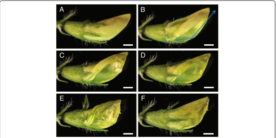
bud, leaving at most one trifoliate leaf adjacent to the chosen flower bud for ease of manipulation. Because M. truncatula flowers and their internal floral organs are small, it was not feasible to perform crosses using the naked eye. Hence, we performed crossing under a dissecting microscope; a magnifying glass or a magnifying binocular headset would also work for this purpose. The flower serving as female was mounted horizontally on its side on a dissecting microscope stage oriented so that the tip of the standard petal faced towards the base of the microscope and the opening of the standard petal faced the dominant hand of the person performing the cross (Figure 4A; Additional file 1: Video S1). The flower bud was secured to the microscope stage using cellophane tape on the pedicel. Subsequently, the flower bud was held in place using a set of forceps on the calyx, while another set of forceps was used to gently lift the standard petal to access the underlying keel petal. Using the sharp tip of a scalpel, the keel petal was gently cut at the bottom third of the flower bud and incision was made along the central ridge of the keel petal all the way to the distal end of the flower (Figure 4B). During this process, care was taken to assure that the tip of the scalpel blade cut only the keel petal and did not extend far inside the flower bud so as Figure 2Position of incisions used to artificially open the
female flower bud to perform genetic crossing. A, Front cut method.B, Side cut method.C, Back cut method.D, Keel petal incision method. Arrows indicate the sites of the cut/incision made on the unopened flower buds to perform anther removal and artificial pollination. Bars = 0.5 mm.
Figure 1M. truncatulaflower structure. A, Unopened flower bud.B, Cut flower bud showing internal reproductive organs. Bars = 0.5 mm.
Veerappanet al. Plant Methods2014,10:11 Page 3 of 10
not to damage the stigma column underneath the keel petal and not to rupture the unopened anthers.
Subsequently, the half of the cut keel petal, facing the investigator, along with the wing and standard petals were pushed upwards with the forceps, allowing visual-ization and access to the anthers and stigma inside the flower bud (Figure 4C). At this point in preparing the flower for pollination, we checked whether the anthers were dehisced. If the anthers were already ruptured, we discarded that flower bud and chose another as the female crossing parent. If the anthers were not yet dehisced, we removed all eight of them by cutting the anther filaments away from the stigma column using sharp forceps tips. Alternatively, anther removal may be accomplished by suction using vacuum applied through a micropipette tip [16]. During anther removal, it is important not to rupture them and to avoid having the anthers contact the stigma. After anther removal, we ascertained that all the anthers were removed (Figure 4D) and visualized the tip of the stigma to verify that there was no accidental deposition of pollen grains on the stigma surface or inside the flower bud during anther removal. In the instances
where we saw that this had occurred, we discarded that flower and started over with a fresh one.
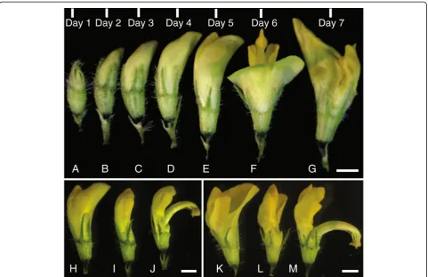
To expose the anthers from a male pollen donor in the cross, we triggered a flower that was about to open (Figure 3E, H) with gentle mechanical pressure until the style and anthers popped open. Alternatively, we have used forceps to remove the petals around the anthers in the pollen donor flower (Figure 3I). We found that using forceps to remove the standard petal first and then the wing and keel petals does not damage the anthers. Once the flower is triggered (Figure 3J) or the petals were removed, we observed the anthers using the dissecting microscope to ascertain that the anther was mature and dehisced with silvery white pollen grains released from ruptured anther sacs visible. This can also be seen using other magnification devices. Already-open flowers also served as pollen donors (Figure 3F, K). In these instances, the standard petal was removed with forceps (Figure 3L) exposing the style and anthers (Figure 3M; flower turned approximately 110° from Figure 3L).
flower multiple times to deposit the pollen grains. At the end of this process, the entire tip of the stigma was surrounded by pollen (Figure 4E). There is variation in pollen quality and quantity from individual flowers; pollen quality may be negatively affected by insect infestation as well. In the case of insufficient pollen
from one flower, we used more than one flower from the male pollen donor plant.
After pollination, the flower was closed by pushing the top of the standard petal back into its original position to cover the wing petal, keel petal and stigma, forming a pouch around the stigma that looks similar to the Figure 4Steps in genetic crossing using the keel petal incision method. A, Unopened optimal female flower bud.B, Flower bud showing the keel petal under the standard petal indicating the site of incision on the central line of the keel petal.C, Flower bud showing the internal floral organs including anthers and stigma underneath the keel petal after making an incision.D, Flower bud showing stigma and style after the removal of anthers.
E, Stigma covered with mature pollen after artificial cross-pollination.F, Closed flower bud after deposition of pollen on stigma. Bars = 0.5 mm.
Figure 5Development of pods from successful crosses. A, Curling of artificially cross-pollinated flower bud 3 days post hybridization.B, Developing pod 6 days post hybridization.C, Developing pod 20 days post-hybridization.D, Developing pod, 35 days post-hybridization wrapped with microperforated polythene sheet.E, Ripening yellow pod, 42 days post-hybridization.F, Completely matured darkened pod fallen off the pedicel, 50 days post-hybridization. Bars = 0.5 mm(A-C). Bars = 5 mm(D-F).
Veerappanet al. Plant Methods2014,10:11 Page 5 of 10
unopened flower bud before crossing (Figure 4F). This prevented pollen from dislodging from the stigma dur-ing subsequent movement of the plant and protected the stigma and pollen from desiccation. The floral petal pouch obviates the need for protecting the pollinated flower with an artificial covering. These coverings promote fungal and bacterial growth on the flowers that may cause the flower to decay and drop. The pouch also does away with the need to place the cross-pollinated flower bud inside humid containers, which may also cause flower damage. After the cross-pollination was completed, the cellophane tape holding the pedicel was carefully removed and the pollinated flower was labeled.
Seed pod development
To distinguish between artificially pollinated and self-pollinated pods, we monitored the artificially self-pollinated flower buds daily and trimmed away new shoots growing adjacent to the pollinated flowers. Successful crosses were visualized by slight curling of pollinated flowers from the 3rd day onwards (Figure 5A). Most of the time, the cross-pollinated flowers continued to curl (Figure 5B) and develop into mature pods (Figure 5C). Once the pods from successful crosses developed into medium size (Figure 5C), we wrapped them using micro-perforated polythene sheets stapled in place (Figure 5D) to prevent the pods from falling off after maturity.
To increase the size of the pods from cross-pollination and number of seeds per cross, we removed all pods on the plant resulting from self-pollination. Removing pods promotes more flower formation and prolongs the dur-ation of flowering. After all the crosses were completed, shoots that do not carry pods from cross-pollination were trimmed. We observed developing pods turning yellow (Figure 5E) and darkening and dropping from the pedicel at maturity (Figure 5F).
We air-dried the pods from successful crosses at room temperature for 2–3 weeks. After this, we collected seeds from the pods and proceeded to confirm the crosses in the F1 generation. We found that larger pods typically
contain 5–6 seeds/pod whereas the smaller pods only carried 1–3 seeds/pod. In general, crosses performed on younger plants resulted in larger pods whereas older plants produced smaller pods.
Cross-pollination confirmation
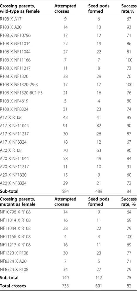
As a first test of the success of cross-pollination by the keel petal incision method, we counted the number of seed pods that formed after cross pollination. We esti-mated an overall success rate of 82%, with slightly higher rates of pod formation when a wild-type parent was the female in the cross (Table 1).
Confirmation of successful crossing relied on pheno-typing and genopheno-typing the progeny. Our labs employ
forward genetics to identify new legume genes that control the development of symbiotic nitrogen-fixing root nodules using the M. truncatula Tnt1 insertion mutant collection in the R108 ecotype background. Tnt1 mutants contain multiple Tnt1 insertions and there are no naturally occurring Tnt1 inserts in any Table 1 Estimation of crossing success via seed pod formation
Crossing parents, wild-type as female
Attempted crosses Seed pods formed Success rate,%
R108 X A17 9 6 67
R108 X A20 14 13 93
R108 X NF10796 17 12 71
R108 X NF11014 22 19 86
R108 X NF11044 27 22 81
R108 X NF11166 7 7 100
R108 X NF11217 11 8 73
R108 X NF1320 38 29 76
R108 X NF1320-29-3 17 17 100
R108 X NF1320-BC1-F3 21 16 76
R108 X NF4619 5 4 80
R108 X NF8324 31 23 74
A17 X R108 43 41 95
A17 X NF11044 91 82 90
A17 X NF11217 30 26 87
A17 X NF8324 18 12 67
A20 X R108 70 63 90
A20 X NF11044 58 49 84
A20 X NF11217 11 10 91
A20 X NF1320 15 9 60
A20 X NF8324 29 21 72
Sub-total 584 489 84
Crossing parents, mutant as female
Attempted crosses Seed pods formed Success rate, %
NF10796 X R108 14 9 64
NF11014 X R108 16 11 69
NF11044 X R108 28 22 79
NF11166 X R108 4 4 100
NF11217 X R108 16 11 69
NF1320 X R108 30 23 77
NF8324 X A20 7 5 71
NF8324 X R108 34 27 79
Sub-total 149 112 75
Total crosses 733 601 82
M. truncatulaecotype [21,34]. To study the mode of in-heritance of mutant phenotypes and to test the co-segregation of candidateTnt1insertions with nodule phe-notypes, we backcrossed two particular mutants with inef-fective, white or brown non-fixing nodules (Nod + Fix-), NF11217 and NF10796, to their wild-type R108 using the keel petal incision method.
For NF11217 backcrosses, the mutant plant was used as the female parent and the wild-type R108 served as the male pollen donor. For NF10796 backcrosses, wild-type served as the female and the mutant plant was used as a male parent. Typically, it is advisable to perform reciprocal crosses by switching male and female parents in separate crosses, because the mutant phenotypes might be governed by maternal or paternal inheritance.
When recessive Tnt1 mutants were used as female, successful crosses were confirmed by wild-type phenotype progeny in the F1population. Nodules of NF11217 mutant
(female parent), wild-type (male parent) and NF11217 × wild-type-BC1F1 progeny were assessed for visual
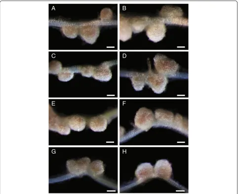
phe-notypes fifteen days after inoculation of roots with Sinorhizobium meliloti. The nodules from wild-type parents are pink (Figure 6A) because of the presence of leghe-moglobin associated with efficient symbiotic nitrogen fixation, whereas the NF11217 mutant plants show white nodules, defective in symbiotic nitrogen fixation (Figure 6B). All the BC1F1 plants tested showed pink
nodules similar to the wild-type (Figure 6C-H), indicat-ing that cross-pollination was successful and suggestindicat-ing the mutation in NF11217 is recessive.
Figure 6Confirmation of successful crosses by nodule phenotypes of the progeny.M. truncatulaplants were grown on an aeroponic chamber, as described in Materials and methods. Five days after germination, plants were starved for nitrogen for five days and then inoculated withSinorhizobium meliloti. Root nodules were examined 15 days post-inoculation.A, Wild-type R108.B, NF11217 mutant.C-H, NF11217 X wild-type R108-BC1F1. The R108
plant(A)has pink wild-type nodules while those of NF11217 are white and ineffective(B). Each plant from the BC1F1progeny(C-H)has pink nodules
indicative of a successful cross. Bars = 250μm.
Veerappanet al. Plant Methods2014,10:11 Page 7 of 10
When wild-type was used as female, success of the crosses was verified by genotyping the F1 plants for the
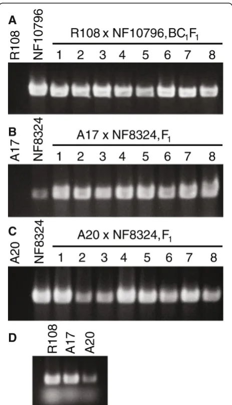
presence of Tnt1 transgene. For the case of NF10796 serving as the male in a cross with a wild-type R108 female, PCR (polymerase chain reaction) genotyping of BC1F1progeny usingTnt1specific primers was performed.
Our results showed the presence of Tnt1insertions in the R108 x NF10796-BC1F1plants (Figure 7A) indicating
cross-ing success. As expected, there was no amplification of Tnt1sequences in the wild-type R108 (Figure 7A).
As a further test of the utility of the keel petal incision method, we attempted out-crossing of Tnt1 Nod+ Fix-mutant NF8324 in the R108 background, to both the A17 and A20 wild-type ecotypes. In these cases, we used the A17 or A20 wild-types as females and NF8324 as a male in the crosses. F1plants arising from the crosses were
genotyped by PCR for the presence of Tnt1sequences in the progeny, indicating a successful cross. As can be seen by the examples (Figure 7B, C), the keel petal incision method also worked for these other M. truncatula eco-types. However, F1progeny from inter-ecotype crosses
involving R108 were observed to have developmental phenotypes (not shown); thus, special care must be taken with these progeny.
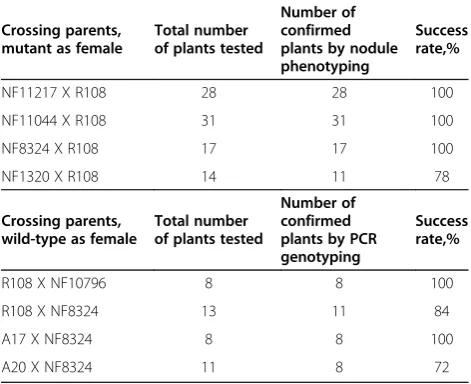
We examined ninety BC1F1plants for nodulation
phe-notypes resulting from backcrosses involving Nod+/Fix-mutants female parents with wild-type R108 as male and found almost all of them had wild-type nodules, indicating successful cross-pollination and absence of self-pollination (Table 2). With the reciprocal situation using the wild-type as female and mutant as male, we tested forty plants from the progeny and obtained suc-cess rates of approximately 87% overall using the R108, A17 and A20 ecotypes, as verified by presence of Tnt1 sequences in the BC1F1and F1progeny (Table 2).
Conclusions
Crossing is essential for genetics. Here we described a new crossing technique for M. truncatula that involves incising the keel petal of the female flower to expose the flower’s stigma and anthers, remove its anthers and permit an artificial cross. After the cross is complete, the standard, wing and keel petals can be arranged similarly to their original positions in the unopened flower bud, protecting the floral organs from desiccation and removing the need for extraneous materials to main-tain humidity for the fertility of the flower and deposited pollen. We demonstrated that this method can be used for the M. truncatula R108, A17 and A20 ecotypes and expect that it will find utility in other ecotypes. This method has been successfully used by investigators of varying experience, including junior graduate students and undergraduates in our labs.
Materials and methods
Plant growth
M. truncatula seeds were scarified with sulfuric acid, treated with bleach, imbibed and vernalized as described [16,28]. Shorter vernalization (1–4 days at 4°C) results in delayed flowering and a larger plant with higher numbers of flowers. Longer vernalization (4–14 days at 4°C) results in earlier flowering accompanied by smaller plant stature and reduced flower numbers. If plants are grown exclu-sively for crossing, longer vernalization is preferred to
Figure 7Confirmation of successful crosses by genotyping. A-C, Wild-type R108, NF10796 and NF8324, R108 x NF10796-BC1F1,
A17 x NF8324-F1and A20 x NF8324-F1plants were PCR genotyped
usingTnt1transposon specific primers.D, Primers for theMtIRE-like
gene were used in the PCR amplifications as positive control for the wild-type genomic DNA.Tnt1DNA is not found in wild-type R108, A17 or A20 plants but can be detected in all F1plants indicating
successful crosses(A-C). Stained gel images have been cropped to show the PCR products of 612 bp fromTnt1and 422 bp from
shorten the time to flowering. Details of the effect of vernalization on flowering time and growth stature can be found in [28]. Different vernalization times were used and did not appear to affect crossing success. After vernalization, seeds were germinated at 25°C in the dark; in our hands, seeds germinated within 12–24 hours. Subsequently, the plants were screened for nodulation phenotypes 15 days post-inoculation withS. melilotiusing an aeroponic chamber [30,35], or by growing the plants on a Turface/vermiculite mixture [23]. After phenotyping, the seedlings were grown in medium size (four inch; ten cm) pots so that they are easy to handle while performing crosses. To avoid waterlogged roots, we grewM. trunca-tulain a peat-based potting medium with Turface (Profile, Buffalo Grove, Ill) mixed in (3:1), at 22°C under16:8 hr light:dark cycles. Plants were irrigated as needed and fertilized with a commercial general purpose fertilizer once a week.
Crosses and PCR confirmation
The crosses were performed under a Nikon model C-PS stereo microscope (Nikon, Tokyo, Japan). Images and video clips ofM. truncatulaflowers and crossing techniques were obtained using Leica MZ10F (Leica, Buffalo Grove, IL) microscope. Two pairs of fine tip forceps (HL-14 #5, www. buyincoins.com) and a straight-edge scalpel (scalpel blade handle 9303 #3, and scalpel blade 9311 #11, both from www.microscopesamerica.com) were used for keel petal in-cision, the removal of anthers from the unopened female flower bud and artificial cross-pollination (Additional file 2: Figure S1). Mature pods from the successful
cross-pollinations were wrapped using micro-perforated poly-thene sheets (MP1120160T, www.prismpak.com).
The following primers were used for PCR genotyping
to confirm crosses: Tnt1-F, GCATTCAAACTAGAAGA
CAGTGCTACC and Tnt1-R, TGTAGCACCGAGATA
CGGTAATTAACAAGA [34] (Tnt1, Genbank:X13777).
MtIRE-like specific primers [36] (MtIRE-like, Genbank: AY770392, Genbank:AC122727) were used as a control to confirm that extracted DNA from wild-type plants was of sufficient quality for PCR using MtIRE-F, CCAAATC
GTTGAAAGCTCGTTCACAACTCC and MtIRE-R, CG
TCTTGACCAGCAAACACGACACG.
Additional files
Additional file 1: Video S1.The keel petal incision crossing method for
M. truncatula. The method is narrated step-by-step: youtu.be/wDtRHWg1LBM.
Additional file 2: Figure S1.Forceps and scalpel used in crossing. Two pairs of fine tip forceps, e.g., HL-14 #5, www.buyincoins.com, and a straight-edge scalpel, e.g., scalpel blade handle 9303 #3, and scalpel blade 9311 #11, both from www.microscopesamerica.com, were used for keel petal incision, the removal of anthers from the unopened female flower bud and cross-pollination. Similar forceps and scalpels are available from other vendors. Bar = 5 cm.
Abbreviations
BC:Back cross; DNA: Deoxyribonucleic acid; EMS: Ethyl methane sulfonate; FNB: Fast neutron bombardment;M. truncatula:Medicago truncatula; PCR: Polymerase chain reaction.
Competing interests
The authors declare that they have no competing interests.
Authors’contributions
VV designed the experiments and made the supplemental video. VV, KK, NA, AS, IK, SS and MT carried out the experiments and edited the manuscript. MU edited the manuscript. VV and RD coordinated the experiments, and wrote and edited the manuscript. All authors read and approved the final manuscript.
Acknowledgements
This work was supported by National Science Foundation grant IOS 1127155 to MU and RD and by USDA-NIFA grant 2010-65115-20384 to MU. AS was partially supported by Department of Education grant P217A120023 to Dr. D Elrod and UNT’s McNair Scholars Program. Video and audio equipment used in the production of the supplemental video for this article were provided by the University of North Texas Media Library.
Author details
1Department of Biological Sciences, University of North Texas, 1155 Union Circle #305220, Denton, Texas 76203, USA.2Plant Biology Division, The Samuel Roberts Noble Foundation, Ardmore, OK 73401, USA.
Received: 9 January 2014 Accepted: 17 March 2014 Published: 16 May 2014
References
1. Cook DR:Medicago truncatula- a model in the making!Curr Opin Plant Biol1999,2:301–304.
2. Kouchi H, Imaizumi-Anraku H, Hayashi M, Hakoyama T, Nakagawa T, Umehara Y, Suganuma N, Kawaguchi M:How many peas in a pod? Legume genes responsible for mutualistic symbioses underground.Plant Cell Physiol2010,
51:1381–1397.
3. Barker DG, Bianchi S, London F, Dattee Y, Duc G, Essad S, Flament P, Gallusci P, Genier G, Muel X, Tourneur J, Denarie J, Huguet T:Medicago truncatula, a Table 2 Confirmed successful crosses
Crossing parents, mutant as female
Total number of plants tested
Number of confirmed plants by nodule phenotyping
Success rate,%
NF11217 X R108 28 28 100
NF11044 X R108 31 31 100
NF8324 X R108 17 17 100
NF1320 X R108 14 11 78
Crossing parents, wild-type as female
Total number of plants tested
Number of confirmed plants by PCR genotyping
Success rate,%
R108 X NF10796 8 8 100
R108 X NF8324 13 11 84
A17 X NF8324 8 8 100
A20 X NF8324 11 8 72
For each cross, the first parent listed in the cross was used as the female. For cases where the mutant served as female in the cross, successful crossing was confirmed by the appearance of pink nodules in the BC1F1progeny. When
wild-type served as female, including inter-ecotype crosses, crosses were confirmed in the BC1F1, or F1progeny by PCR genotyping usingTnt1specific
primers, as in Figure7.
Veerappanet al. Plant Methods2014,10:11 Page 9 of 10
model plant for studying the molecular genetics of theRhizobium-legume symbiosis.Plant Mol Biol Report1990,8:40–49.
4. Dixon RA, Liu C, Jun JH:Metabolic engineering of anthocyanins and condensed tannins in plants.Curr Opin Biotechnol2013,24:329–335. 5. Verdier J, Zhao J, Torres-Jerez I, Ge S, Liu C, He X, Mysore KS, Dixon RA,
Udvardi MK:MtPAR MYB transcription factor acts as an on switch for proanthocyanidin biosynthesis inMedicago truncatula.Proc Natl Acad Sci USA2012,109:109.
6. Zhao J, Dixon RA:The‘ins’and‘outs’of flavonoid transport.Trends Plant Sci2010,15:72–80.
7. Uppalapati SR, Ishiga Y, Doraiswamy V, Bedair M, Mittal S, Chen J, Nakashima J, Tang Y, Tadege M, Ratet P, Chen R, Schultheiss H, Mysore KS:Loss of abaxial leaf epicuticular wax inMedicago truncatula irg1/palm1mutants results in reduced spore differentiation of anthracnose and nonhost rust pathogens. Plant Cell2012,24:353–370.
8. Uppalapati SR, Marek SM, Lee H-K, Nakashima J, Tang Y, Sledge MK, Dixon RA, Mysore KS:Global gene expression profiling duringMedicago truncatula– Phymatotrichopsis omnivorainteraction reveals a role for jasmonic acid, ethylene, and the flavonoid pathway in disease development.Mol Plant Microbe Interactions2009,22:7–17.
9. Tadege M, Lin H, Bedair M, Berbel A, Wen J, Rojas CM, Niu L, Tang Y, Sumner L, Ratet P, McHale NA, Madueño F, Mysore KS:STENOFOLIA regulates blade outgrowth and leaf vascular patterning inMedicago truncatulaandNicotiana sylvestris.Plant Cell2011,23:2125–2142. 10. Lin H, Niu L, McHale NA, Ohme-Takagi M, Mysore KS, Tadege M:Evolutionarily
conserved repressive activity of WOX proteins mediates leaf blade outgrowth and floral organ development in plants.Proc Natl Acad Sci USA2013,110:366–371.
11. Ge L, Peng J, Berbel A, Madueño F, Chen R:Regulation of compound leaf development byPHANTASTICAinMedicago truncatula.Plant Physiol2014,
164:216–228.
12. Zhu H, Choi H-K, Cook DR, Shoemaker RC:Bridging model and crop legumes through comparative genomics.Plant Physiol2005,137:1189–1196. 13. Benedito VA, Torres-Jerez I, Murray JD, Andriankaja A, Allen S, Kakar K,
Wandrey M, Verdier J, Zuber H, Ott T, Moreau S, Niebel A, Frickey T, Weiller G, He J, Dai X, Zhao PX, Tang Y, Udvardi MK:A gene expression atlas of the model legumeMedicago truncatula.Plant J2008,55:504–513. 14. Young ND, Debellé F, Oldroyd GED, Geurts R, Cannon SB, Udvardi MK,
Benedito VA, Mayer KFX, Gouzy J, Schoof H, Van de Peer Y, Proost S, Cook DR, Meyers BC, Spannagl M, Cheung F, De Mita S, Krishnakumar V, Gundlach H, Zhou S, Mudge J, Bharti AK, Murray JD, Naoumkina MA, Rosen B, Silverstein KAT, Tang H, Rombauts S, Zhao PX, Zhou P,et al:TheMedicagogenome provides insight into the evolution of rhizobial symbioses.Nature2011,
480:520–524.
15. Tadege M, Wang TL, Wen J, Ratet P, Mysore KS:Mutagenesis and beyond! Tools for understanding legume biology.Plant Physiol2009,151:978–984. 16. Penmetsa RV, Cook DR:Production and characterization of diverse
developmental mutants ofMedicago truncatula.Plant Physiol2000,
123:1387–1398.
17. Wang HL, Li G, Chen R:Fast Neutron Bombardment (FNB) Mutagenesis for Forward and Reverse Genetic Studies in Plants.InFloriculture, Ornamental and Plant Biotechnology: Advances and Topical Issues (1st Edition). Volume 1.
Edited by Silva JT. Isleworth, UK: Global Science Books; 2006:629–639. 18. Rogers C, Wen J, Chen R, Oldroyd G:Deletion-based reverse genetics in
Medicago truncatula.Plant Physiol2009,151:1077–1086. 19. Rogers C, Oldroyd GED:Fast Neutron Mutagenesis for Functional
Genomics.InThe Handbook of Plant Functional Genomics.Edited by Kahl G, Meksem K. Weinheim, Germany: Wiley-VCH; 2008:291–306.
20. Tadege M, Ratet P, Mysore KS:Insertional mutagenesis: a Swiss Army knife for functional genomics ofMedicago truncatula.Trends Plant Sci2005,
10:229–235.
21. Tadege M, Wen J, He J, Tu H, Kwak Y, Eschstruth A, Cayrel A, Endre G, Zhao PX, Chabaud M, Ratet P, Mysore KS:Large-scale insertional mutagenesis using theTnt1retrotransposon in the model legumeMedicago truncatula.Plant J2008,54:335–347.
22. Cheng X, Wen J, Tadege M, Ratet P, Mysore KS:Reverse genetics in Medicago truncatulausingTnt1insertion mutants.Methods Mol Biol2011,
678:179–190.
23. Pislariu CI, Murray JD, Wen J, Cosson V, Muni RRD, Wang M, Benedito VA, Andriankaja A, Cheng X, Jerez IT, Mondy S, Zhang S, Taylor ME, Tadege M, Ratet P, Mysore KS, Chen R, Udvardi MK:AMedicago truncatulatobacco
retrotransposon insertion mutant collection with defects in nodule development and symbiotic nitrogen fixation.Plant Physiol2012,
159:1686–1699.
24. Floss DS, Levy JG, Lévesque-Tremblay V, Pumplin N, Harrison MJ:DELLA proteins regulate arbuscule formation in arbuscular mycorrhizal symbiosis.Proc Natl Acad Sci USA2013, doi:10.1073/pnas.1308973110. 25. Yeoh CC, Balcerowicz M, Zhang L, Jaudal M, Brocard L, Ratet P, Putterill J:
Fine mapping links theFTa1flowering time regulator to the dominant Spring1locus in Medicago.PLoS One2013,8:e53467. doi:53410.51371/ journal.pone.0053467.
26. Jaudal M, Yeoh CC, Zhang L, Stockum C, Mysore KS, Ratet P, Putterill J:
Retroelement insertions at the MedicagoFTa1locus inspringmutants eliminate vernalisation but not long-day requirements for early flowering. Plant J2013,76:580–591.
27. Pathipanawat W, Jones RAC, Sivasithamparam K:An improved method for artificial hybridization in annualMedicagospecies.Aust J Agric Res1994,
45:1329–1335.
28. Chabaud M, Lichtenzveig J, Ellwood S, Pfaff T, Journet E-P:Vernalization, Crossings and Testing for Pollen Viability.InThe Medicago Truncatula Handbook.Edited by Mathesius U, Journet E-P, Sumner LW. 2006. http:// www.noble.org/MedicagoHandbook/. ISBN 0-9754303-1-9.
29. Taylor M, Blaylock L, Nakashima J, McAbee D, Ford J, Harrison M, Udvardi M:
Medicago TruncatulaHybridization–Supplemental Videos.InThe Medicago Truncatula Handbook, Version 2011.Edited by Mathesius U, Journet E-P, Sumner LW. Ardmore, Oklahoma, USA: The Samuel Roberts Noble Foundation; 2011:1–5. vol. http://www.noble.org/MedicagoHandbook/. pp. 1–5. 30. Veereshlingam H, Haynes JG, Sherrier DJ, Penmetsa RV, Cook DR, Dickstein
R:nip, a symbioticMedicago truncatulamutant that forms root nodules with aberrant infection threads and plant defense-like response. Plant Physiol2004,136:3692–3702.
31. Schnabel E, Mukherjee A, Smith L, Kassaw T, Long S, Frugoli J:Thelss supernodulation mutant ofMedicago truncatulareduces expression of theSUNNgene.Plant Physiol2010,154:1390–1402.
32. Schnabel EL, Kassaw TK, Smith LS, Marsh JF, Oldroyd GED, Long SR, Frugoli JA:
TheROOT DETERMINED NODULATION1gene regulates nodule number in roots ofMedicago truncatulaand defines a highly conserved, uncharacterized plant gene family.Plant Physiol2011,157:328–340. 33. Penmetsa RV, Frugoli JA, Smith LS, Long SR, Cook DR:Dual genetic
pathways controlling nodule number inMedicago truncatula. Plant Physiol2003,131:998–1008.
34. D’Erfurth I, Cosson V, Eschstruth A, Lucas H, Kondorosi A, Ratet P:Efficient transposition of theTnt1tobacco retrotransposon in the model legume Medicago truncatula.Plant J2003,34:95–106.
35. Journet E-P, de Carvalho-Niebel F, Andriankaja A, Huguet T, Barker DG:Rhizobial Inoculation and Nodulation ofMedicago Truncatula.InThe Medicago Truncatula Handbook.Edited by Mathesius U, Journet E-P, Sumner LW. 2006. http://www. noble.org/MedicagoHandbook/. ISBN 0-9754303-1-9.
36. Pislariu CI, Dickstein R:AnIRE-like AGC Kinase Gene,MtIRE, has unique expression in the invasion zone of developing root nodules inMedicago truncatula.Plant Physiol2007,144:682–694.
doi:10.1186/1746-4811-10-11
Cite this article as:Veerappanet al.:Keel petal incision: a simple and
efficient method for genetic crossing inMedicago truncatula.Plant Methods201410:11.
Submit your next manuscript to BioMed Central and take full advantage of:
• Convenient online submission
• Thorough peer review
• No space constraints or color figure charges
• Immediate publication on acceptance
• Inclusion in PubMed, CAS, Scopus and Google Scholar
• Research which is freely available for redistribution