International Journal of Applied And Pure Science and
Agriculture
www.ijapsa.com
SYNOVIAL FLUID ANALYSIS IN DOGS WITH ELBOW, HIP AND
STIFLE JOINT DISORDERS
Anirudh. A1 and L. Ranganath2
1
Ph.D Scholar, Department of Veterinary Surgery and Radiology, KVAFSU, Veterinary College, Bangalore-24, India. 2
Professor and Head, Department of Veterinary Surgery and Radiology, KVAFSU, Veterinary College, Bangalore-24, India.
Abstract
The study consisted of 18 dogs divided into Three groups. Each group consisted of Six dogs with elbow, hip and stifle joint disorders respectively. All the dogs were subjected to synovial fluid evaluation. The physico-chemical parameters recorded in these dogs included synovial fluid volume, color, viscosity, transparency, protein concentration, total nucleated cell count and differential cell count respectively. In the present study the mean synovial fluid volume increased compared to the normal average values. The color of synovial fluid was variable and varied from red tinged to yellow. The Viscosity and transparency of the synovial fluid decreased compared to normal average values. The mean values of protein concentration and total nucleated count of synovial fluid were higher compared to normal average values. The mean mononuclear cell count was lower than normal values whereas the mean synovial fluid differential cell count (Polymorphs) was relatively higher as compared to normal values. The variations in the values of different parameters of synovial fluid within the groups were statistically non- significant (P≤0.05)
Keywords: Synovial fluid, Arthrocentesis, synovial fluid volume, inflammatory arthropathies, synovial membrane.
I. Introduction
II. Materials and Methods
A total of 18 dogs with elbow, hip and stifle joint disorders were subjected to arthrocentesis and synovial fluid analysis. Dogs showing signs of lameness involving elbow, hip and stifle joint were selected and subjected to routine physical examination. A total of 18 dogs with joint disorders were selected and were divided into three groups. Group-I consisted of six dogs with congenital or acquired elbow joint disorders. Group-II consisted of six dogs with congenital or acquired hip joint disorders. Group-III consisted ofsix dogs with congenital or acquired stifle joint disorders.
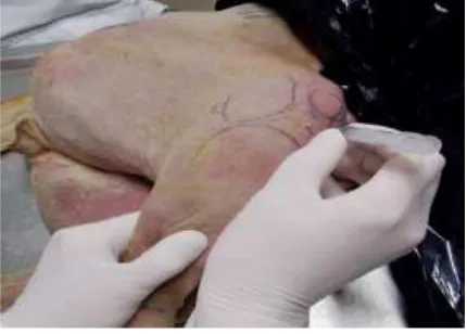
Arthrocentesis of the elbow joint was done with the animal on lateral recumbency. The patient was positioned in lateral recumbency with the affected limb above the table. A 1.5 inch 18G needle was inserted in the caudo dorsal portion of the joint between the anconeal process and the olecranon fossa. Synovial fluid was aspirated using a 3 milliliter syringe. (Fig 1) For arthrocentesis of the hip joint, An 18 to 22-gauge spinal needle (2.5- to 3-inch) was used. The hip was flexed slightly, and the femur was positioned parallel to the table to maximize the width of the joint space. The hip was palpated, and the needle was inserted at the midpoint of the proximal edge of the greater trochanter. To ensure that the needle was placed within the joint, a syringe was attached to the needle and synovial fluid was aspirated using a 3 milliliter syringe. (Fig 2). For arthrocentesis of stifle joint, the patient was positioned in dorsal recumbency with the pelvis placed at the end of the operating table to facilitate unobstructed access to both sides of the stifle. A cranio-medial approach was chosen. An 18 G hypodermic needle was inserted into the supra patellar pouch and synovial fluid was withdrawn. (Fig 3).
Fig 1: Arthrocentesis of elbow joint Fig 2: Arthrocentesis of Hip joint
The data was subjected to statistical analysis as per Snedecor and Cochran (1996) to arrive at a conclusion (P≤ 0.05). Differences between groups and period means were calculated by a two way analysis of variance (ANOVA) using computer based statistical programme (Graph pad prism).
III. RESULTS AND DISCUSSION
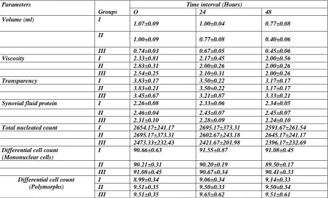
In the present study the mean synovial fluid volume ranged between 0.40±0.06 and 1.00±0.09 ml (Table 1). The volume of synovial fluid was found to be increased in dogs with joint disorders when compared to the synovial fluid volume in normal dogs which is in accordance with the earlier reports of where 5-20 ml of synovial fluid was recorded in dogs affected with elbow or stifle pathology[6]. During joint disease, synovial fluid volume is usually increased. Synovial fluid volume is considered increased in small animals when more than 0.5 milliliters of fluid can be easily collected from the small, distal joints (i.e., carpus, tarsus) or when more than 1.0 milliliters can be easily collected from the larger, proximal joints (i.e., stifle, hip, shoulder) [3]. An increase in synovial fluid volume is a nonspecific finding and can be seen with any cause of non-inflammatory or inflammatory joint disease. In many cases of degenerative joint disease, synovial fluid analysis will be essentially normal. However, in a few cases the findings, particularly those seen on cytologic examination, will confirm a low-grade but ongoing inflammatory process. In the present study, the color of synovial fluid in dogs with joint disorders was variable and varied from red tinged to yellow.
The synovial fluid from inflammatory joints shows variable discoloration [2, 7, and 10]. Discoloration may be because of dilution from excess serum[7]. The colour of the normal synovial fluid is clear to colourless [4,6,9].The red tinge of synovial fluid of in a few joint affected dogs recorded in the present study may possibly attributed to haemorrhage during collection. [4, 9, 12].In the present study, the mean viscosity of synovial fluid varied between 2.33±0.21 and 2.17± 0.31 (table 1). The Viscosity of the synovial fluid was found to be decreased in joint affected dogs. The significant decrease in the viscosity of synovial fluid of joint affected dogs was attributed to dilution of synovial fluid by hyaluronidase and lysozymal enzymes [6, 8]. Those subjects with non-injured stifle, hip or elbow joint displayed slightly higher viscosity compared to those dogs with injured joints.
This observation in joints is most likely due to local inflammation and swelling which increases fluid content in the joint and therefore would dilute the synovial fluid. In chronic conditions, it is possible that the joint has moved beyond the initial inflammation seen in the acute condition and therefore the decreased viscosity is due to mechanical and biochemical destruction of hyaluronic acid, secondary to osteoarthritis. This observation is in agreement to earlier reports in dogs with different joint lesions [5, 12]. In the present study, the mean Transparency of synovial fluid varied between 3.17±0.17 and 3.83± 0.21 (table 1).The mean value of synovial fluid transparency decreased relatively in joint affected dogs.
of joint inflammation [9,12].The mean total nucleated count ranged between 2450±122.80 and 2902.67±243.18 (Table 1).
The present findings suggest that, in joint affected dogs, the total nucleated cell count was relatively higher in joint affected dogs compared to the normal average total nucleated count. Further, it has been reported that the cause for increased nucleated cell count may include non-inflammatory arthropathies, which have a normal to mildly increased nucleated cell count, and inflammatory arthropathies [9]. In another study, the normal synovial fluid WBC/μL ranged from 0– 2900 with an average value of 430 WBC/μL [1].The mean values of synovial fluid differential cell count (mononuclear) ranged between 89.50±0.17 and 91.55±0.87 (Table 1) and the mean values of synovial fluid differential cell count (polymorphs) ranged between 8.99±0.34 and 9.65±0.62 (table 1).
The present findings suggest that, in joint affected dogs, the mean synovial fluid differential cell count (mononuclear) was lower than normal values. In normal synovial fluid, mononuclear cells, when present, are in large numbers; usually representing 95% of nucleated cells [3, 8,10]. Normal synovial fluid mononuclear cell count range between 88 to 100 [1]. Therefore, the observation made in the present study as well as the observations of made in the previous studies seems to suggest that mononuclear cell count may not be a sensitive indicator of joint disorders in dogs. The mean synovial fluid differential cell count (polymorphs) was relatively higher as compared to normal values. In normal synovial fluid, neutrophils, when present, are in small number; usually representing less than 5% of nucleated cells [8,10]. The normal synovial fluid neutrophil count range between 0–12 percent [1]. Therefore, the observation made in the present study as well as the observations of made in the previous studies seems to suggest that neutrophil count may not be a sensitive indicator of joint disorders in dogs. Significant increase in neutrophil count of synovial fluid of polyarthritis dogs was noticed in the previous studies and these observations suggests that in inflammatory arthropathies, neutrophil count may increase [2,7]
Table 1: Mean ± SE Values of synovial fluid parameters in dogs of groups I, II and III at different intervals
Parameters
Groups
Time interval (Hours)
O 24 48
Volume (ml) I
1.07±0.09 1.00±0.04 0.77±0.08
II
1.00±0.09 0.77±0.08 0.40±0.06
III 0.74±0.03 0.67±0.05 0.45±0.06
Viscosity I 2.33±0.81 2.17±0.45 2.00±0.56
II 2.83±0.31 2.00±0.26 2.00±0.26
III 2.54±0.25 2.10±0.31 2.00±0.26
Transparency I 3.83±0.17 3.50±0.22 3.17±0.17
II 3.83±0.21 3.50±0.22 3.17±0.17
III 3.45±0.67 3.21±0.87 3.33±0.21
Synovial fluid protein I 2.26±0.08 2.33±0.06 2.34±0.05
II 2.46±0.04 2.43±0.07 2.45±0.07
III 2.31±0.10 2.28±0.09 2.24±0.10
Total nucleated count I 2654.17±241.17 2695.17±373.31 2591.67±261.54
II 2695.17±373.31 2602.67±243.18 2645.17±241.17
III 2473.33±232.43 2421.67±201.98 2396.17±232.69
Differential cell count (Mononuclear cells)
I 90.66±0.63 91.55±0.87 91.08±0.45
II 90.21±0.31 90.20±0.19 89.50±0.17
III 91.08±0.45 90.67±0.34 90.41±0.33
Differential cell count (Polymorphs)
I 8.99±0.34 9.06±0.34 9.14±0.33
II 9.51±0.35 9.50±0.33 9.50±0.34
III 9.51±0.35 9.65±0.62 9.51±0.61
IV. Conclusion
Synovial fluid analysis of dogs with elbow, hip and stifle joint disorders revealed physico-chemical changes in the synovial fluid parameters which is of diagnostic importance in dogs with various joint disorders. Arthrocentesis has both diagnostic and therapeutic application. Diagnostic procedures include synovial fluid analysis, cytologic examination, and microorganism culture, as well as intra articular injection of contrast material for arthrography. Synovial fluid analysis should be an integral part of any diagnostic evaluation of an animal with Lameness, especially the animal with joint effusion.
Bibliography
[1] Alleman, A. R., Denicola, D. B. and Wamsley, H. L., 2007. Synovial Fluid Analysis: 79th Western Veterinary
Conference: 10.
[2] Bennett, D. B. and May, C., 1995. Joint diseases of dogs and cats, in Ettinger SJ, Feldman EC (eds): Textbook of Veterinary Internal Medicine (ed 4). Philadelphia, PA, Saunders: 2032-2077.
[3] Boon, D., 1997. Synovial fluid analysis: A guide for small-animal practitioners. Vet. Med., 92: 443-451. [4] Coles, E.H., 1986. Veterinary Clinical Pathology. 4th Edn., W.B. Saunders Company. Philadelphia. Pp: 256-260. [5] Conrad, B. P., Canapp, S. O., Cross, A. R., Levy, C. E., Galecki, B., Horodyski, M. and Transontay, R., 2003. Can
Synovial Fluid Viscosity be used as a Physical marker for Osteoarthritis Severity? Summer Bioengineering
Conference. Sonesta Beach Resort In Key Biscayne, Florida: 1157-1158.
[6] Fernandez, F.R., Grindem, C.B., Lipowitz, A.J. and Perman, V., 1983. Synovial fluid analysis: Preparation of smears for cytologic examination of canine synovial fluid. J. Am. Anim. Hosp. Assoc., 19: 727-734.
[7] Hopper, P. E., 1993. Immune-mediated joint diseases, in Slatter D: Textbook of Small Animal Surgery (Ed 2). Philadelphia, A, Saunders. 1928-1937.
[8] Houlton, J.E.F., 1994. Ancillary aids to the diagnosis of joint disease, in Collinson RW, Houlton JEF (Eds): Manual
of Small Animal Arthrology. Cheltenham, UK, British Small Animal Veterinary Association. 22-38.
[9] Jacques, D., Cauzinille. L., Bouvy, B. and Dupre, G., 2002. A Retrospective Study of 40 Dogs with Polyarthritis.
Vet. Surg., 31: 428-434.
[10] Parry, B.W., 1999. Synovial fluid, in Cowell RL, Tyler RD, Meinkoth JH (Eds). Diagnostic Cytology and
Hematology of the Dog and Cat (Eds 2). St Louis, MO, Mosby, 104-119.
[11] Snedecor, C. W. and Cochran, W. G., 1996. In: Statistical Analysis. Eighth edition. Oxford and IBH publishing co. New Delhi, pp. 335-345.