R E S E A R C H A R T I C L E
Open Access
Beta-hydroxy-beta-methyl-butyrate blunts
negative age-related changes in body
composition, functionality and myofiber
dimensions in rats
Jacob M Wilson
1,2, Samuel C Grant
3, Sang-Rok Lee
1, Ihssan S Masad
3,5, Young-Min Park
1, Paul C Henning
1,4,
Jeffery R Stout
6, Jeremy P Loenneke
7, Bahram H Arjmandi
1, Lynn B Panton
1and Jeong-Su Kim
1*Abstract
Purpose:To determine the effects of 16 wk. of beta-hydroxy-beta-methylbutyrate (HMB) administration on age-related changes in functionality and diffusion tensor imaging (DTI) determined myofiber dimensions.
Methods:Twelve young (44 wk.), 6 middle-aged (60 wk.), 10 old (86 wk.), and 5 very old (102 wk.) male Fisher-344 rat’s body composition and grip strength were assessed at baseline. Following, 6 young, 6 middle-aged, 5 old and 5 very old rats were sacrificed for baseline myofiber dimensions and gene transcript factor expression in the soleus (SOL) and gastrocnemius (GAS). The remaining 6 young and 5 old rats were given HMB for 16 wk. and then sacrificed.
Results:Fat mass increased in the middle-aged control condition (+49%) but not the middle-aged HMB condition. In addition, fat mass declined (-56%) in the old HMB condition but not the old control condition. Normalized strength declined and maintained respectively in the control and HMB conditions from 44 to 60 wk. and increased (+23%) (p < 0.05) from 86 to 102 wk. in only the HMB condition. Declines occurred in myofiber size in all muscles from 44 to 102 wk. in the control condition(-10 to -15%), but not HMB condition. Atrogin-1 mRNA expression in the SOL and GAS muscles was greater in the 102-wk control condition than all other conditions: SOL (+45%) and GAS (+100%). This elevation was blunted by HMB in the 102 wk. old SOL. There was a condition effect in the SOL for myogenin, which significantly increased (+40%) only in the 102-wk. HMB group relative to the 44-wk. group. Conclusions:HMB may blunt age-related losses of strength and myofiber dimensions, possibly through attenuating the rise in protein breakdown.
Keywords:Beta-hydroxy-beta-methylbutyrate, Aging, Fat-free mass, Strength, Sarcopenia
Backgrounds
In the 20th century, the United States experienced a 57% increase in lifespan (from 49.2 to 76.5 years) [1]. With continued growth per annum life expectancy is projected to rise to approximately 80 and 84 years of age in women and men, respectively, by the year 2050 [1]. It has been shown that there is a 30% loss of muscle tissue that occurs from the 5th to 8th decade of life [2].
This progressive age-related loss of muscle tissue, strength, and function is termed sarcopenia [3]. Sarco-penia is associated with a greater likelihood of disability, functional impairment in activities of daily living [4,5], increased incidence of falls, insulin resistance [6], and hip fractures [7]. Each of these factors appears to contri-bute to a projected doubling of 65 year olds becoming limited to nursing homes by 2020 [1]. It is projected that as individuals aged 65 years or older increase from 13% to 20% of the population from 2000 to 2020, a par-alleled 2 to 6 billion dollar increase in hip fracture expenditures is projected to occur [7]. Therefore, a * Correspondence: [email protected]
1
Department of Nutrition, Food and Exercise Sciences, The Florida State University, Tallahassee, FL, USA
Full list of author information is available at the end of the article
better understanding of the factors that cause slow or possibly reverse sarcopenia is critical for improving the quality of life in elderly populations, as well as blunting the estimated increase in health care costs.
Within the last decade, long-term essential amino acid (EAA) supplementation has been demonstrated to serve as a possible treatment and/or prevention for the muscle loss associated with aging [8-13]. Leucine has been found to be a crucial component within the EAA com-plex to possibly attenuate the progression of muscle wasting [10,12]. One of reasons that leucine may attenu-ate muscle wasting comes from its conversion to beta-hydroxy-beta-methylbutyrate (HMB) [14]. However, only 5% of leucine is metabolized into HMB [15]. Thus, an individual would need to consume 60 to 120 g of leucine in order to obtain the most frequently adminis-tered dosages (3 to 6 g, respectively) for this supplement in research studies. HMB has attenuated muscle wasting in numerous clinical situations including those involving cancer [16-19], human caloric restriction [20], and limb immobilization [21]. HMB also has been found to coun-ter age-related losses in limb circumference [9], upper and lower body strength [8], and functionality in activ-ities of daily living [9]. Moreover HMB has been demon-strated to signal the simultaneous increase and decrease in protein synthesis and proteolysis in both aging and clinically cachexic conditions [16,22]. Given HMB’s capacity to subsequently enhance and depress anabolic and catabolic pathways [16,22], HMB would be a good candidate as a dietary supplement to partially reverse deficits in net anabolism in sarcopenic muscle following RET.
To our knowledge, no research has investigated the effects of HMB on age-related changes in muscle cell (myofiber) size. Moreover, no study to date has com-pared and contrasted if differential responses exist between young and older individuals to HMB consump-tion. Therefore, the primary aim of this study was to determine the effects of 16 wk. of HMB administration in young and old rats on age-related changes in body composition, functionality, and myofiber dimensions
using advanced ex vivomagnetic resonance (MR)
ima-ging techniques and the potential molecular mechan-isms mediating these effects.
Methods
Animals and overview of experiment
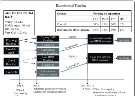
All procedures in this study were approved by our insti-tutions Animal Care and Use Committee. Fourteen young (44 wk.), 7 middle aged (60 wk.), 14 old (86 wk.), and 7 very old (102 wk.) male Fisher 344 rats were used in the study. However, death due to the aging process as well as general anesthesia during various imaging processes resulted in a remainder of 12 young (44 wks.),
6 middle aged, which served as the control (60 wk.), 10 old (86 wk.), and 5 very old, which served as the control (102 wk.) animals that completed the study (see Figure 1 for timeline), which still met the criteria for our origi-nal sample size determination (see power aorigi-nalysis below). Each animal was assessed for functionality (grip strength and motor performance using incline plane) as well as lean, fat, and total body mass using dual-energy X-ray absorptiometry (DXA) pre- and post-treatment (see Figure 1 for experimental design). After baseline measures, 6 young, 6 middle aged control, 5 old, and 5 very old control rats were anesthetized and their right gastrocnemius (GAS) and soleus (SOL) muscles were isolated, blotted, and quickly frozen in liquid nitrogen for laterin vitromolecular analysis. After isolating mus-cles from the right hind limb, a cardiac perfusion proto-col was implemented to drain blood from the rat’s body. Following, the left GAS and SOL muscles of the rats were harvested and directly immersed in 4% paraformal-dehyde for an ex vivoanalysis of myofiber dimensions. Remaining young (44 wk.) and old (86 wk.) rats were given HMB (0.46 g/kg/d) for 16 wk. After the supple-mentation period, the remaining rats were assessed for post-treatment measures in body composition and func-tionality and then sacrificed for in vitro molecularand ex vivoMR analyses.
HMB administration
All animals were raised in our laboratory prior to experimentation, therefore giving us a strong basis for how much HMB should be added to their food. Typi-cally, daily food consumption values ranged from 15-25 g/day for a 250 g rat (or 60-100 g feed/kg BW). Based on their average diet, the HMB dosage was calculated as ~1% CaHMB (Metabolic Technologies Inc., Ames, Iowa, USA), to achieve an ~0.50 g HMB/kg BW/daily dose
Day 1: DXA & Functionality
Measures AGE OF FISHER 344 RATS
Young: 44 wks Middle Aged: 60 wks Old: 86 wks Very Old: 102 wks
Groups Feeding Composition
CHO PRO FAT HMB2
Control 54% 16% 30% 0 %
Intervention: HMB-Treated 54% 16% 30% 1 %
Day 2:
- Treatment groups receive HMB - Sacrifice for molecular analysis
Day 111-112: (16 wks)
Experimental Timeline
12 young
Baseline 6 sacrificed
6 young HMB Condition
6 sacrificed as
Control Condition 6 middle
aged
10 old
Baseline 5 sacrificed
5 very old 5 sacrificed as
Control Condition
5 old HMB Condition
6 young, now middle aged HMB treatment
(aging)
(aging) 5 old, now very old HMB treatment
sacrificed
sacrificed
-DXA / Functionality -Immediate sacrifice for cellular & molecular analysis
a
Compared to
[20]. Based on previous human studies, and assuming a rodents metabolism are at least 6 times more than humans, we chose a 6 gram metabolic equivalent HMB intervention (the upper limit given to humans in research [23]) and calculated a human-to-rodent conver-sion to provide an appropriate, and safe dosage for each animal [20]. Daily food consumption of rats was mea-sured every 6th day by weighing the food remaining and subtracting it from the amount that was administered. Upon termination of this study, the average kilocalories (kcals) for total food consumed, as well as for each macronutrient, were calculated.
Body composition
Dual-energy X-ray absorptiometry (DXA) was per-formed using a Lunar QDR system (iDXA, Lunar Corp., Madison, Wisconsin, USA) with specific software (ver-sion V8-19a) and an internal standard adapted for small animal scans. Total body mass (TBM), lean body mass (LBM), and fat mass (FM) were measured on all ani-mals’pre and post 16 wk. of HMB administration.
Functionality measures
The grip strength test was used as a measure of limb strength [24]. In this procedure, the rats were positioned in front of a force gauge (DFS-101 Force gauge, AME-TEK TCI, CA, USA) so that they could grasp the ten-sion sensitive steel bar of the device with their forelimbs. After visual observation of gripping, the researcher gently pulled back on the rat’s tail until it released its hold on the bar. Force produced was mea-sured in grams. Three trials were performed by the same experienced investigator for each rat throughout the study for consistency and the greatest force was recorded as maximum grip strength, which was then normalized to body mass of each rat.
The inclined plane test was used to assess sensory motor function and hind limb strength [25]. Perfor-mance was determined as the rats’ability to maintain their body position for 5 sec on an inclined plane, while the angle of the surface was changed from 20° to 60° at 2° intervals, with a rest period of at least 5 min.
Muscle isolation
Both right and left hind limb muscles were collected in the National High Magnetic Field Laboratory (NHMFL): one forin vitromolecular analysis and the other for MR analysis. Following anesthesia, precise surgical methods were used to excise the GAS and SOL muscles from the hind limb. Muscles were then frozen in liquid nitrogen. Prior to removing the left calf muscles, a cardiac perfu-sion protocol was implemented to drain blood from the rat’s body since it could interfere with the clarity of the imaging process.
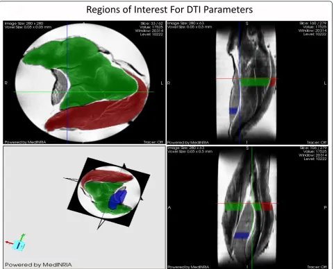
Diffusion tensor imaging (DTI) analysis for myofiber dimensions
For this study we were able to utilize the MR technique termed Diffusion Tensor Imaging (DTI) analysis to study muscle cell architecture at the NHMFL. DTI is based on the principle that the cellular diffusion of water corre-sponds to cell geometry in muscle. The advantage of DTI concerns the ability of random diffusion of water mole-cules to probe with far greater detail then general ima-ging techniques [26,27]. Unlike biopsy techniques, DTI is able to provide the average myofiber dimensions of an entire muscle, as opposed to a small sample of the mus-cle. Part of the DTI analysis involves calculating the mean diffusion of water within a muscle fiber (termed apparent diffusion coefficient, ADC), fractional aniso-tropy (FA) and the 3 principle directions of water diffu-sion denoted as Eigen vectors 1, 2 and 3, representative of the local fiber coordinate system [26,27]. The diffusive transport along the 3 principle directions are denoted as eigenvalues 1, 2, and 3 (l1,l2, andl3) which correspond to diffusive transport along the long axis, as well as the long and short cross-sectional axes of the muscle fibers, respectively [28] (Figure 2). FA is a general measure of the differences in the magnitude of diffusion between the 3 principle directions of diffusion. With smaller cross sectional areas (CSA), FA increases while larger cross sectional areas decrease FA. Thus, FA is inversely pro-portional to myofiber size [26,27].
DTI datasets of the muscles in 7-noncollinear gradient directions were acquired using a widebore 11.75-T verti-cal magnet with a Bruker Avance console and Micro2.5 gradients. Using a 15-mm birdcage coil, spin echo DTI scans were acquired with b values of 0, 500, and 1000 s/ mm2 at an in-plane resolution of 50 × 50 μm2 and a slice thickness of 500 μm. The DTI acquisition para-meters were as follows: TE = 20.5 ms, TR = 2.75 s,Δ= 12.7 ms and δ = 2.1 ms. Also, a high resolution
(40-μm3) 3D gradient-recalled echo (GRE) image was
acquired (TE/TR = 10/150 ms) for anatomical and volu-metric measurements. After acquisition, the images were processed with MedINRIA http://wwwsop.inria.fr/ asclepios/software/MedINRIA/ to calculate diffusion tensor parameters such as: FA, andl1,l2 and l3. The region of interest (ROI) was chosen in the widest region of the GAS and SOL muscle for processing as shown in Figure 3.
Semi-quantitative reverse transcription polymerase reaction (RT-PCR)
7 software. All primer sets have been previously tested for optimal conditions. For each PCR reaction, 18S (with a 324-bp product) was co-amplified with each tar-get cDNA (mRNA) to express each as a ratio of tartar-get mRNA/18S. Images were captured under UV, and mRNA expressions were analyzed via the Bio-Rad
Che-miDoc™XRS imaging system and the Bio-Rad
Quanti-tyOne® software (Bio-Rad Laboratories, Hercules, CA, USA) as described previously [29].
mRNA expression of 4EBP1 was used as a negative marker of protein synthesis, while the E3 ligase atrogin-1 was used as a positive regulator of protein degrada-tion. Mitogenic factors, IGF-IEa and its isoform IGF-IEb (mechano growth factor (MGF)), were used as positive regulators of mitogenesis and myogenesis. Myostatin and its receptor activin IIB were measured as negative regulators of myogenesis. Muscle cell regeneration was
analyzed by transcriptional levels of the myogenic regu-latory factors (MRFs): myogenin and myogenic differen-tiation factor (MyoD).
Statistical analysis
Lean body mass, FM, TBM, functionality (grip strength and incline plane, MR-determined myofiber dimensions and target genes associated with myofiber size were ana-lyzed using one way ANOVA across six groups includ-ing 1 young baseline (44 wks), 2 middle aged (60 wks, control and HMB), 1 old (86 wks.), and 2 very old (102 wks. control and HMB) groups using Statistica (Stat-Soft®, Tulsa, OK, USA) (Figure 1). Significance was set at p ≤0.05, and a tukey post hoc analysis was used to determine which specific mean values differed from others for each variable. The overarching goal of this project was to use MR to examine the impacts of age
and HMB on skeletal muscle cells during the aging pro-cess. Myofiber size was therefore one of the primary outcome measures in this project and provided the basis for the sample sizes as determined by the G*Power ana-lysis software [30,31]. Our rationale for sample size was based on a study by Payne et al. [32]. These investiga-tors found that Fisher 344 rats 102 wks of age demon-strated significant atrophy in the soleus than young adult animals (Effect size (ES) of 3.7). Based on an alpha level of 0.05, a power of 80 and an ES of 3.7, a total of 30 rats (5 per experimental group) were needed to have sufficient power to detect age related changes in myof-ber dimensions.
Results
Food and HMB consumption
All values for food consumed are presented in Table 1. Average total Kcals and Kcals for carbohydrates, protein, and fat were not different between groups.
Body composition
There were no condition effects for LBM. In regards to FM, there were significant condition (p≤0.05, ES = 0.5)
effects, with greater FM (g) in the middle aged (60-wk) control (+49%) but not in the middle aged HMB condi-tion, compared to the baseline young animals (Figure 3). Moreover, FM was significantly lower (-56%) in the very old HMB (102-wk) but not in the control condition compared to the 86 wk. old baseline animals.
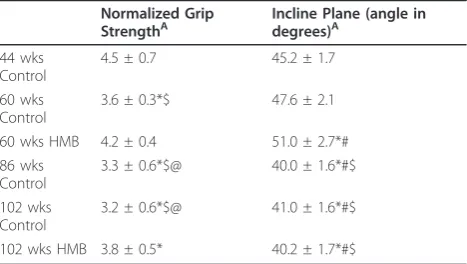
Functionality measures
All test reliability scores for functionality were above .9. There were significant condition (p≤ 0.05, ES = 0.7) effects for normalized grip strength in which strength was lower in the control condition, but was maintained in the HMB condition when comparing 44 to 60 wks. of age animals (Table 2). In old animals, normalized strength increased by 23% (p < 0.05) when comparing 86 to 102 wks. of age with HMB, with no change in the control condition. There was a condition effect (p ≤
0.05, ES = 0.4) for incline plane performance, which was greater in the 60 wk hmb condition than 44 wk condi-tion, but was not different than baseline in the 60 wk control condition. Both old groups declined in incline plane performance relative to the 44 wk baseline group of animals.
Diffusion tensor imaging determined myofiber dimensions
We analyzed the GAS and SOL muscles and calculated the DTI parameters for those muscles (Figure 4). Frac-tional anisotropies (FA), apparent diffusion coefficients (AP), and eigenvalues [33] 1, 2, and 3 were investigated. There was a main condition effect for FA for the GAS (Figure 4A) (p≤ 0.05, ES = 0.5) and SOL (Figure 4B) (p≤0.05, ES = 0.5) muscles (Figure 4). Post hoc analysis revealed that while FA was significantly greater in the 102-wk control from both 44 and 86 wk., the 102-wk HMB condition only differed from 44 wk. No changes
Table 1 Average Kcal consumption for among conditions
Kcals Kcals (CHO) Kcals (PRO) Kcals (Fat)
44 wks Baseline 67.3 ± 4.1 38.9 ± 2.4 19.2 ± 1.2 9.0 ± 0.6 60 wks Control 66.8 ± 1.8 38.7 ± 1.1 19.0 ± 0.5 8.9 ± 0.3 60 wks HMB 65.9 ± 1.5 38.2 ± 0.9 18.7 ± 1.2 8.8 ± 0.6 86 wks Baseline 62.3 ± 6.5 35.5 ± 3.64 17.4 ± 2.0 8.2 ± 0.9 102 wks Control 62.5 ± 5.8 36.1 ± 2.4 17.8 ± 1.0 8.4 ± 0.5 102 wks HMB 63.2 ± 6.19 36.8 ± 3.6 18.1 ± 1.8 8.5 ± 0.8
Table 2 The Effects of Aging and HMB on Neuromuscular Function
Normalized Grip StrengthA
Incline Plane (angle in degrees)A
44 wks Control
4.5 ± 0.7 45.2 ± 1.7
60 wks Control
3.6 ± 0.3*$ 47.6 ± 2.1
60 wks HMB 4.2 ± 0.4 51.0 ± 2.7*#
86 wks Control
3.3 ± 0.6*$@ 40.0 ± 1.6*#$
102 wks Control
3.2 ± 0.6*$@ 41.0 ± 1.6*#$
102 wks HMB 3.8 ± 0.5* 40.2 ± 1.7*#$
A indicates a main condition effect. * indicates p < 0.05, significantly different from 44 wks, $ indicates p < 0.05, significantly different from 60 wks HMB, # indicates p < 0.05, significantly different from 60 wks control, @ indicates p < 0.05, significantly different from 102 wks HMB
in FA occurred from 44 to 60 wk. in any of the condi-tions. There was a main condition effect for the GAS (p
≤0.05, ES = 0.4) and SOL (p≤0.05, ES = 0.4) muscles for l 2, indicative of myofiber CSA. There was also a main condition effect in the GAS (p≤ 0.05, ES = 0.4)
and SOL (p ≤ 0.05, ES = 0.4) muscles for l 3, also
indicative of myofiber CSA. Post hoc analysis revealed that l 2 was lower (p≤ 0.05) in the SOL and GAS in
the 86-wk and 102-wk control group. In addition l 3
declined in the SOL of the 86-wk old condition, and in all muscle groups in the 102-wk control group. How-ever, no changes occurred in the 102-wk HMB
condition or any of the 60-wk conditions for any muscle analyzed. In the GAS, bothl 2 and 3 were greater in the 102-wk HMB than non-HMB condition. No condi-tion effects were found for ADC, or l1, representative of diffusion in the longitudinal axis of the myofibers in any of the muscles analyzed.
Semi-quantitative reverse transcription polymerase reaction
Regulators of protein turnover
No significant condition effects were found for either the SOL or GAS muscles for 4EBP-1 mRNA expression (Figure 5). However, there were significant condition effects for both the soleus (p≤0.05, ES = 0.5) and gas-trocnemius muscles (p≤ 0.05, ES = 0.6) for atrogin-1 mRNA expression. There were condition effects for all muscles for atrogin-1, which was greater in the 102-wk control than all other groups in both the soleus (+ 45%) and gastrocnemius (+100%) muscles. However, the rise was blunted in the soleus in the 102-wk HMB condition.
Positive and negative regulators of mitogenesis
Myostatin mRNA expression was too low in the soleus to process data. For the remaining data sets, no main effects were found for IGF-I, MGF, myostatin, or activin RIIB in any muscles analyzed (Figure 6).
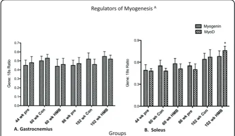
Regulators of myogenesis
There were no main effects in the soleus or gastrocne-mius for MyoD, or for the gastrocnegastrocne-mius in myogenin (Figure 7). However, there was a main group effect in
the soleus for myogenin (p ≤ 0.05, ES = 0.3) which
while approaching significance in the 102-wk control group (p = 0.056) only significantly increased in the 102-wk HMB group relative to the 44-wk group.
Discussion
The primary aim of the present study was to determine the effects of 16 wk. (approximately 15-16% of F344 rats
normal lifespan) of HMB administration in young and old rats on age-related changes in body composition, myofiber dimensions, strength, and incline plane func-tion. The major findings of this study were that HMB blunted negative age-related changes in body composi-tion and muscle cellular dimensions.
Body composition
Results indicated no changes in LBM when comparing young to old rats. Our results agreed with Yu et al. [34] who also found that LBM did not change from young to old age in F344 rats. However, it is possible that the DXA measure of LBM in rats was not sensitive enough to detect age-related sarcopenia, and it’s possible that the cross sectional design underestimates these changes. In general, both human and rodent models have shown to underestimate age-related changes in muscle mass when done in cross sectional designs relative to longitu-dinal designs [35-37]. Our old animals were raised in our laboratory from 44 to 86 weeks of age. While the HMB group continued (16-wk administration) until very old age (102 wk.), the control group was sacrificed at 86
Figure 5Regulators of protein balance in the gastrocnemius and soleus muscles. A indicates a main group effect (p < 0.05), * indicates a significant difference from the 44-wk group (p < 0.05).
Figure 6Regulators of Mitogenesis in the gastrocnemius and soleus muscles. * indicates a significant difference from the 44-wk group (p < 0.05).
wk. of age. Therefore, we performed a quazi-longitudinal comparison between the groups, in which a separate group of 5 control animals were used at 102 wk. in place of those 5 sacrificed at 86 wks. Intriguingly, both groups significantly declined in LBM from 44 to 86 wks. of age, and while this loss was maintained in the old control group, the 102-wk HMB group was no longer significantly lower in LBM than when they were 44 wk. of age (Figure 8). Baier et al. [38] also performed a long-itudinal analysis in over 70 elderly women with an aver-age aver-age of 76 years of aver-age. These subjects were randomly divided into either a cocktail containing HMB or placebo supplemented groups for a 12-month dura-tion. Their results indicated that LBM progressively increased over a 12-month time span when supplement-ing with the nutrition cocktail with no change occurrsupplement-ing in the placebo condition.
Fat mass (FM)
In both humans and the Fisher 344 rat model, FM increases up to 70% of the lifespan, and then plateaus or decreases thereafter [39,40]. In our control rats, FM increased from young to middle age, with no changes occurring from old to very old age. Perhaps the most intriguing finding of our study was that HMB prevented fat gain from young to middle age, and significantly low-ered body fat after the 16-wk HMB administration from the old to very old age. Our results also concur with past animal research, which demonstrated significantly lower hindlimb fat pad weight following HMB
administration in both healthy and dystrophic mice [41]. Interestingly enough, these changes were independent of food intake, which agreed with past research indicating that grams of food consumed may not significantly change with age in the F344 rat model [42], nor with HMB supplementation. To date, the underlying mechanisms that HMB exerts its effects on adipose remain to be elucidated. It may be that HMB directly increases oxidative capacity in myofibers, as exposure of cultured myotubes to the leucine metabolite increased palmitate oxidation by 30% [43].
Muscle strength and sensory motor function
The present study employed a direct measure of grip strength [24], as well as the incline plane test, which has been previously utilized to study both sensory motor function and whole body strength [25]. Sensory motor function is a combination of not only muscle strength, but motor unit recruitment and rate of muscle contrac-tion [44]. For example, recovery of balance following sudden perturbations requires a quick and powerful reflex response to overtake the falling momentum [45]. There was an overall decline in grip strength from 44 to 102 wk. of age. When normalized to body mass how-ever, grip strength declined from 44 to 60 wk. only in the control, but not in the HMB condition. Moreover, normalized grip strength increased by 23% in the old HMB condition from 86 to 102 wk. of age.
In addition, incline plane performance increased from young to middle aged rats that were administered HMB. Our results on overall functionality concur with Flakoll et al. [9] who previously demonstrated that 12 wk. of a cocktail containing HMB (also contained Arginine and Lysine) significantly increased grip strength, leg exten-sion force, as well as get up-and-go performance in older adults. Finally, changes in functionality and strength without detectable changes in LBM may indi-cate an increase in muscle quality. However, this is cur-rently speculative and would need to be verified by future research.
Myofiber dimensions
Previous research with HMB supplementation has been restricted to indirect measures of muscle tissue which include caliper measurements [46,47], DXA analysis [38,48], and limb circumference measures [9]. However, the hallmark of sarcopenia is a decline in muscle mass and then ultimately in myofiber dimensions. To our knowledge, our study is unique as we are the first to view actual changes in muscle cellular dimensions fol-lowing HMB administration throughout senescence. In particular, we employed the diffusion tensor imaging (DTI) technique, which uses a powerful magnet at the NHMFL. This technique has been validated for studying
changes in myofiber dimensions including myofiber length and cross sectional area (CSA) following ischemia reperfusion injury [26,49,50]. As predicted, no changes occurred in myofiber dimensions from 44 to 60 wk. of age. While sarcopenia was evident in the 86-wk and 102-wk control conditions, bothl2 and l3, indicative of myofiber CSA were relatively maintained in the soleus and gastrocnemius muscles of rats consuming HMB. Our results are consistent with previous work from Flakoll [9] and Bair et al. [38] who found that a cocktail containing HMB was able to counter age-related losses in limb circumference. These results are also consistent with several additional muscle wasting models which demonstrated HMB could blunt muscle loss during sepsis [51], cancer [16], limb immobilization [21], and in critically ill trauma patients [52].
Transcript factors associated with myofiber size
Perhaps the most studied aspect of HMB is its effects on protein breakdown. The first research conducted was in humans, which demonstrated that HMB could signifi-cantly lower 3-methylhistadine following strenuous bouts of exercise [23]. However, only recently have its mechanisms of action been elucidated. The current study analyzed atrogin-1, an E3 ligase in the Ubiquitin pathway, which is commonly elevated in muscle wasting conditions such as aging [53,54]. We found that HMB was able to attenuate the age-related rise in atrogin-1 mRNA expression in the soleus muscle. This is impor-tant as atrogin-1 mRNA expression has been demon-strated to be a predictor of long-term changes in proteolysis and muscle wasting [55-57]. Moreover past research has found gene expression of atrogin-1 to be elevated in aging muscle tissue [55,56]. While our research analyzed HMB’s effects on transcription of components of the Ubiquitin pathway, researchers in the Tisdale laboratory have studied direct activity of the Ubiquitin pathway [16,22]. These researchers found that HMB decreased proteasome activity, expression of both alpha and beta subunits of the 20s chamber, and the ATPase subunits of the 19 s caps.
Previous research from Baier and colleagues [38] found that whole body protein synthesis increased up to 14% during a 12-month period when subjects consumed an HMB containing cocktail. We looked at the effects of HMB directly in skeletal muscle on 4EBP-1 gene expres-sion, the inhibitory binding protein that prevents forma-tion of the eukaryotic initiaforma-tion factor 2F complex which is rate limiting to translation initiation [58]. We did not see any aging or supplement effects on 4EBP-1. Our results agreed with Kovarik et al. [51] who found that HMB was able to attenuate a sepsis induced protein catabolic state in rat skeletal muscle primarily by blunt-ing an increase in proteolysis, without preventblunt-ing a
decline in protein synthesis. However, a more recent study by Pimentel et al. [59] found that while HMB sup-plementation increased total mTOR protein expression, and phosphorylation of ribosomal protein s6 kinase (p70s6k) in healthy rats, that it was not able to increase the total protein expression of p70S6K. Thus the com-bined results from protein and gene changes from Pimental et al. [59] and our current study, respectively, may indicate that HMB does not directly regulate the expression of these two downstream targets of mTOR.
Positive and negative regulators of mitogenesis and myogenesis
In our previous research with old female rats, we found that IGF-IEa mRNA expression was increased in a group administered HMB during 10-wk resistance train-ing [60]. The current study found no significant main effects for myostatin, MGF, or IGF. However, past research found that the addition of HMB to serum-starved myoblasts increased IGF-I mRNA in a dose dependent manner. It is possible that the more robust effects seen in cell culture are due to a greater overall direct exposure of myocytes to HMB, as this study con-firmed that HMB’s effects on IGF-I were dose depen-dent. However, future research will need to be conducted to examine if higher doses elicit differential responses in animal studies.
MyoD and myogenin were taken as early and late reg-ulators of satellite cell differentiation, respectively [61]. Our results showed a main group effect for myogenin in the soleus. However, this regulator of differentiation only significantly increased in the 102-wk. HMB condi-tion, and not in the 102-wk. control condition. While it is tempting and certainly possible to suggest that HMB was at least partially responsible for this increase, it is more easily explained by a compensatory process accompanying the aging process [62] as the control con-dition very closely approximated a significant rise as well (p = 0.07).
Conclusions
our study only began to elucidate the mechanisms this supplement works through, we did find that it lowered the E3 ligase atrogin-1, which is involved in a rate-limit-ing step in Ubiquitination of target substrates for degra-dation. It is suggested that future studies look directly at changes in myofiber growth with an in vivo MR DTI technique on the same animals over time concurrently analyzing changes in protein content of its regulators.
Acknowledgements
We would like to thank Dr. John A. Rathmacher, Metabolic Technologies Inc., Ames, Iowa for supplying us with CaHMB and Dr. Neema Bakhshalian, Kenneth Leonard, and Michael Zourdos for their great contributions on the present study. Special thanks to Ryan P Lowery for his contributions on our manuscript.
Author details
1Department of Nutrition, Food and Exercise Sciences, The Florida State
University, Tallahassee, FL, USA.2Department of Health Sciences and Human Performance, The University of Tampa, Tampa, FL, USA.3The National High Magnetic Field Laboratory & Department of Chemical & Biomedical Engineering, The Florida State University, Tallahassee, FL, USA.4Military Performance Division, United States Army Research, Institute of Environmental Medicine, Natick, MA, USA.5Biomedical Engineering Department, College of Engineering, King Faisal University, Al-Ahsa, Saudi Arabia.6Sport and Exercise Science, College of Education, University of Central Florida, Orlando, FL.7Department of Health and Exercise Science, University of Oklahoma, Norman, Oklahoma, USA.
Authors’contributions
J-SK was a PI for the present study responsible for funding, providing resources, study design, supervising data collection and tissue analysis, and manuscript preparation. JMW was responsible for study design, data collection, molecular and gene analysis, and manuscript preparation. SCG and IM assisted in study design, data collection and conducted the myofiber dimension analysis. S-rL, Y-mP and PCH assisted in data collection/analysis for the study, and harvesting of tissues. BHA and LBP assisted in funding, providing resources, and manuscript preparation. JRS and JPL helped extensively in manuscript preparation. All authors read and approved final manuscript.
Competing interests
The authors declare that they have no competing interests.
Received: 7 December 2011 Accepted: 18 April 2012 Published: 18 April 2012
References
1. Kuczmarski RJOC, Gummer-Strawn LM, Flegal KM, Guo SS, Wei R, Mei Z, Curtin LR, Roche AF, Johnson CL:CDC growth charts: United States.
Advance data from vital and health statistics.National Center for Health
Statistics; 2000314(1).
2. Larsson L, Grimby G, Karlsson J:Muscle strength and speed of movement
in relation to age and muscle morphology.J Appl Physiol1979,
46(3):451-456.
3. Volpi E, Sheffield-Moore M, Rasmussen BB, Wolfe RR:Basal muscle amino acid kinetics and protein synthesis in healthy young and older men. JAMA2001,286(10):1206-1212.
4. Avlund KSM, Davidsen M, Lovborg B, Rantanen T:Maximal isometric muscle strength and functional ability in daily activities among
75-year-old men and women.Scand J Med Sci Sports1994,4(1):32-40.
5. Janssen I, Heymsfield SB, Ross R:Low relative skeletal muscle mass (sarcopenia) in older persons is associated with functional impairment
and physical disability.J Am Geriatr Soc2002,50(5):889-896.
6. Dela F, Kjaer M:Resistance training, insulin sensitivity and muscle
function in the elderly.Essays Biochem2006,42:75-88.
7. Langlois JA, Visser M, Davidovic LS, Maggi S, Li G, Harris TB:Hip fracture risk in older white men is associated with change in body weight from
age 50 years to old age.Arch Intern Med1998,158(9):990-996.
8. Vukovich MD, Sharp RL, Kesl LD, Schaulis DL, King DS:Effects of a low-dose amino acid supplement on adaptations to cycling training in untrained individuals.Int J Sport Nutr1997,7(4):298-309.
9. Flakoll P, Sharp R, Baier S, Levenhagen D, Carr C, Nissen S:Effect of beta-hydroxy-beta-methylbutyrate, arginine, and lysine supplementation on strength, functionality, body composition, and protein metabolism in
elderly women.Nutrition2004,20(5):445-451.
10. Katsanos CS, Kobayashi H, Sheffield-Moore M, Aarsland A, Wolfe RR:A high proportion of leucine is required for optimal stimulation of the rate of
muscle protein synthesis by essential amino acids in the elderly.Am J
Physiol Endocrinol Metab2006,291(2):E381-387.
11. Combaret L, Dardevet D, Rieu I, Pouch MN, Bechet D, Taillandier D, Grizard J, Attaix D:A leucine-supplemented diet restores the defective postprandial inhibition of proteasome-dependent proteolysis in aged rat skeletal muscle.J Physiol2005,569(Pt 2):489-499.
12. Fujita S, Volpi E:Amino acids and muscle loss with aging.J Nutr2006,
136(1 Suppl):277S-280S.
13. Kim JS, Wilson JM, Lee SR:Dietary implications on mechanisms of
sarcopenia: roles of protein, amino acids and antioxidants.J Nutr
Biochem2010,21(1):1-13, doi:10.1016/j.jnutbio.2009.06.014. 14. Wilson GJ, Wilson JM, Manninen AH:Effects of
beta-hydroxy-beta-methylbutyrate (HMB) on exercise performance and body composition
across varying levels of age, sex, and training experience: A review.Nutr
Metab (Lond)2008,5:1.
15. Van Koevering M, Gill DR, Smith RA, Owens F, Nissen S, Ball R:Effect ofβ
-hydroxy-β-methyl butyrate on the health and performance of
shipping-stressed calves.Oklahoma State Univ Res Rep; 1993, 312-331.
16. Smith HJ, Mukerji P, Tisdale MJ:Attenuation of proteasome-induced proteolysis in skeletal muscle by {beta}-hydroxy-{beta}-methylbutyrate in
cancer-induced muscle loss.Cancer Res2005,65(1):277-283.
17. Smith HJ, Lorite MJ, Tisdale MJ:Effect of a cancer cachectic factor on protein synthesis/degradation in murine C2C12 myoblasts: modulation
by eicosapentaenoic acid.Cancer Res1999,59(21):5507-5513.
18. Eley HL, Russell ST, Baxter JH, Mukerji P, Tisdale MJ:Signaling pathways initiated by beta-hydroxy-beta-methylbutyrate to attenuate the depression of protein synthesis in skeletal muscle in response to
cachectic stimuli.Am J Physiol Endocrinol Metab2007,293(4):E923-931.
19. May PE, Barber A, D’Olimpio JT, Hourihane A, Abumrad NN:Reversal of cancer-related wasting using oral supplementation with a combination
of beta-hydroxy-beta-methylbutyrate, arginine, and glutamine.Am J Surg
2002,183(4):471-479.
20. Cohen DD:The effect ofβ-hydroxy-β-methylbutyrate (HMB) and resistance training on changes in body composition during positive and
negative energy balance - a randomized double-blind study.London:
Queen Mary and Westfield College, University of London; 1997. 21. Soares JMC, Póvoas S, Neuparth MJ, Duarte JA:The effects of
beta-hydroxy-beta-methylbuturate (HMB) on muscle atrophy induced by
immobilization.Med Sci Sports Exerc2001,33(5), supp 140.
22. Smith HJ, Wyke SM, Tisdale MJ:Mechanism of the attenuation of proteolysis-inducing factor stimulated protein degradation in muscle by
beta-hydroxy-beta-methylbutyrate.Cancer Res2004,64(23):8731-8735.
23. Cabe PA, Tilson HA, Mitchell CL, Dennis R:A simple recording grip
strength device.Pharmacol Biochem Behav1978,8(1):101-102.
24. Rivlin AS, Tator CH:Objective clinical assessment of motor function after
experimental spinal cord injury in the rat.J Neurosurg1977,47(4):577-581.
25. Heemskerk AM, Drost MR, van Bochove GS, van Oosterhout MF, Nicolay K, Strijkers GJ:DTI-based assessment of ischemia-reperfusion in mouse
skeletal muscle.Magn Reson Med2006,56(2):272-281.
26. Heemskerk AM, Strijkers GJ, Drost MR, van Bochove GS, Nicolay K:Skeletal muscle degeneration and regeneration after femoral artery ligation in
mice: monitoring with diffusion MR imaging.Radiology2007,
243(2):413-421.
27. Andersen JL:Muscle fibre type adaptation in the elderly human muscle. Scand J Med Sci Sports2003,13(1):40-47.
28. Kim JS, Cross JM, Bamman MM:Impact of Resistance Loading on Myostatin Expression and Cell Cycle Regulation in Young and Older
29. Faul F, Erdfelder E, Lang AG, Buchner A:G*Power 3: a flexible statistical power analysis program for the social, behavioral, and biomedical
sciences.Behav Res Methods2007,39(2):175-191.
30. Faul F, Erdfelder E, Buchner A, Lang AG:Statistical power analyses using
G*Power 3.1: tests for correlation and regression analyses.Behav Res
Methods2009,41(4):1149-1160, doi:10.3758/BRM.41.4.1149.
31. Payne AM, Dodd SL, Leeuwenburgh C:Life-long calorie restriction in Fischer 344 rats attenuates age-related loss in skeletal muscle-specific
force and reduces extracellular space.J Appl Physiol2003,95(6):2554-2562.
32. FAO/WHO/UNU:Energy and Protein Requirements. Technical Report
Series.World Health Organization, Switzerland Geneva; 1989724.
33. Yu BP, Masoro EJ, Murata I, Bertrand HA, Lynd FT:Life span study of SPF Fischer 344 male rats fed ad libitum or restricted diets: longevity,
growth, lean body mass and disease.J Gerontol1982,37(2):130-141.
34. Lushaj EB, Johnson JK, McKenzie D, Aiken JM:Sarcopenia accelerates at
advanced ages in Fisher 344 × Brown Norway rats.J Gerontol2008,
63(9):921-927.
35. Lexell J, Taylor CC, Sjostrom M:What is the cause of the ageing atrophy? Total number, size and proportion of different fiber types studied in
whole vastus lateralis muscle from 15- to 83-year-old men.J Neurol Sci
1988,84(2-3):275-294.
36. Frontera WR, Hughes VA, Fielding RA, Fiatarone MA, Evans WJ, Roubenoff R:
Aging of skeletal muscle: a 12-yr longitudinal study.J Appl Physiol2000,
88(4):1321-1326.
37. Baier S, Johannsen D, Abumrad N, Rathmacher JA, Nissen S, Flakoll P: Year-long changes in protein metabolism in elderly men and women supplemented with a nutrition cocktail of
beta-hydroxy-beta-methylbutyrate (HMB), L-arginine, and L-lysine.JPEN J Parenter Enteral
Nutr2009,33(1):71-82.
38. Bertrand HA, Lynd FT, Masoro EJ, Yu BP:Changes in adipose mass and cellularity through the adult life of rats fed ad libitum or a
life-prolonging restricted diet.J Gerontol1980,35(6):827-835.
39. Prentice AM, Jebb SA:Beyond body mass index.Obes Rev2001,
2(3):141-147.
40. Payne ET, Yasuda N, Bourgeois JM, Devries MC, Rodriguez MC, Yousuf J, Tarnopolsky MA:Nutritional therapy improves function and complements corticosteroid intervention in mdx mice.Muscle & nerve2006,33(1):66-77. 41. Black BJ Jr, McMahan CA, Masoro EJ, Ikeno Y, Katz MS:Senescent terminal
weight loss in the male F344 rat.Am J Physiol Regul Integr Comp Physiol
2003,284(2):R336-342, doi:10.1152/ajpregu.00640.2001.
42. Ransone J, Neighbors K, Lefavi R, Chromiak J:The effect of beta-hydroxy beta-methylbutyrate on muscular strength and body composition in collegiate football players.J Strength Cond Res2003,17(1):34-39. 43. Skelton DA, Greig CA, Davies JM, Young A:Strength, power and related
functional ability of healthy people aged 65-89 years.Age Ageing1994,
23(5):371-377.
44. Toraman A, Yildirim NU:The falling risk and physical fitness in older people.Arch Gerontol Geriatr2010,51(2):222-226, doi:10.1016/j. archger.2009.10.012.
45. Panton LB, Rathmacher JA, Baier S, Nissen S:Nutritional supplementation of the leucine metabolite beta-hydroxy-beta-methylbutyrate (hmb) during resistance training.Nutrition2000,16(9):734-739.
46. Portal S, Zadik Z, Rabinowitz J, Pilz-Burstein R, Adler-Portal D, Meckel Y, Cooper DM, Eliakim A, Nemet D:The effect of HMB supplementation on body composition, fitness, hormonal and inflammatory mediators in elite adolescent volleyball players: a prospective randomized,
double-blind, placebo-controlled study.Eur J Appl Physiol2011, doi:10.1007/
s00421-011-1855-x.
47. Vukovich MD, Stubbs NB, Bohlken RM:Body composition in 70-year-old adults responds to dietary beta-hydroxy-beta-methylbutyrate similarly to
that of young adults.J Nutr2001,131(7):2049-2052.
48. Galban CJ, Maderwald S, Uffmann K, Ladd ME:A diffusion tensor imaging analysis of gender differences in water diffusivity within human skeletal
muscle.NMR Biomed2005,18(8):489-498.
49. Zaraiskaya T, Kumbhare D, Noseworthy MD:Diffusion tensor imaging in
evaluation of human skeletal muscle injury.J Magn Reson Imaging2006,
24(2):402-408.
50. Kovarik M, Muthny T, Sispera L, Holecek M:Effects of beta-hydroxy-beta-methylbutyrate treatment in different types of skeletal muscle of intact
and septic rats.J Physiol Biochem2010,66(4):311-319,
doi:10.1007/s13105-010-0037-3.
51. Kuhls DA, Rathmacher JA, Musngi MD, Frisch DA, Nielson J, Barber A, MacIntyre AD, Coates JE, Fildes JJ:Beta-hydroxy-beta-methylbutyrate
supplementation in critically ill trauma patients.J Trauma2007,
62(1):125-131, doi:10.1097/TA.0b013e31802dca93. discussion 131-122.
52. Nissen S, Sharp R, Ray M, Rathmacher JA, Rice D, Fuller JC Jr, Connelly AS, Abumrad N:Effect of leucine metabolite
beta-hydroxy-beta-methylbutyrate on muscle metabolism during resistance-exercise training.J Appl Physiol1996,81(5):2095-2104.
53. Edstrom E, Altun M, Hagglund M, Ulfhake B:Atrogin-1/MAFbx and MuRF1
are downregulated in aging-related loss of skeletal muscle.J Gerontol
2006,61(7):663-674.
54. Gomes MD, Lecker SH, Jagoe RT, Navon A, Goldberg AL:Atrogin-1, a muscle-specific F-box protein highly expressed during muscle atrophy. Proc Natl Acad Sci USA2001,98(25):14440-14445.
55. Clavel S, Coldefy AS, Kurkdjian E, Salles J, Margaritis I, Derijard B: Atrophy-related ubiquitin ligases, atrogin-1 and MuRF1 are up-regulated in aged
rat Tibialis Anterior muscle.Mech Ageing Dev2006,127(10):794-801.
56. Pattison JS, Folk LC, Madsen RW, Booth FW:Selected Contribution: Identification of differentially expressed genes between young and old rat soleus muscle during recovery from immobilization-induced atrophy. J Appl Physiol2003,95(5):2171-2179.
57. Sacheck JM, Ohtsuka A, McLary SC, Goldberg AL:IGF-I stimulates muscle growth by suppressing protein breakdown and expression of
atrophy-related ubiquitin ligases, atrogin-1 and MuRF1.Am J Physiol Endocrinol
Metab2004,287(4):E591-601.
58. Anthony JC, Yoshizawa F, Anthony TG, Vary TC, Jefferson LS, Kimball SR:
Leucine stimulates translation initiation in skeletal muscle of
postabsorptive rats via a rapamycin-sensitive pathway.J Nutr2000,
130(10):2413-2419.
59. Pimentel GD, Rosa JC, Lira FS, Zanchi NE, Ropelle ER, Oyama LM, Oller do Nascimento CM, de Mello MT, Tufik S, Santos RV: beta-Hydroxy-beta-methylbutyrate (HMbeta) supplementation stimulates skeletal muscle
hypertrophy in rats via the mTOR pathway.Nutr Metab (Lond)2011,
8(1):11, doi:10.1186/1743-7075-8-11.
60. Park Y-M, Lee S-R, Wilson JM, Henning P, Grant S, Rathmacher J, Kim J-S:
Effects ofβ-hydroxy-β-methylbutyrate (HMB) on Muscle IGF-I and MGF
mRNA Expression in Aged Female Rats during 10-Week Resistance Training.FASEB2010,21:621-624.
61. Kim JS, Kosek DJ, Petrella JK, Cross JM, Bamman MM:Resting and load-induced levels of myogenic gene transcripts differ between older adults
with demonstrable sarcopenia and young men and women.J Appl
Physiol2005,99(6):2149-2158.
62. Marsh DR, Criswell DS, Carson JA, Booth FW:Myogenic regulatory factors
during regeneration of skeletal muscle in young, adult, and old rats.J
Appl Physiol1997,83(4):1270-1275.
doi:10.1186/1550-2783-9-18
Cite this article as:Wilsonet al.:Beta-hydroxy-beta-methyl-butyrate blunts negative age-related changes in body composition, functionality and myofiber dimensions in rats.Journal of the International Society of Sports Nutrition20129:18.
Submit your next manuscript to BioMed Central and take full advantage of:
• Convenient online submission
• Thorough peer review
• No space constraints or color figure charges
• Immediate publication on acceptance
• Inclusion in PubMed, CAS, Scopus and Google Scholar
• Research which is freely available for redistribution