R E S E A R C H A R T I C L E
Open Access
Essential infantile esotropia: postoperative motor
outcomes and inferential analysis of strabismus
surgery
Adriano Magli
1,4*, Roberta Carelli
1, Francesco Matarazzo
2and Dario Bruzzese
3Abstract
Background:The aim of this retrospective study is a long-term evaluation of postoperative motor outcomes and the inferential analysis of strabismus surgery in infant eyes with essential infantile esotropia.
Methods:576 patients were compatible with the criteria: confirmed EIE diagnosis, angle≥30 pD, absence of associated ocular anomalies, onset by 6 months of age, absence of hyperopia > 3 Diopters, operation before age 4. Preoperative deviation classes (30–40 pD, 41–59 pD,≥60 pD) were established, different types of surgery were performed. Follow-up was conducted for 5 years after surgery. Longitudinal data were analyzed using general linear mixed models stratified according to the class of pre-operative deviation. A random intercept and a random slope with time (in months) was assumed with an unstructured within subject correlation structure for repeated measurements. Results:In patients with preoperative angle≤40 pD, a significant interaction effect for intervention by time (F5,155.9=
3.56, p = 0.004) and a significant intervention effect (F5,226.1= 6.41, p < 0.001) on residual deviation were observed; only
the intervention 5 showed a residual deviation inside the limits of a partial success. In Class 41-59, a significant interaction effect for intervention by time (F4,166.7= 5.16, p = 0.001), intervention (F4,178.1= 2.48, p = 0.046) and time
(F1,174.6= 9.99, p = 0.002) on residual deviation were observed; intervention 7 had the highest degree of stability
showing an outcome within the range of a partial success. In Class≥60 pD no significant effect for intervention (F4,213.9= 0.74, p = 0.567), time (F1,169.5= 0.33, p = 0.569) or intervention by time (F4,160.9= 1.08, p = 0.368) on residual
deviation was observed; intervention 3,6 and 7 resulted in a residual deviation within the range of a partial success. Conclusions:We suggest, where possible, a two-horizontal muscles approach in small angle EIE, while a multiple muscles surgery in large angle EIE.
Keywords:Essential infantile esotropia, Motor outcomes, Surgical techniques, Long-term follow-up, Inferential analysis
Background
Essential Infantile Esotropia (EIE) typically onsets within the first 6 months of age in absence of central nervous system abnormalities, exhibiting strabismus angle >30 pD, slight hyperopia, medium grade amblyopia, dissoci-ated vertical deviation, latent nystagmus, oblique muscle dysfunction, abduction restriction and excessive adduc-tion, cross-fixation and reduced or absent binocular vision [1-4].
The incidence of EEI is 1%, according to some authors [5-7]; Archer et al. [8] estimate an incidence at around 0.5% while Helveston and co-workers [9] found it nearer to 0.1%.
Ophthalmological and orthoptic examinations are car-ried out in the strabismus patient mainly in order to: diagnose or rule out the presence of amblyopia, establish strabismus type, evaluate and define deviation angle, analyze sensory state (for unilateral or alternating exclu-sion, central suppresexclu-sion, anomalous retinal correspond-ence, VBS) [10] and stereopsis.
Without doubt, EIE therapy is centred on surgery, however surgical intervention is subordinate to several conditions: horizontal deviation stability, estimation of * Correspondence:[email protected]
1Department of Ophthalmology, Pediatric Unit, University of Salerno, Salerno,
Italy
4Gi.Ma Center, Via Mergellina 44, 80100 Naples, Italy
Full list of author information is available at the end of the article
vertical deviation (if any), absence of accommodative factors, presence of alternating fixation (spontaneous or re-educated), sufficient patient cooperation to set up an appropriate operative program.
Methods
The aim of the study carried out by our Paediatric Oph-thalmic Department, is the long-term evaluation of post operative follow-up of EIE patients, operated within the first 4 years of life.
A retrospective review was undertaken of all EIE sur-geries encoded in our database performed by the senior author at is private practice and at the University hos-pital. Charts of 1248 patients with infantile esotropia who underwent different surgical procedures from 1980 through 2007 were reviewed.
We evaluated patient history, clinical presentation, workup, treatment and outcomes. Between one and three pre-surgical examinations were carried out, in order to es-tablish angle stability. Patients were followed up at 3 months, 6 months, 1 year and 5 years after surgery.
The study inclusion criteria are: confirmed EIE diagno-sis, angle≥30 pD, absence of associated ocular anomal-ies, EIE onset by 6 months of age, absence of hyperopia > 3 Diopters.
All children included in our study were alterning pre-operatively; surgical intervention was subordinate to the presence of alternating fixation (spontaneous or re-educated). Patients with only one follow-up or without reliable pre and postoperative measurements were ex-cluded. Patients were also excluded if any CNS abnor-mality was diagnosed.
No surgery was performed after age 4. All patients had never undergone extraocular muscle surgery before our study.
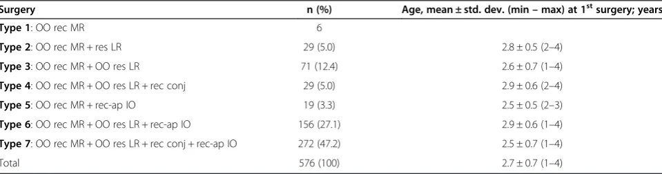
7 types of surgery were performed, identified by num-bers 1 to 7 (Table 1). Type 1 surgery (OO rec RM) pa-tients were excluded from the data analysis, due to too small sample for statistical evaluation.
Finally, our statistical sample included 576 patients, compatible with all the above mentioned criteria.
Several parameters were estimated both pre-operatively and post-operatively:
Presence/absence of familiarity and systemic anomalies; Type of esotropia;
Horizontal deviation (estimated after the complete correction of the refractive error when present; the mean deviation between near and distant is measured by Krimsky test, or, when possible, with cover-test);
Deviation variability (variability was defined when the horizontal deviation angle showed a difference ≥ 15 pD between the first, second and third pre-operative visit; patients presenting variability were excluded from the statistical data analysis);
Ductions and versions, including Inferior Oblique (IO), Superior Oblique (SO), Lateral Rectus (LR), Super-ior Rectus (SR) and InferSuper-ior Rectus (IR) muscles hyper/ hypo functions and LR Pseudoparalysis;
Hyperopia; Up-shoot (an arbitrary scale from (+) to (++++) was set up, where the greatest number of + indi-cated the greatest up-shoot; both RE and LE up-shoots were reported, thus bringing to light any differences be-tween the two eyes);
Alphabetical variations (V or A variations), DVD, anomalous head position, nystagmus, cross-fixation and sensory state evaluation.
Complete eye examination was performed, with Visual Acuity (UCVA and BCVA) and Amblyopia as-sessment. Classification of amblyopia degree is variable among different authors. We consider mild amblyopia as a BCVA greater than 0.2 logMAR, medium grade or moderate amblyopia a BCVA between 0.2 and 0.5 logMAR, severe amblyopia a BCVA lower than 0.5 logMAR.
Follow-ups were carried out at 3 months, 6 months, 1 year, 5 years. All patients were operated under general anaesthesia.
All patients were discharged day after surgery. Patients were treated with antibiotic-corticosteroids eye drops for 10 days following operation. Informed consent was obtained prior to surgery from the parents, after inform-ing of all peri and post-operative risks. All surgical
Table 1 Different surgical approaches
Surgery n (%) Age, mean ± std. dev. (min–max) at 1stsurgery; years
Type 1: OO rec MR 6
Type 2: OO rec MR + res LR 29 (5.0) 2.8 ± 0.5 (2–4)
Type 3: OO rec MR + OO res LR 71 (12.4) 2.6 ± 0.7 (1–4)
Type 4: OO rec MR + OO res LR + rec conj 29 (5.0) 2.9 ± 0.6 (2–4)
Type 5: OO rec MR + rec-ap IO 19 (3.3) 2.5 ± 0.5 (2–3)
Type 6: OO rec MR + OO res LR + rec-ap IO 156 (27.1) 2.9 ± 0.6 (1–4)
Type 7: OO rec MR + OO res LR + rec conj + rec-ap IO 272 (47.2) 2.5 ± 0.7 (1–4)
Total 576 (100) 2.7 ± 0.7 (1–4)
procedures were conducted in accordance with the Helsinki Declaration.
Patients were divided into classes according to degree of deviation, surgical approach and age at surgery.
Regarding deviation, patients were grouped in three classes. Class 1: 30–40 pD, Class 2: 41–59 pD, Class 3:≥ 60 pD.
Post-operative success was defined by the following criteria: totally successful (0 pD deviation); partially suc-cessful (deviation > 0 and≤+10 pD or deviation < 0 and≥ -10 pD), unsuccessful (deviation > + 10 pD and < -10 pD). Concerning statistical analysis, SPSS software (version 20.0; SPSS Inc., Chicago, IL, USA) and R (version 2.5.0; The R Foundation for Statistical Computing) were used. Analyses included only available data and missing values were not imputed.
Data were summarized as means ± standard deviation for continuous variables and as frequencies (%) for cat-egorical variables.
Longitudinal data were analyzed using general linear mixed models stratified according to the class of pre-operative deviation. A random intercept and a random slope with time (in months) was assumed with an unstructured within subject correlation structure for re-peated measurements. All the models included interven-tion type, time period, and the interacinterven-tion between intervention and time, and were further adjusted for the following covariates: preoperative deviation (pD), pres-ence of nistagmus (Yes/No),prespres-ence of Lateral Recti muscles Hypofunctions (Yes/No), presence of Inferior oblique muscles hyperfunctions (Yes/No), age at inter-vention (years) and subsequent interinter-vention (Yes/No).
Results
The sample included 576 patients (249 F and 327 M). Mean age at surgery was 2.7 ± 0.7 (1–4); 68 (11.8%) pa-tients were operated after 3 years of age.
Regarding the sample distribution: 210 patients (36.5%) had a deviation between 30 and 40 pD, 181 (31.4%) between 41 and 59 pD and 185 children (32.1%) had a pre-operative angle≥60 pD. The mean pre-operative deviation was 49.8 ± 13.2 pD, ranging from 30 to 100 pD.
According to surgery type (Table 1) our patients showed greater frequency of type 7 (47.2%) and type 6
(27.1%) interventions while intervention 5 was the least frequent (3.3%), with different distribution in the differ-ent deviation classes. Mean age at first surgery varied slightly according to surgical approach.
Table 2 describes the different deviations found during follow-up visits (3 months, 6 months, 1 year and 5 years). 13.0% of patients achieved 1 to 10 pD esotropia at 5 years follow-up; 18.4% of patients had persistent exotropia less than 10 pD and 34.9% had orthotropia. Number of pa-tients under examination decreases during follow-up, due to patient drop-out for various reasons. Frequency distribution of the 576 subjects, classified according to surgery and according to pre-operative deviation (3 types) is shown in Table 3.
Data concerning entity of recessions and resections in terms of mm are reported in Table 4.
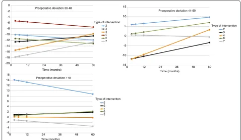
Longitudinal analysis. Mean post-operative residual deviations, with 95% confidence intervals (CIs), for each intervention type at 3 months, 6 months, 1 year and 5 years, classified according to the class of pre-operative deviation are presented in Figure 1, Tables 5, 6 and 7.
Class 30–40 pD. A significant interaction effect for intervention by time (F5,155.9= 3.56, p = 0.004) and a
sig-nificant intervention effect (F5,226.1= 6.41, p < 0.001) on
residual deviation were observed. No significant effect for time emerged (F1,155.8= 1.9, p = 0.168). The covariate
of a second intervention was also observed to have a significant impact on residual deviation (F1,161.4= 80.44,
p < 0.001) with an average reduction of 17.4 pD (95% C.I. 13.6 pD; 21.3 pD).
Class 41–59 pD. A significant interaction effect for intervention by time (F4,166.7= 5.16, p = 0.001),
interven-tion (F4,178.1= 2.48, p = 0.046) and time (F1,174.6= 9.99,
p = 0.002) on residual deviation were observed. The fol-lowing covariates also had a significant impact on re-sidual deviation: Nistagmus (F1,144.4= 6.34, p = 0.013)
and Lateral Recti Hypofunction (F1,149.6= 28.93, p <
0.001) causing, during 5 years follow up after the inter-vention, an average increase in the residual deviation of 4.8 pD (95% C.I. 1.1 pD; 8.5 pD) and 7.0 pD (95% C.I. 4.4 dP; 9.5 pD) respectively.
Class≥60 pD. No significant effect for intervention (F4,213.9= 0.74, p = 0.567), time (F1,169.5= 0.33, p = 0.569)
or intervention by time (F4,160.9= 1.08, p = 0.368) on
re-sidual deviation was observed. The covariate Lateral
Table 2 Angle deviation (pD) during follow-up
Follow-up Ocular alignment (pD)
<−10Δ From−10Δto−1Δ 0Δ From +1Δto +10Δ > + 10Δ Missing
3 months n (%) 111 (19.3) 150 (26.0) 131 (22.7) 114 (19.8) 64 (11.1) 6 (1.1)
6 months n (%) 99 (17.2) 144 (25.0) 140 (24.3) 123 (21.4) 59 (10.2) 11 (1.9)
1 year n (%) 95 (16.5) 137 (23.8) 184 (31.9) 68 (11.8) 74 (12.9) 18 (3.1)
Recti Hypofunctions was also observed to have a signifi-cant impact on residual deviation (F1,152.8= 7.07, p =
0.009) with an average increase of 4.9 pD (95% C.I. 1.3 pD; 8.6 pD).
Concerning reoperations, 57 patients out of 576 (9.9%) underwent a second surgery. Type 5 surgery lead to highest reoperation rate (52% of patients who underwent type 5).
Discussion
Aims of all EIE surgery are the early or near recovery of orthotropy with the lowest possible number of surgeries.
The traditional surgical approach, maintained by sev-eral authors, is medial recti bilatsev-eral recession [10-16].
In cases of small deviation angles, majority of authors suggest the double recession of the medial recti, possibly associated with unilateral lateral recti resection.
Optimal surgical technique for large-angle esotropia correction is still controversial. Surgery can be per-formed on 3 muscles, with double medial recti recession and unilateral lateral rectus resection [17] or on 4 mus-cles (double medial recti recession with bilateral resec-tion of the lateral recti) [18].
According to Vroman et al., the success rate for ocular realignment in patients with EIE by using bilateral med-ial rectus muscle recession did not appear to diminish when applied to deviations greater than 50 pD, as com-pared with smaller angle deviations. In their opinion, surgery on 3 or 4 horizontal rectus muscles may be un-necessary in the treatment of patients with very large an-gles [19]. Some surgeons prefer a large medial recti recession [20-22]. A seven-millimeter bilateral medial rectus recession is considered by some authors a pos-sible approach in large angle congenital esotropia [23,24], as alternative to multiple muscle procedures in the initial treatment of large-angle EIE, leaving the lat-eral rectus muscles unoperated for future surgeries, if necessary. Those who support large recession [25,26], such as Helveston and Prieto-Diaz, claim that in their cases [26], this surgical choice led to a reduction in re-interventions number, obtaining eye alignment in over 70% of cases. On the other hand, some authors suggest in large-angle infantile esotropia (≥60 PD) a three hori-zontal muscle surgery, appearing to have a good long-term success rate without leading to high rates of either residual esotropia or consecutive exotropia [27,28].
In our opinion, in essential infantile esotropia, surgery on fewer muscles might lead to an increase in surgical dosage (supramaximal recessions) with greater incomi-tances in lateral gaze positions. Therefore, we rarely Table 3 Distribution of the 576 subjects used in
longitudinal analysis classified according to the surgery type and the pre-operative deviation class
Class of pre-operative deviation Type of intervention 30-40 41-59 ≥60
2 23 (79.4) 3 (10.3) 3 (10.3)
3 27 (38.1) 16 (22.5) 28 (39.4)
4 16 (55.2) 6 (20.7) 7 (24.1)
5 19 (100.0) 0 (0.0) 0 (0.0)
6 76 (48.7) 30 (19.2) 50 (32.1)
7 49 (18.0) 126 (46.3) 97 (35.7)
Total 210 (36.5) 181 (31.4) 185 (32.1)
Table 4 Descriptive analysis of millimeters (mm) of intervention, mean, median and range according to the different surgery types
Type of intervention
mm of intervention
RE LE RE LE RE LE RE LE
rec MR rec MR res LR res LR rec conj rec conj rec anter IO rec anter IO
2 Mean ± Std. Dev. 4.22 ± 0.51 4.41 ± 0.57 8 ± 0.53 7.38 ± 0.74
Median [min; max] 4 [3; 5] 4 [3; 5] 8 [7; 9] 7 [6; 9]
3 Mean ± Std. Dev. 4.02 ± 0.59 4.01 ± 0.65 6.65 ± 0.86 6.7 ± 0.85
Median [min; max] 4 [3; 6] 4 [3; 7] 6 [5; 9] 7 [5; 9]
4 Mean ± Std. Dev. 4.59 ± 0.61 4.53 ± 0.6 7.22 ± 0.64 7.12 ± 1.05 3.83 ± 0.54 3.85 ± 0.53
Median [min; max] 5 [3; 5.5] 5 [3; 5.5] 7 [6; 9] 7 [4; 9] 4 [2; 4] 4 [2; 4]
5 Mean ± Std. Dev. 6 ± 0 5.95 ± 0.23 6.95 ± 0.23 6.95 ± 0.23
Median [min; max] 6 [6; 6] 6 [5; 6] 7 [6; 7] 7 [6; 7]
6 Mean ± Std. Dev. 4.38 ± 0.54 4.36 ± 0.52 6.53 ± 0.59 6.47 ± 0.74 6.59 ± 1.13 6.66 ± 0.81
Median [min; max] 4 [3; 7] 4 [3; 6] 6 [5; 8] 6 [4; 9] 6 [2; 9] 7 [5; 8]
7 Mean ± Std. Dev. 4.6 ± 0.57 4.73 ± 0.54 6.81 ± 0.51 6.85 ± 0.55 3.98 ± 0.16 3.98 ± 0.16 6.56 ± 1.23 7.3 ± 0.89
Median [min; max] 5 [3; 6] 5 [3; 6] 7 [6; 9] 7 [5; 9] 4 [3; 5] 4 [3; 5] 7 [4; 9] 7 [5; 9]
perform only medial recti bilateral recession (type 1). In our cohort, 6 cases who had undergone this approach, operated before age 4, were fulfilling all the inclusion criteria. Nevertheless these cases were not included in the analysis, due to too small group sample. Accordingly, type 2 and type 5 surgeries have been used by us less fre-quently, compared to other surgical options.
On the other hand, we believe that a multiple muscles approach can give the opportunity to reduce recessions and distribute them amongst all horizontal recti. A min-imal and harmonious dosage may reduce the incidence of incomitances, with excellent results even in large eso-deviations. When oblique muscle hyperfunction is also present, it is essential to carry out an inferior oblique
muscle recession with anteroposition, in addition to horizontal surgery. No myotomies, myectomies o disin-sertions were performed in our cohort for cases with ob-lique dysfunction.
In order to correct hypertropia, in patients with DVD, a superior rectus recession can also be carried out. In our cohort, entity of DVD, when present, did not require an approach during first surgical procedure.
Furthermore, all types of surgery can also be associ-ated with conjunctival recession.
Our inferential analysis results show the distribution of confidence intervals during follow-up in the different deviation groups. Regarding pre-operative deviations ranging between 30–40 pD, it is seen that during the -20
-18 -16 -14 -12 -10 -8 -6 -4 -2 0
0 12 24 36 48 60
2 3 4 5 6 7
Type of intervention
Time (months)
-15 -10 -5 0 5 10 15
0 12 24 36 48 60
2 3 4 6 7 Type of intervention
Time (months)
-6 -4 -2 0 2 4 6 8 10 12 14 16
0 12 24 36 48 60
2 3 4 6 7 Type of intervention Preoperative deviation 30-40
Preoperative deviation 41-59
Preoperative deviation ≥ 60
Time (months)
Figure 1Estimated marginal means (with 95% C.I) of residual deviation during follow-up in the different groups.
Table 5 Estimated marginal means (with 95% C.I) of residual deviation for all intervention types over time in the class of pre-operative deviation 30 pD–40 pD
Follow up
Type of intervention 3 months 6 months 1 year 5 years
2 −10.1 [−15.1;−5.1] −10.2 [−15.1;−5.3] −10.4 [−15.1;−5.6] −11.9 [−16.8;−7]
3 −12.6 [−17.5;−7.7] −12.5 [−17.3;−7.7] −12.3 [−16.9;−7.6] −10.5 [−15.8;−5.3]
4 −15.4 [−21.5;−9.4] −15.1 [−21.1;−9.2] −14.5 [−20.3;−8.8] −9.8 [−15.8;−3.7]
5 −5.4 [−10.6;−0.1] −5.5 [−10.6;−0.3] −5.7 [−10.7;−0.7] −7.5 [−12.6;−2.4]
6 −11.5 [−15.5;−7.4] −11.5 [−15.6;−7.5] −11.7 [−15.7;−7.7] −13.2 [−17.2;−9.2]
5 year follow up, only the intervention 5 showed a re-sidual deviation inside the limits of a partial success while intervention 7 showed the worse results leading to a secondary exotropia. Concerning deviations between 41 and 59 pD, intervention 7 had the highest degree of stability during the 5 year follow up, showing an out-come always within the range of a partial success. Partial success 5 years after surgery was also observed for inter-vention 3, despite showing exotropia during follow-up. Intervention 6 on the other hand, presented stability during the first year after surgery but was associated with residual esotropia at last follow-up. In patients presenting preoperative angle≥60 pD, during the whole follow-up period, intervention 3,6 and 7 resulted in a re-sidual deviation within the range of a partial success. The covariate Lateral Recti Hypofunctions was also ob-served to have a significant impact on residual deviation, increasing, when present, the residual deviation.
Thus, our inferential analysis allows us to suggest, in deviations≤40 pD, a surgery performed only on two medial recti muscles (associated in our series with treat-ment of inferior oblique overaction). On the other hand, in small angle esotropia, an approach on multiple hori-zontal muscles might lead to an increased risk of post-operative exotropia.
Furthermore, in our data, type 7 surgery was the most valid therapeutic approach in children with a pre-operative deviation≥60 pD (the so-called “large angle esotropia”), showing greater stability of residual ocular deviation results over time.
We consider it essential, therefore, to recommend, where possible, at first surgery in large angle esotropic
children, a multiple muscle approach and a less invasive approach in small angle deviations.
Concerning reoperations, 9.9% of patients underwent re-operation. The frequency of re-intervention on patients operated with type 5 is higher, relatively to operation type. This suggests that an approach including only MR and IO, without LR, might increase re-operative rate. Never-theless these data are considered preliminary data and should be further analyzed in a statistical model.
Another important result concerns the importance of follow-up. In the past, authors suggested that residual deviation results, detectable in later stages, could be foreseen at 6 weeks follow-up. In a recent study, the alignment at distance was recorded with a mean of 10.9 years after first surgery, forty-five percent of pa-tients being within 8Δ of orthotropia [29]. Previous studies have reported higher rates of postoperative microtropia, with shorter postoperative durations [30,31]. Reports with longer follow-up periods have fewer micro-tropic patients at the final follow-up examination. This finding underscore the importance of longterm follow-up, in order to define the evolution of congenital esotropia after surgical intervention.
Our study amply demonstrates that our data show broad oscillations at different post-operative examinations, even though some values keep within defined intervals, underlining the importance of long-term follow-up.
The main limitations of this study are the scarceness of analyzable data after 5 years postoperatively, the difficul-ties of comparing data at superimposable biological ages and the absence of analysis concerning visual acuity during follow-up.
Table 6 Estimated marginal means (with 95% C.I) of residual deviation for all intervention types over time in the class of pre-operative deviation 41-59 pD
Follow up
Type of intervention 3 months 6 months 1 year 5 years
2 6 [−9.8; 21.8] 6.2 [−9.1; 21.4] 6.6 [−7.7; 20.8] 9.7 [−2.1; 21.5]
3 −11.8 [−19.6;−3.9] −11.3 [−19;−3.6] −10.4 [−17.7;−3.1] −3.4 [−9.6; 2.9]
4 −11.9 [−23.5;−0.2] −11.1 [−22.3; 0.2] −9.5 [−20; 1] 3.3 [−5.2; 11.8]
6 1.2 [−6.1; 8.4] 1.5 [−5.7; 8.6] 2.1 [−4.8; 9] 7 [0.6; 13.4]
7 0.6 [−5.2; 6.5] 0.6 [−5.2; 6.4] 0.4 [−5.3; 6.2] −0.5 [−6.1; 5.2]
Table 7 Estimated marginal means (with 95% C.I) of residual deviation for all intervention types over time in the class of pre-operative deviation≥60 pD
Follow up
Type of intervention 3 months 6 months 1 year 5 years
2 14.1 [−2; 30.2] 13.8 [−1.8; 29.4] 13.3 [−1.4; 27.9] 8.8 [−4.2; 21.7]
3 1 [−5.2; 7.1] 1 [−5.1; 7.1] 1.1 [−4.7; 6.9] 1.8 [−3.3; 7]
4 0.1 [−10.3; 10.6] 0.1 [−10; 10.2] 0.1 [−9.4; 9.6] −0.1 [−7.9; 7.6]
6 0.6 [−5.2; 6.4] 0.6 [−5.1; 6.4] 0.8 [−4.7; 6.4] 2.2 [−3; 7.4]
Conclusions
In conclusion, in our study, the most successful surgery for large angle EIE patients, in terms of ocular deviation stability over the long period of observation, is double medial recti recession associated with double lateral recti resection, together with inferior oblique recession and anteroposition. Our results agree with other authors [27,28] suggesting this approach in large angle esotropia, for the low rate of either residual esotropia or consecu-tive exotropia and the good long-term success rate.
Further studies are necessary to extend our results, overcome the limitations of the study and carry out im-provements suggested by the results of other authors.
Competing interests
The authors declare that they have no competing interests.
Authors' contribution
AM and RC planned the study, FM and RC collected the data and drafted the manuscript. DB performed the statistical analysis. AM reviewed the final paper. All authors read and approved the final manuscript.
Author details 1
Department of Ophthalmology, Pediatric Unit, University of Salerno, Salerno, Italy.2Department of Ophthalmology, University Federico II, Naples, Italy. 3
Department of Preventive Medical Sciences, Federico II University, Naples, Italy.4Gi.Ma Center, Via Mergellina 44, 80100 Naples, Italy.
Received: 9 June 2013 Accepted: 10 March 2014 Published: 25 March 2014
References
1. Costenbader FD:Infantile esotropia.Trans Am Ophthalmol Soc1961,
59:397–429.
2. Von Noorden GK:Bowman lecture. Current concepts of infantile esotropia.Eye1988,2:343–347.
3. Von Noorden GK, Campos E:Binocular Vision and ocular motility. Theory and management of Strabismus.6th edition. Mosby; 2002:311–349.
4. Von Noorden GK, Campos E, Lennerstrand G:Strabismus and amblyopia. Experimental basis for advances in clinical management. Proceedings of an International Symposium 1987; June 24–26.Wenner-Gren Center, Stockholm: Springer; 1988.
5. Friedman Z, Neumann E, Hyams SW, Peleg B:Ophthalmic screening of 38000 children age 1 to 2 ½ years in child welfare clinics.J Pediatr Ophthalmol Strabismus1980,17:261–267.
6. Vasseneix C, Retout A, Ducrotte D, Brasseur G:Infantile esotropia: comparison of surgery results when the intervention takes place before or after 30 months of age.J FrOphtalmol2005,287:743–748.
7. Meyer K, Breitschwerdt H, Kolling GH, Simonsz HJ:The early vs late infantile strabismus surgery study: do sources for bias exist in this non– randomised trial? Early vs late infantile strabismus surgery study group. Br J Ophthalmol1998,82:934–938.
8. Archer SM, Sondhi N, Helveston EM:Strabismus in infancy.Ophthalmology
1989,96:133–137.
9. Nixon RB, Helveston EM, Miller K, Archer SM, Elllis FD:Incidence in strabismus in neonates.Am J Ophthalmol1985,100:798–801. 10. Wright KW, Spiegel PH:Pediatric Ophtalmology and Strabismus.
Springer-Verlag; 2003.
11. Nucci P, Serafino M, Trivedi RH, Saunders RA:One-muscle surgery in small-angle residual esotropia.JAAPOS2007,11:269–272.
12. Ing M, Costenbader FD, Parks MM, Albert DG:Early surgery for congenital esotropia.Am J Ophthalmol1966,61:1419–1427.
13. Helveston EM, Patterson JH, Ellis FD:En bloc recession of medial recti for concomitant Esotropia. Proceedings of the New Orleans Academy of Ophthalmology.St Luis, C.V: Mosby Co; 1978:130–243.
14. Keskinbora KH, Pulur NK:Long-term results of bilateral medial rectus recession for congenital esotropia.J Pediatric Ophthalmol Strabismus2004,
41:351–355.
15. Tran HM, Mims JL, Wood RC:A new dose–response curve for bilateral medial rectus recessions for infantile esotropia.AAPOS2002,6:112–119. 16. Fisher N, Flom MC, Jampolsky A:Early surgery of congenital esotropia.
Am J Ophthalmol1968,65:439–443.
17. Edwards W, Moran CT, Askew M:Statistical analysis of esotropia surgery. J Pediatr Strabismus1973,10:256–266.
18. Vroman DT, Hutchinson AK, Saunders RA, Wilson ME:Two-muscle surgery for congenital esotropia: rate of reoperation in patients with small versus large angles of deviation.JAAPOS2000,4:267–270.
19. Damanakis A, Arvanitis P, Ladas I, Theodossiadis GP:8 mm bimedial rectus recession in infantile esotropia of 80–90 prism dioptres.Br J Ophthalmol
1994,78:842–844.
20. Tolun H, Dikici K, Ozkiris A:Long-term results of bimedial rectus recessions in infantile esotropia.J Pediatr Ophthalmol Strabismus1999,
36:201–205.
21. Altintas AK, Yilmaz GF, Duman S:Results of classical and augmented bimedial rectus recession in infantile esotropia.Strabismus1999,7:227–236. 22. Weakley DR, Parks MM:Results from 7-mm bilateral recessions of the
medial rectus muscles for congenital esotropia.Opthalmic Surg1990,
21:827–830.
23. Weakley DR Jr, Stager DR, Everett ME:Seven-millimeter bilateral medial rectus recessions in infantile esotropia.J Pediatric Ophthalmol Strabismus
1991,28:113–115.
24. Prieto-Diaz J:Large bilateral medial rectus recession in early esotropia with bilateral limitation of abduction.J Pediatr Ophthalmol Strabismus
1980,17:101–105.
25. Helveston EM, Ellis FD, Schott J, Mitchelson J, Weber JC, Taube S, Miller K:
Surgical treatment of congenital esotropia.Am J Ophthalmol1983,
96:218–228.
26. Forrest MP, Finnigan S, Finnigan S, Gole GA:Three horizontal muscle squint surgery for large angle infantile esotropia.Clin Experiment Ophthalmol2003,31:509–516.
27. Camuglia JE, Walsh MJ, Gole GA:Three horizontal muscle surgery for large-angle infantile esotropia: validation of a table of amounts of surgery.Eye2011,25:1435–1441.
28. Louwagie CR, Diehl NN, Greenberg AE, Mohney BG:Long-term follow-up of congenital esotropia in a population-based cohort.J AAPOS2009,
13:8–12.
29. Teller J, Savir H, Yelin N, Cohen R, Leviav A, Elstin R:Late results of surgery for congenital esotropia.Metab Pediatr Syst Ophthalmol1988,11:115–118. 30. Maruo T, Kubota N, Sakaue T, Usui C:Esotropia surgery in children: long
term outcome regarding changes in binocular alignment; a study of 956 cases.Binocul Vis Strabismus Q2000,15:213–220.
31. Shauly Y, Prager TC, Mazow ML:Clinical characteristics and long-term postoperative results ofinfantile esotropia.Am J Ophthalmol1994,
117:183–189.
doi:10.1186/1471-2415-14-35
Cite this article as:Magliet al.:Essential infantile esotropia: postoperative motor outcomes and inferential analysis of strabismus surgery.BMC Ophthalmology201414:35.
Submit your next manuscript to BioMed Central and take full advantage of:
• Convenient online submission
• Thorough peer review
• No space constraints or color figure charges
• Immediate publication on acceptance
• Inclusion in PubMed, CAS, Scopus and Google Scholar
• Research which is freely available for redistribution