Iranian Biomedical Journal 11 (2): 101-111 (April 2007)
*Corresponding Author; E-mail: [email protected]; Abbreviations: CNS, Central Nervous System; TNZ, Thermo-Neutral
Study of Changes in Some Pathophysiological Stress Markers in
Different Age Groups of an Animal Model of Acute
and Chronic Heat Stress
Rakesh Kumar Sinha
Dept. of Biomedical Instrumentation, Birla Institute of Technology, Mesra, Ranchi, Jharkhand 835215, India
Received 26 March 2006; revised 28 October 2006; accepted 30 October 2006
ABSTRACT
Background: This study demonstrates the changes in six different pathophysiological parameters such as
body weight, body temperature, fecal pellet count, blood-brain barrier (BBB) permeability, plasma corticosterone level and emergence of hemorrhagic peptic ulcer spots due to exposure to high environmental heat in three different age groups of freely moving rats. Methods: Each age group of rats was sub divided into
three groups: (i) acute heat stress-subjected to a single exposure for four hours in the Biological Oxygen Demand incubator at 38°C; (ii) chronic heat stress-exposed for 21 days daily for one hour in the incubator at 38°C, and (iii) handling control groups. The data were recorded for the analyses of the changes in different parameters just after the heat exposure from acute stressed rats and on 1st, 3rd, 6th, 9th, 12th, 15th, 18th and 21st
day on chronic stressed rats for body temperature, body weight, fecal pellets count. For the analysis of changes in three other parameters, BBB permeability, plasma corticosterone level and peptic ulcer spots following chronic exposure to high environmental heat, data were recorded on 22nd day for the analysis.
Results: Analysis of variance (ANOVA-1) of the observations demonstrates a significant increase in body
temperature, fecal pellet count, BBB permeability (except in adult group), plasma corticosterone level and emergence of hemorrhagic peptic ulcer spots in all three different age group of rats due to exposure to acute heat stress. However, chronic heat was found responsible for the significant reduction in body weight in weaning and young rats, increase in body temperature, number of fecal pellets excreted (in early days of chronic stress) and number of peptic ulcer spots in all three age groups of rats. At the same time, BBB extravasations were not observed in rats except very mild in weaning group. Conclusion: The results of the
present study indicate that the acute as well as chronic exposure to hot environment significantly alters the physiology of different organs of the body. Iran. Biomed. J. 11 (2): 101-111, 2007
Keywords: Heat stress, Body temperature, Body weight, Blood-brain barrier (BBB), Plasma corticosterone.
INTRODUCTION
nvironmental heat is one of the well-known stressor to the mankind. Although, the problems of heat-afflicted illness are receiving increased importance in view of the current estimates of global warming and its impact on biological systems, the etiological factors that lead to heat exhaustion and heat stroke have not been well established. However, the failure of cardiovascular system had been thought to be an
important factor. Inadequate acclimatization also appears to be a significant factor predisposing to the onset of heat stroke.
Review of literature revealed that the afflictions and damages to the central nervous system (CNS) imposed by high environmental temperature have largely been ignored as the likely cause of heat induced mortality, although it is well known that neurochemical and cellular mechanisms of neural tissues are highly temperature sensitive [1]. But, after death reports due to hot environmental from different part of world,
E
102 Sinha Iran. Biomed. J., April 2007
studies of brain function in stressful conditions have attracted the attention [2]. The hypothalamus is the chief center in the brain having regulatory action over the body temperature. It utilizes sensory information from core, muscle, skin and chemoreceptors to control sweating mechanisms, vasomotor changes in blood vessels and motor neurons of the muscles, which in turn affect the temperature in the body itself. The hypothalamus is responsible for further heat exchange with the environment by increasing the heart rate in order to increase the blood flow to skin and sweating is initiated in order to enhance evaporative heat loss. The strain of the heat exposure is related to hypothalamus quantitatively in the equilibrium temperature attained and in the increase in thermal conductance and output of sweat for evaporation loss. Reestablishment of body temperature in the face of heat gain depends only to a minor extent on depression of metabolic heat production [3]. Lechin et al. [4] gave additional support favoring
the diagnosis of stress in acute heat conditions, which is based on some findings, such as increased plasma corticosterone level, platelet aggregability and changed noradrenaline/adrenaline ratio. The balance of body fluid and salts play a major role in heat-related illness called as heat disorders, which are a group of physically related illness caused by exposure to high temperatures, restricted fluid intake or failure of temperature regulation mechanism of the body. However, on the basis of describing the threshold values for exposure to hot environment, it was observed that the specific changes of body temperature of ± 4ºC from normal could impair both physical and mental task [5]. Studies on heat stress also suggest that the physiological responses to the thermal stress vary widely among individuals and within an individual as well.
The effect of ambient temperature on metabolic rate of homeothermic animals is one of the well-studied responses in thermal physiology and it has been suggested that metabolism is a function of ambient temperature in the homeotherm. There is a range of environmental temperature, where metabolic rate is minimal and body temperature is regulated primarily through the modulation of peripheral vasomotor tone and the concomitant control of dry or sensible heat loss; this range of ambient temperature is termed as thermo-neutral zone (TNZ) [6]. Within a species, the TNZ varies widely, depending on a variety of biological factors like health, age, thermal adaptation and so
on. The ambient temperature is only one of the several physical factors determining the heat exchange with the environment, which occurs via ‘dry’ (conduction, convection or irradiation) and ‘wet’ (evaporation) mechanism [7]. In addition to being dependant on ambient temperature, each mechanism also depends on one or more of the following factors: 1) air humidity, 2) air velocity, 3) barometric pressure 4) contact with housing structure and 5) effective radiant field [6]. Depending on these factors, an animal’s exposure to the exact same ambient temperature can be qualified as exposure to either cold or heat. The effects of extreme changes in environmental temperature on the brain function, growth and thermoregulatory drives of the animals have been very well documented in past [8, 9].
Literatures suggest that very few studies related with heat-induced pathophysiological changes are clinically justified. Further, the majority of the reports are involved in the study of pathophysiology either under hyperthermia or under acute heart stress [2, 8]. Age is another important factor influencing stress response and it has already been demonstrated that age of the subjects has strong correlations with the hot environment [2]. Therefore, in the present paper an effort has been made to study the changes in six well-established pathophysiological stress markers on the three different age groups of rats subjected to the clinically valid model of acute as well as chronic heat stress.
MATERIALS AND METHODS
Subjects. Experiments were carried out on male
Charles Foster rats of three age groups (Weaning, 4-5 weeks; Young, 9-11 weeks; Adult, above 6 months). All three age groups of rats were divided into acute and chronic heat stress groups and further subdivided into stressed and control groups. Each of 12 animal groups contains five rats. Rats were individually housed in polypropylene cages (30 cm × 20 cm × 15 cm) with drinking water and food ad libitum. The
animal room was artificially illuminated with a 12 hours light cycle (light during 7.00 A.M. to 7.00 P.M.) at 24 ± 1°C.
Heat stress model. The stress was produced in the
rats by subjecting them in the Biological Oxygen Demand (BOD) incubator at preset temperature of 38
± 1°C and relative humidity of 45-50% simulated with the environmental conditions of Varanasi (India) in the months of May and June [10-14].
Iran. Biomed. J., April 2007 Pathophysiology of Heat Stress 103
Acute heat stress. Each rat of this group was
subjected to a single exposure in the incubator at the temperature of 38 ± 1°C for four hours from 8.00 A.M. to 12.00 P.M. (Indian Standard Time).
Chronic heat stress. Rats were subjected to a
chronic heat exposure in the BOD incubator for one hour daily for 21 days from 8.00 A.M. to 9.00 A.M. at 38 ± 1°C.
Control. Rats were handled and processed as the
acute and chronic stressed rats, respectively, but at controlled incubator temperature of 24 ± 1°C (same as the room temperature). These groups of rats were treated as controls for acute and chronic heat stressed groups.
Stress markers studied. Changes in six
well-known stress markers were observed after acute as well as chronic heat exposure to confirm the stress:
1) Body temperature. Change in body
temperature was used as an important criterion for stress reaction in animal [1]. Therefore, core temperature was recorded for both acute and chronic stress group of animals through the thermistor probe, connected to 6-channel telethermometer (Aplab, India). The marked probe at 4 cm was inserted to the rectum of the animal and kept static for 1 minute to record the body temperature. For the chronic stress groups, the body temperature was recorded on every third day just before putting them into the incubator for chronic heat stress. For acute stress group, recording the body temperature was done before and after the heat exposure to monitor the change in body temperature.
2) Body weight. The loss in body weight was
recognized as a particular characteristic response to the stress [15]. For chronic stress groups of animals, body weight was noted on every third day and compared with the respective control groups of rats of same age. Weights were recorded from the acute stressed group of animals just before the stress and again just after the removal of rats from incubator.
3) Fecal pellets. Fecal pellets excreted during the
exposure period were counted as an index of stress. For chronic stress, fecal pellet excreted during one hour of incubation period was recorded on every third day of stress and for the acute stress, the fecal pellets excreted during the incubation
period was counted on the day of experiment.
4) Blood-brain barrier (BBB). The change in BBB
after acute and chronic heat stress was tested in all three age groups of animals according to the method described earlier [8]. Evans blue dye was used as barrier tracer in the present study, which forms Evans blue-albumin complex in circulation. This tracer gives a direct visual impression of increased barrier permeability; therefore, it is widely used as a BBB tracer substance.
5) Plasma corticosterone. The change in the level
of corticosterone, assayed in an all three age group of animals was estimated according to the spectrofluorometric method as described earlier [16]. Animals were decapitated without anesthesia and trunk blood was collected into polypropylene tubes containing heparin. Plasma was separated by centrifugation and assayed immediately. The change in plasma corticosterone level was compared with their respective control groups of animals.
6) Gastric ulcers. Almost all kinds of stress stimuli
produce hemorrhagic spots in stomach and duodenum. Therefore, after stress, the animals were sacrificed and the stomach was dissected out. The hemorrhagic spots in the mucosal part were examined with the help of magnifying glass and numbers of hemorrhagic spots were counted.
Statistical analysis. All the statistical analyses were performed in the laboratory with the help of software package (MS EXCEL-98). The F-Test and the one way Analysis of Variance (ANOVA-1) were performed to compare data of different parameters following heat exposure with their respective controls.
RESULTS
Body temperature. The change in body temperature following acute heat stress is shown in Figure 1. In stress groups, the body temperature recorded after four hours of incubation was compared with body temperature recorded before the incubation at high temperature. The results showed that acute heat exposure significantly increased the body temperature in all three groups of rats such as in weaning (F1,8 =
158.36, P<0.01), young (F1,8 = 86.72, P<0.01) and
adult (F1,8 = 135.52, P<0.01), respectively. It was also
observed that the increased body temperature of the animals returned to the control level after four to five hours of the incubation.
104 Sinha Iran. Biomed. J., April 2007
Fig. 1. Data expressed in mean ± S.E. The body temperature of all three age groups of animals were recorded before and after the acute heat exposure compared to control groups, **P<0.01.
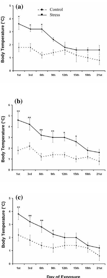
The rectal temperatures of chronically heat stressed rats were measured on every third day, just before daily exposure to 38 ± 1°C for one hour (Fig. 2). No change in body temperature was recorded in all the three groups during chronic stress till 3rd day.
The body temperature was found increased in both weaning and young rats from 6th day onwards. But
in adult group of rats, the rise in body temperature was recorded only on 18th and 21st day. The result
from one-way ANOVA showed significant increase in the mean rectal temperature of all three groups i.e. in weaning (F1,8 = 40.16, P<0.01), young (F1,8 =
114.04, P<0.01) and adult (F1,8 = 11.70, P<0.01) rats
on 21st day of chronic heat stress.
Body weight. The statistical analyses of the results
of body weight measurement in rats subjected to acute heat stress suggested that there was no
Fig. 2. Data expressed in mean ± S.E. The body temperature of (a) weaning group, (b) young group and (c) adult group of rats were recorded on every third day just before the rats were subjected to chronic heat stress compared to their respective control groups. *P<0.05 and **P<0.01.
Animal Groups
Control Stress
Body
Te
mpe
ra
tute
(
°
C)
Control Stress
(a)
(b)
(c)
Body
Te
mpe
ra
ture
(
°
C)
Body
Te
mpe
ra
ture
(
°
C)
Body
Te
mpe
ra
ture
(
°
C)
Iran. Biomed. J., April 2007 Pathophysiology of Heat Stress 105
Fig. 3. Data given in mean ± S.E. The body weights were recorded in (a) weaning group, (b) young group and (c) adult group of rats on every third day during chronic heat stress compared to their respective control groups. *P<0.05 and
**P<0.01.
significant change in the body weights in any group of animals due to four hours of acute heat exposure. In weaning and young groups of rats, reduction in body weights was observed due to chronic exposure to hot environment, while the adult rats did not show any significant change in body weight as compared to the control group (Fig. 3). Observations from weaning group show significant decrease in body weight from 6th day onwards. While on 21st day, the
decrease in body weight was observed maximum (F1,8 = 21.34, P<0.01). On the other hand, the young
rats show a significant change in body weight only on 18th and 21st day (F
1,8 = 9.84, P<0.05) of the
chronic stress.
Pellets count during heat stress. One way
ANOVA conducted on the pellets released during four hour of acute heat exposure at 38 ± 1°C, showed that the pellet count was significantly high in all groups of rats such as in weaning (F1,8 = 22.00,
P<0.01), young (F1,8 = 24.50, P<0.01) and adult rats
(F1,8 = 23.67, P<0.01) in comparison to their
respective control groups (Fig. 4). The pellets released during chronic heat stress at 38 ± 1°C by all three groups of rats are shown in Figure 5. The results indicate a highly significant rise in pellet released in weaning (F1,8 = 8.10, P<0.05), young
(F1,8 = 11.20, P<0.01) and adult group (F1,8 = 12.05,
P<0.01) on first day of chronic stress. However, the
increase in pellet released was gradually reduced latter on. In chronic stress groups, the reduced release of pellets noted during the end stage of chronic heat stress was probably due to adaptation of animals to the emotional stress.
Fig. 4. Data given in mean ± S.E. The Fecal Pellets released were recorded in all three groups of rats following acute heat stresscompared to respective control groups; **P<0.01.
Day of Exposure
(a)
(b)
(c)
Body
Te
mpe
ra
ture
(
°
C)
Body
Te
mpe
ra
ture
(
°
C)
Body
Te
mpe
ra
ture
(
°
C)
Control Stress
Animal Groups
Body
Te
mpe
ra
tute
(
°
C)
Control Stress
106 Sinha Iran. Biomed. J., April 2007
Fig. 5. Data given in mean ± S.E. The fecal pellets released by (a) weaning group, (b) young group and (c) adult group of rats were recorded on every third day during chronic heat stress compared to their respective control groups of rats. *P<0.05 and **P<0.01.
BBB permeability. The patterns of extravasations
of Evans blue dye following acute as well chronic heat stress as evaluated with naked eye observation are given in Figure 6. The increase in permeability of BBB following heat stress was observed dependent on the age of the subjects. The acute heat stress led to high permeability of Evans blue dye in weaning rats, while, mild to moderate amount of permeability of the tracer was observed in young rats. The observations showed that chronic heat stress caused relatively low extravasations of dye in brain cortex of weaning and young rats in comparison to the acute stress groups. In adult group of rats, very low or no extravasations of Evans blue dye was noted following either acute or chronic heat exposure.
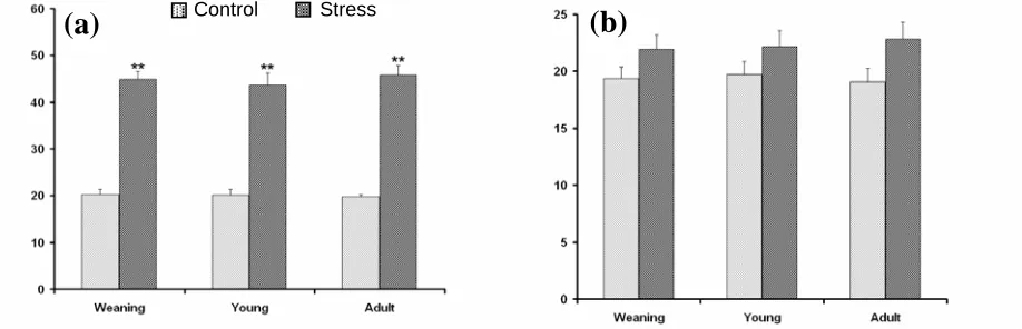
Plasma corticosterone level. One way ANOVA
showed that after acute heat stress, plasma corticosterone level stress was significantly increased in all the three age group of rats such as in weaning (F1,8 = 98.32, P<0.01), young (F1,8 =
168.12, P<0.01) and adult (F1,8 = 132.05, P<0.01) as
shown in Figure 7. The high plasma corticosterone level following acute heat stress represents the stressful condition of the rats. On the other hand, the analyses of data present no significant change in the corticosterone level in blood plasma in any group of rats following chronic heat exposure to high environmental heat in comparison to their respective control group of rats.
Hemorrhagic ulcers spots. Post mortem of
stomach showed that macroscopic gastric ulcers (pinpoint to hemorrhagic) were present in all the rats of all three age groups after acute as well as chronic exposure to hot environmental stress. However, a low count of the ulcer spots was observed following chronic heat stress in comparison to the acute heat stress.
DISCUSSION
Results from the observations of stress markers following heat stress, either acute or chronic, provide evidences of the stressful conditions in all three different age groups of rats. The increase in body temperature is one of the main characteristics of the stress, induced by acute exposure of the high environmental heat. The body temperature of all groups of rats was significantly increased by acute heat stress similar to the findings of Sinha [11]. The Day of Exposure
(a)
(b)
(c)
Body
Te
mpe
ra
ture
(
°
C)
Body
Te
mpe
ra
ture
(
°
C)
Body
Te
mpe
ra
ture
(
°
C)
Control
Stress
Iran. Biomed. J., April 2007 Pathophysiology of Heat Stress 107
Fig. 6. The dorsal view of the rat brain and distribution (shaded area) of Evans blue dye in all three groups of rats following acute and chronic heat stress. CC, Cerebral cortex; CB, Cerebellum; BS, Brain stem.
review of literature suggests that the immediate rise in the body temperature plays an essential role in the stimulation of the mechanisms necessary for heat dissipation. However, following the 21 days of chronic exposure to the high environmental heat, the body temperature of the rats of all three age groups was found to set at the higher temperature similar to the results obtained by Dey [1].
Normally the body temperature of the mammals remains constant except for a daily or circadian temperature variation of ± 1.00 to 1.50ºC. The hypothalamus of the mammalian brain contains thermosensitive neurons that sense not only the changes in core temperature, but also integrates
central and peripheral thermal information to elicit the most appropriate response for the given internal and external thermal condition [17]. It has been suggested that if temperature of the hypothalamus rises above the normal range, various heat loss responses are initiated to lower body temperature and return the hypothalamic temperature to its set point temperature. However, it is proposed that ‘set point’ is not static one, rather it is flexible and peripheral inputs can alter it. Thermal physiologists have generally accepted the concept of set point temperature. However, Kanosue et al. [18] have
raised some questions regarding the existence of set point temperature. Though other researchers now
Fig. 7. Data shown in mean ± S.E. Histogram shows the plasma corticosterone level in all three age groups of rats following (a) acute heat stress and (b) chronic heat stress compared to their respective control groups; **P<0.01.
CC
CB BS
Acute-Weaning Acute-Young Acute-Adult
Chronic-Weaning Chronic-Young Chronic-Adult
(a) (b) (c)
(d) (e) (f)
Control Stress
(a)
(b)
108 Sinha Iran. Biomed. J., April 2007
justified the existence of a set point temperature. They suggested that the set point theory is based on the fact that thermoregulatory system operates continuously to maintain core temperature and to maintain a balance between heat gain and heat loss thermoeffectors [9]. Some preoptic neurons of hypothalamus not only sense local temperature but also receive synaptic input from different pathways of thermoceptors in the skin, spinal cord and other locations throughout the body, and integrate central and peripheral thermal signals to control the particular body temperature by shifting this hypothalamic set point temperature as found in the chronic exposure to high environmental heat [17]. In the light of the present findings, it can be stated that the increase in body temperature by ambient heat affects the thermoregulatory responses to counter the hyperthermia. However, along with the temperature regulation of the body, thermoregulatory center of the brain is also known to be responsible for the adjustment of sleep and behavior of mammals [13, 14, 19]. This is according to the earlier suggestion that the hypothalamic temperature is a major feedback signal in the thermoregulatory system in mammals, and the activity of the thermoregulatory network have a direct influence in the state of sleep and behavior of the mammals [20]. Further, it can also be suggested that chronic heat stress influences the complex brain function that involves various neuronal and non-neuronal systems, which finally is responsible for changes in different physiological activities including sleep and behavior of the subjects. The change in set point of body temperature following chronic heat stress, and disturbed thermoregulatory systems of hypothalamus might be the main factor responsible these effects. The changes due to chronic exposure to high environmental heat can be monitored in all experimental age groups.
The single acute heat exposure did not alter the body weight of the rats of any age group, while the chronic heat stress significantly decreased the body weights of weaning and young rats. It has already been reported that the growth axis is inhibited at many levels of stress [14]. An acute elevation of growth hormone concentration in plasma is usually noted during the onset of the stress response in man. However, the prolonged activation of stress system has been reported to suppress the growth hormone secretion, leads to the decrease in the body weights. The growth and feeding are under complex and very redundant control mechanisms [15]. Changes in body weight, due to heat exposure are mainly
consequences of the well-known effects of stress on CNS and peripheral mechanism. Though, we have not recorded the food intake of the rats. However, the results from different laboratories show that reduced food intake following chronic exposure to high environmental heat, may be one of the reason in reduction of the body weights of the rats of weaning and young groups [15].
The reduction in growth rate in chronic heat exposure has been documented as the main feature displayed in rats. The present data confirm that chronic exposure to heat stress resulted in adaptation of hypothalamo-pituitary-adrenal (HPA) system. This adaptation may represent psychological and/or physiological mechanisms acting on the HPA system responsiveness [15]. Contributions of lateral hypothalamic area (LHA) to feeding behavior have been well established. On the basis of their responsiveness to electrophoretically applied glucose, neurons of the LHA have been characterized as either glucose sensitive or insensitive. Substances found to be effective in suppressing food intake mimic the action of glucose and inhibit LHA glucose sensitive neuron activity [21]. Altered hypothalamic activities due to changed set point temperature of the body may be one of the major causes of reduction in body weight of the rats in the present study. In the view of the involvement of hypothalamus in the feeding behavior and further in the reduction in the weights of the subjects, the involvements of different neurotransmitters and neurohormones cannot be ruled out. However, extensive review of literature and study is needed to establish their role in the growth and the reduction in the body weight of the subjects due to any stressful event. The results of the effect of heat stress in the growth and body weight on different age groups of rats suggest that the lower age of the subjects are more susceptible to the stressful event. The probable cause of this may be the immature neuronal membranes in these groups. This immature membrane can cause greater extravasations of neurotransmitters and neurohormones across the membrane that affect the body weight of the subjects.
The count of the number of fecal pellets excreted was significantly increased during four hour of acute heat stress in all three age groups of rats in comparison with their respective control animals. While in chronic heat stress conditions all three groups of rats exhibited a significant increase in the fecal pellets excreted during the first day of stress, the change in the number of pellets excreted was
Iran. Biomed. J., April 2007 Pathophysiology of Heat Stress 109
found to be decreased with time and returned to the control value on 21st day, similar to the findings of Sharma [2]. The result of pellet counts during chronic heat stress demonstrates the adaptation of the animals to the adverse environmental conditions. The hot environmental stress stimulates the pelvic nerves from spinal cord, which carry parasympathetic innervations to the large intestine. This causes peristaltic motion of the colon. Therefore, during extreme parasympathetic stimulation, caused by emotional disturbances in stressful conditions, the peristaltic motion of colon was increased that influences the increase in pellets excretion.
The acute heat exposure increased extravasations of the Evans blue dye in weaning and young groups, while the chronic heat stress induced only a mild increase in the permeability of Evans blue dye in the brain cortex of weaning rats. Increased permeability in the BBB in acute and chronic heat stress has already been reported by Sharma et al. [8], which
verify our results. It has been considered that the acute heat stress influences the release of serotonin in brain and plasma and that may be the main contributing factor to the breakdown of BBB permeability either directly or indirectly. Though the contribution of other factors released during high environmental heat stress cannot be ruled out in influencing the BBB permeability. This is further evident from the experiments that animals subjected to chronic heat stress show adaptation to high environmental heat as BBB permeability of Evans blue was found to be decreased as compared to acute exposure.
It has already been demonstrated that hyperthermia influence the brain function by modifying the permeability of BBB that was mediated by neurochemicals mainly by serotonin [8]. Findings of the chronic exposure of the present work also suggest that hot environmental exposure for 21 days daily for one hour induces heat tolerance and play an important role in heat adaptation and in heat-adapted rats; BBB break down was not observed [2, 8]. Age is an important factor influencing stress and the results showed that young rats were more susceptible to heat stress-induced changes in permeability of BBB in comparison to adult rats [8]. The highest permeability in BBB in weaning group compared to young-adult and older group of rats following acute and chronic heat stress may have occurred due to immature membrane properties of neuronal circuit [22]. It has been suggested that in contrast to adult neurons, whose
energy is provided almost entirely by glucose, the weaning brain obtains most part of its energy from ketone bodies and other products of the lipid metabolism [23]. Due to these factors, the oscillations within the intrathalamic and thalamocortical circuits are found changed in weaning rats [24] with respect to young-adult and older rats, and thus produce age dependent changes in their pathophysiology following exposure to acute as well as chronic heat stress.
The plasma corticosterone level was significantly increased in all age group of rats following acute heat stress but it was found unchanged after chronic heat stress. It is believed that increased secretion of corticosterone in acute heat conditions cause rapid mobilization of amino acids and fats, and responsible for gluconeogenesis, which immediately make available both energy and other compounds to the tissues that is essential to fulfill the body requirement in stressful conditions. There are several reports published in the past that support the hypersecretion of corticosteroids in acute stress conditions and homotypic stress results in adaptation accompanied by a declined corticosterone level [15, 25]. Similar to the present study in chronic heat stress, the results obtained by previous works [11] suggest that after acclimatization to the high environmental heat, the plasma corticosterone level was unaltered. Similar to the past report, it has been demonstrated that the chronic exposure to the environmental heat did not stimulate pituitary-adrenal system [26].
Heat stress is known to stimulate limbic areas of brain and pathways projecting from these areas to the hypothalamus, stimulates corticotropin releasing hormone (CRH) secretion into HPA axis. Besides, CRH not only stimulates pituitary to increases the corticotropin secretion, but also increase the sympathetic outflow to adrenals resulting in increased output of both cortical and medullary hormones that may cause behavioral abnormalities in the subjects [27]. At the same time, it has also been agreed that an exposure to 1 hour per day for 21 days resulted in physiological adjustment and adaptation to hot environment, which also reflects in the observations of corticosterone in the present study.
The results of peptic ulcer count shows that a large number of hemorrhagic ulcer spots were found in stomach following acute heat stress, while relatively less ulcer spot count was observed following chronic exposure to high heat. The emergence of hemorrhagic peptic ulcers in stressful conditions was
110 Sinha Iran. Biomed. J., April 2007
mainly caused due to protein malnutrition and protein degeneration that can be considered as a major risk factor for the peptic ulcers. The peptic ulcers are considered as one of the non-specific response among the general triad to any type of stress reaction as described by Selye [28].
The gastrointestinal tract is considered as the most sensitive among all other organs that is influenced mostly by any kind of stressor. The gastric surface mucosal cells are the first line of defense against insults derived from the stress stimuli. Peptic ulcers are described as one of the confirmed stress conditions, discussed in the general stress triads in the scientific research papers on biological stress [28]. Experiments in hyperthermia and other hot environmental stress such as acute and chronic heat stress are also reported to produce stress-induced ulcers in the stomach of the rats [8]. However, cultures of gastric surface mucous cells of guinea-pig fundic glands, after exposure to elevated ambient temperature exhibited to acquire resistance to long term or chronic stress. But the sudden extended exposure of stress was found producing hemorrhagic spots due to protein degradation in stomach [29]. The above findings indicate that rats exposed to 38ºC exhibits stress symptoms. Some of the symptoms of the present findings also resemble to the clinical symptom and established that animals were under severe stress [8]. Sharma and his co-workers [8] have studied extensively a similar animal model to find out the brain dysfunction under heat stress. On the other hand, chronic heat exposure indicates behavioral and physiological adaptations [1, 28]. It has been suggested that following acute exposure to high environmental heat, as cooling was resumed; the pathophysiological changes are either totally or partially reversed. Some of the physiological functions did not return to the control level and that may occur due to CNS damage, which attributed to anorexia, dehydration, metabolic imbalance, energy failure or cellular changes following heat stress [30]. However, chronic heat stress is responsible for producing relatively more durable or permanent pathophysiological changes due to adaptations of animals' physiological systems to the new environment. Further, in this study the changes in six different stress markers have been discussed mainly on the basis of neuronal and brain function. The results of the present study may also help in developing a new approach in understanding the thermoregulatory activities of the mammals under acute as well as chronic exposure to high environmental heat.
REFERENCES
1. Dey, P.K. (1998)Modification of dopamine receptor agonist mediated behavioral responses in rats following exposure to chronic heat stress.
Biomedicine 18: 41-47.
2. Sharma, H.S. (2006) Hyperthermia induced brain oedema: Current status & future perspectives. Ind. J.
Med. Res. 123: 629-652.
3. Szymusiak, R. and McGinty, D. (1990) Control of slow wave sleep by thermoregulatory mechanisms.
Prog. Clin. Biol. Res. 365: 53-64.
4. Lechin, F., Van Der Dijs, B. and Mireya, B. (1996) Stress versus depression. Prog.
NeuroPsycho-pharmacol. Biol. Psychiatry 20: 899-944.
5. Hanson, M.A. (1999) Development of a draft British standard: the assessment of heat strain for workers wearing personal protective equipment. Ann. Occup.
Hyg. 43: 309-319.
6. Romanovsky, A.A., Andrei, I.I. and Shimansky, Y.P. (2002) Ambient temperature for experiments in rats: a new method for determining the zone of thermal neutrality. J. Appl. Physiol. 92: 2667-2679.
7. Werner, J. (1998) Biophysics of heat exchange between body and environment. In: Physiology and
Pathophysiology of Temperature Regulation.
(Blatteis, C.M. ed.) World Scientific, Singapore. pp. 25-45.
8. Sharma, H.S., Westman, J. and Nyberg, F. (1998) Pathophysiology of brain edema and cell changes following hyperthermic brain injury. In: Progress in
Brain Research 115.(Sharma, H.S. and Westman, J.
eds.) Elsevier, Amsterdam. pp. 351-412.
9. Gordon, C.J. (2001) The therapeutic potential of regulated hypothermia. Emerg. Med. J. 18: 81-89.
10. Sinha, R.K. (2003) Artificial neural network detects changes in electro-encephalogram power spectrum of different sleep-wake states in an animal model of heat stress. Med. Biol. Eng. Comput. 41: 595-600.
11. Sinha, R.K. (2004) Electro-encephalogram
disturbances in different sleep-wake states following exposure to high environmental heat. Med. Biol.
Eng. Comput. 42: 282-287.
12. Sinha, R.K. and Ray, A.K. (2004) Effect of acute and chronic heat exposure on frequency of EEG components in different sleep-wake state in young rats. Iran. Biomed. J. 8: 69-75.
13. Sinha, R.K. and Ray, A.K. (2004) An assessment of changes in open-field and elevated plus-maze behavior following heat stress in rats. Iran. Biomed.
J. 8: 127-133.
14. Sinha, R.K. and Ray, A.K. (2006) Sleep-wake study in an animal model of acute and chronic heat stress.
Physiol. Behav. 89: 364-372.
15. Cure, M. (1989) Plasma corticosterone response in continuous versus discontinuous chronic heat exposure in rat. J. Physiol. Behav. 45: 1117-1122.
16. Nicholson, W.E. and Vanloom, G.R. (1973) Some
Iran. Biomed. J., April 2007 Pathophysiology of Heat Stress 111
practical innovations in the biological assay of adrenocorticotropic hormone (ACTH). J. Lab. Clin.
Med. 81: 803-808.
17. Boulant, J.A. (1991) Thermoregulation. In: Fever:
Basic Mechanism and Management (Mackowlak, P.
ed.) Raven Press, New York. pp. 1-22.
18. Kanosue, K., Romanovsky, A.A., Hosono, T. Chen, X.M. and Zhang Y.Z. (1997) “Set Point” revisited.
In: Thermal Physiology (Johannsen, B.N. and
Neilsen, R. eds.), Copenhagen, pp. 39-43.
19. Sinha, R.K. (2006) p-CPA pretreatment reverses the changes in sleep and behavior following acute immobilization stress. J. Physiol. Sci. 56: 123-129.
20. Thomas, T.C. and Kumar, V.M. (2002) Effect of ambient temperature on brain temperature and sleep-wakefulness in medial preoptic area lesioned rats.
Ind. J. Physiol. Pharmacol. 46: 287-297.
21. Shimizu, N., Oomura, Y. and Kai, Y. (1989) Stress-induced anorexia in rats mediated by serotonergic mechanisms in the hypothalamus. Physiol. Behav.
46: 835-841.
22. Frank, M.G., Morrissette, R. and Heller, H.C. (1998) Effects of sleep deprivation in neonatal rats. Am. J.
Physiol. 275: R148-R157.
23. Folbergrova, J. (1995) Glycogen phosphorylase activity in the cerebral cortex of rats during development: effect of homocysteine induced seizures. Brain Res. 694: 128-132.
24. Steriade, M., McCormick, D.A. and Sejnowski, T.J. (1993) Thalamocortical oscillations in the sleeping and aroused brain. Science 262: 679-685.
25. Djordjević, J., Cvijić, G. and Davidović, V. (2003) Different activation of ACTH and corticosterone release in response to various stressors in rats.
Physiol. Res. 52: 67-72.
26. Kant, G.J., Bunnell, B.N., Mougey, E.H., Pennington, L.L. and Meyerhoff, J.L. (1983) Effects of repeated stress on pituitary cyclic AMP, and plasma prolactin, corticosterone and growth hormone in male rats.
Pharmacol. Biochem. Behav. 18: 967-971.
27. Monteiro, F., Abraham, M.E., Sahaksri, S.D. and Mascarenhas, J.F. (1989) Effect of immobilization stress on food intake, body weight and weights various organs in rat. Ind. J. Physiol. Pharmacol.
33: 186-190.
28. Selye, H. (1984) The stress concept: Past, Present and Future. In: Stress Research (Cooper, C.L. ed.) John
Wiley & Sons Ltd., pp. 1-34.
29. Rokutan, K. (2000) Role of heat shock proteins in gastric mucosal protection. J. Gastroenterol.
Hepatol. 15 Suppl: D12-D19.
30. Dubois, M., Sato, S., Lees, D.E., Bull, J.M., Smith, R., White, B.G., Moore, H. and Macnamara, T.E. (1980) Electroencephalographic changes during whole body hyperthermia in humans. Electroenceph.
Clin. Neurophysiol. 50: 486-495.