Iranian Rehabilitation Journal, Vol. 12, No. 19, March 2014
Original Article
Scapular Position and Orientation during Abduction, Flexion and
Scapular Plane elevation Phase
Roshanak Keshavarz*
Tehran University of Medical Sciences, Tehran, Iran
Hassan Shakeri, PhD.; Amir Massoud Arab, PhD.
University of Social Welfare and Rehabilitation Sciences, Tehran, Iran
Hossein Ashrafi, PhD. University of Kashan, Kashan, Iran
Ailin Talim Khani
University of Social Welfare and Rehabilitation Sciences, Tehran, Iran
Objectives: The purpose of this study was to compare scapular kinematics during elevation phase of abduction, flexion, and scapular plane elevation phase between fifteen persons with shoulder impingement syndrome (SIS) and thirteen persons without it .
Methods: Values of scapular kinematics include scapular superior and lateral translations, upward rotation, external rotation, and posterior tipping were statistically tested with mixed model analysis of variance .
Results: Scapular upward rotation during 30˚, 60˚, and 90˚ of abduction, and initial angle of scapular plane elevation were significantly different between groups (P < 0.05). Posterior tipping was significantly decreased in patients with SIS at the initial angle of flexion (P =0.015). Lateral translation at 90˚, and 110˚ of abduction (P =0.015, and P=0.012, respectively) were lesser in patients .
Discussion: It seems that scapular kinematics during arm elevation in different movement planes is different, especially upward rotation between persons with and without SIS .
Keywords: Scapular kinematics, different humorous angles, shoulders movement planes
Submitted: 5 January 2014 Accepted: 27 February 2014
Introduction
Shoulder impingement syndrome (SIS) first was described subacromial bursitis (1), but knowledge in SIS pathology was greatly explained by Neer (2) and after by Hawkins (3), as an internal or external compression force on rotator cuff tendons, subacromial bursa, and long head of the biceps tendon under the subacromial arch during arm elevation (4). One of the most investigated different segmental movement patterns in clinical findings and scientific experiments which have been associated with SIS is abnormal pattern of the scapular kinematics (5-8).
Several studies illustrated abnormal three dimensional (3-D) scapular kinematics in patients with SIS and put forth a decrease in the scapular upward rotation, and posterior tipping during arm
elevation phase of abduction and scapular plane elevation (5, 7, 9, 10). Increased scapular superior translation and decreased scapular external rotation have been also reported in patients with SIS during abduction (1, 5, 11). During flexion, some investigators found greater upward rotation and clavicular elevation in these patients(7, 8); in contrast, there is the study which demonstrated lesser upward rotation (12). These changes may reduce the subacromial space, resulting in impingement of the subacromial structures. Thus, assessment and restoration of scapular movement has been emphasized in the clinical evaluation and rehabilitation program for SIS (13-15).
Review of the literature revealed that 3- D scapular kinematics has been assessed in different homeruns angles of arm elevation (15, 16) but just in the
scapular and frontal planes, apart (12, 14, 17, 18). Scapular position and orientation are different during arm elevation in various movement planes and humorous angles. Therefore, it is necessary to know scapular kinematics differences in doing the best exercise therapy and treatment protocols based on the movement plane and homeruns angles. Moreover, frontal, sagittal and scapular planes are the most functional shoulder movement planes. There is no study to evaluate differences of scapular kinematics during abduction, flexion and scapular plane elevation at various humorous angles. The purpose of this study was to collectively establish and compare the 3-D scapular kinematics during arm elevation phase of abduction, flexion and scapular plane elevation between persons with and without SIS.
Methods
Twenty-eight participants were recruited and categorized through their signs and there was a sample of convenience made up of subjects who were between the ages of 21 and 70 years. Fifteen patients (female: 8 and male: 7) with SIS who thirteen of them were right handed, were recruited from Shahid Moayeri and Shahid Modarres Hospitals (mean age: 46.6 14.2 (yrs) and mean BMI: 27.43 4.4) to the symptomatic group (SG). In this regard, every patient had X-ray of shoulder and was referred by the physiatrist. Thirteen subjects (female: 6 and male: 7) (mean age: 47.46 14.3 (yrs) and mean BMI: 27.76 5.12) who ten of them were right handed, were initially recruited from University of Social Welfare and Rehabilitation Sciences` workers and did not have any shoulder pathology experience as well as cervical radiculopathy for asymptomatic group (AG). The study was approved by the institutional university research committee on human right and informed consent was obtained.
Patients were included if they showed positive sign in X-ray, two or more shoulder impingement screening items and in at least one of the specific subacromial impingement tests. The shoulder impingement screening items were: (1) a history of proximal anterior or lateral shoulder pain persisted for more than one week during the last six months and the pain intensity was more than three visual analogue scale(5, 11); (2) painful arc with active shoulder elevation; (3) tenderness to palpation of rotator cuff tendons; (4) pain with resisted isometric shoulder abduction; (5) shoulder abduction of at least 130˚ relative to the thorax with no sign of
partial or total rotator cuff tearing and the specific subacromial impingement tests consisted: (6) positive Neer and Hawkin's test and (7) positive Yocum test (1, 5, 11, 15).
Subjects of SG were excluded if any of the followings was found: (1) a history of dislocation or subluxation and traumatic injuries on the tested shoulder complex; (2) a history of shoulder surgery within the last one year; (3) reproduction of symptoms in the cervical screaming examination (active and passive range of motion, and overpressure); (4) acromioclavicular degenerative joint disease(1, 5, 11); (5) clavicular osteolysis; (6) failure to complete two testing sessions; (7) thick tearing of rotator cuff muscles (1, 5, 11) and (8) hooked acromial morphology through X-ray (19-22). The Vicon motion analyser (460 Oxford, UK) and acromion marker cluster (AMC) were used to collect the 3-D kinematic data at a sampling rate of 100 Hz. To describe spatial position, a global coordinates system and a local coordinates system were used. The spatial positions of spherical reflective markers were placed at anatomical references according to recommendations of the International Society of Biomechanics (ISB) (13, 23-26).
After obtaining informed consent, subjects completed the demographic questionnaire. The subject was positioned sitting with their arms relaxed on both sides, with feet at a comfortable width apart and, looking forward, without back and arm supports. The measurements of scapular motions were performed in both groups. Thirteen skin markers were placed on the symptomatic arm in patients and on the matched arm in healthy ones, using double-sided tape (5, 11, 21).
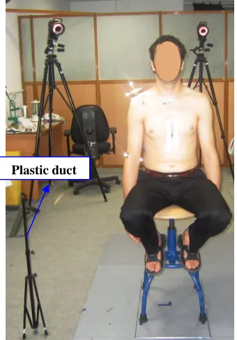
Calibration was done prior each test. The testing movement in this study was abduction, flexion and scapular plane elevation, which involved elevating the humerus in the frontal, sagittal and the scapular planes (30˚ anterior to the frontal plane) (11), guided by a plastic duct (Figure 1). Each cycle of movement took 4 s to complete (4-s elevation), marched by a metronome rhythm (5, 11, 34). Before testing, subjects practiced several arm elevations in three movement planes (frontal, sagittal and scapular planes). When the subject was able to equal the movement rhythm, he would then perform three successive movements with one kilogram weight in hand while the continuous kinematic data were concurrently gathered. One kilogram weight hand was selected, in that scapular kinematics is near to functional activities. Moreover, the loaded condition has significant effect on the scapular kinematics (35, 36).
Figure 1. 30˚ anterior to the frontal plane guided by a plastic duct.
The kinematic data from the Vicon motion analyzer and AMC were recorded then put them on prepared MATLAB program which was established according to the ISB protocol, and used to define the anatomical coordinate systems. The Euler angles of the rotational matrices of the humerus and scapula with respect to the thorax were then calculated (5). Scapular rotations were represented as rotation about the Y-axis of the scapula (scapular internal/external rotation), about the Z-axis of the scapula (upward/downward rotation), and about the X-axis of the scapula (posterior/anterior tilt). The displacement of the scapula relative to the thorax was calculated by the distance of AA to IJ in the directions of X (+: lateral), Y (+: superior) and Z (+: posterior) of the thorax coordinate system (5, 32). The position and orientation data of the humorous and scapula at 30º, 60º, 90º and 110º of arm elevation were obtained for further comparisons of differences in scapular kinematics during elevation phase of abduction, flexion and scapular plane elevation between groups.
Dynamic 3-D scapular kinematics differences in two groups were examined by a mixed model analysis of variance (ANOVA; [3 * 2 * 4]) was used to test the interaction effect and main effect of planes (frontal, sagittal and scapular), group (patient or healthy group), and homeruns angles (30º, 60º, 90º and 110º). A criterion level of P < 0.05 was considered statistically significant for the overall analysis. The within-day reliability of the kinematic data has been established in the pilot studies. Reliability tests contained inter correlation coefficient (ICC) and standard error of measurement (SEM). Results of the ICCs revealed the Vicon motion analyzer and AMC had very good to moderate intra-rater reliability when measuring dynamic scapular kinematics at 30˚, 60˚, 90˚ and 110˚ (ICC= 0.64 - 0.94) and SEM results were between 3.2-5.7 mm and 2.9˚-8.8˚.
Results
Fifteen participants with SIS and thirteen healthy persons completed the tests Table (1). The duration of shoulder symptoms ranged from 21 to 720 days (median = 120 days).
Table 1. Demographics tables
Variables Healthy group (n=13) Symptomatic group (n=15)
Age (years) 47.461514.30976 46.6 14.241 Weight (kg) 71.2308 10.77925 74.06676.92270 Height (cm) 160.9231 11.30974 165.2000 9.25203
BMI 27.75855.13814 27.43514.40752
Male (n=6) % (n=7) 46.7% Sex Female (n=7) % (n=8) 53.3% Right (n=10) % (n=13) 86.7% Dominate side
Left (n=3) % (n=2) 13.3%
The total testing time was about an hour for each session. All testing of our study was conducted. No subject complained of fatigue and pain during and after the test.
Interaction of homeruns angle, plane and group in scapular kinematics - The changing scores of 3-D scapular kinematics between the two groups were
analyzed by ANOVA with mixed model. Scapular superior translation, lateral translation, upward rotation, external rotation, and posterior tipping were various in different planes, humors angles and interaction of plane and humorous angles which the detailed significant scapular kinematics amounts Table (2).
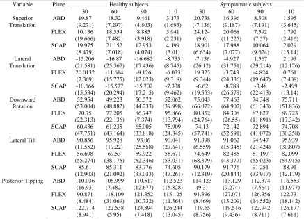
Table 2. Scapular kinematics measurements during abduction, flexion and scaption
Healthy subjects Symptomatic subjects Variable Plane
30 60 90 110 30 60 90 110 ABD 19.87 (9.271) 18.32 (7.297) 9.461 (4.803) 3.173 (1.693) 20.738 (-7.136) 16.396 (9.187) 8.308 (7.191) 1.595 (3.645) FLEX 10.136 (19.666) 18.554 (7.482) 8.885 (3.918) 3.941 (2.231) 24.124 (9.6) 20.068 (11.225) 7.592 (7.57) 1.792 (2.416) Superior Translation SCAP 19.975 (8.479) 21.152 (7.018) 12.953 (4.074) 4.199 (3.01) 18.901 (6.634) 17.988 (7.077) 10.064 (9.624) 2.029 (13.14) ABD -15.206 (21.581) -16.87 (25.367) -16.682 (17.436) -8.735 (8.745) -7.136 (26.12) -4.927 (31.751) 1.567 (29.214) 2.193 (12.176) FLEX 20.0132 (7.369) -11.614 (15.775) -9.126 (12.023) -6.033 (9.318) 19.325 (9.344) -3.743 (24.336) -4.824 (19.647) 0.761 (7.408) Lateral Translation SCAP -10.666 (15.534) -15.577 (20.294) -15.702 (17.215) -7.338 (9.462) -6.62 (19.553) -8.788 (26.579) -3.48 (22.413) -2.499 (13.14) ABD 52.954 (53.004) 49.223 (48.882) 50.572 (44.233) 52.062 (39.998) 75.043 (66.072) 77.463 (64.907) 74.348 (61.343) 75.711 (51.836) FLEX 70.75 (22.313) 77.205 (22.136) 86.747 (7.374) 95.866 (13.794) 80.852 (24.764) 84.308 (26.55) 87.827 (11.891) 89.723 (17.342) Downward Rotation SCAP 60.436 (47.751) 61.235 (43.164) 65.005 (33.818) 75.909 (24.345) 74.13 (57.741) 72.142 (52.591) 72.894 (41.072) 74.708 (30.258) ABD 90.856 (11.552) 95.928 (19.22) 93.06 (25.558) 90.041 (27.641) 91.398 (6.559) 91.062 (15.345) 94.947 (21.424) 95.939 (30.807) FLEX 56.698 (55.274) 69.53 (38.175) 59.922 (52.346) 58.671 (53.031) 74.649 (68.379) 82.485 (43.377) 81.197 (55.023) 82.099 (54.915) Lateral Tilt SCAP 85.61 (12.903) 85.311 (21.092) 83.776 (33.033) 74.605 (43.261) 90.179 (12.319) 91.776 (20.844) 91.251 (33.917) 88.91 (42.179) ABD 110.036 (16.93) 108.999 (7.482) 110.517 (12.677) 112.523 (15.828) 114.123 (9.3) 113.129 (9.274) 112.374 (7.564) 116.553 (11.977) FLEX 90.871 (8.484) 118.109 (31.069) 121.352 (10.732) 115.125 (11.364) 91.396 (8.469) 127.071 (13.209) 126.356 (14.552) 122.731 (18.142) Posterior Tipping SCAP 122.714 (8.941) 122.538 (5.95) 124.394 (7.418) 126.244 (13.045) 119.65 (8.756) 119.516 (9.436) 122.942 (8.711) 126.177 (7.611)
Figure 2. Scapular upward rotation (degree) differences during abduction (A) and scaption (B)
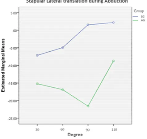
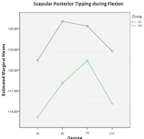
Also in scapular lateral translation, interaction of group and plane (F = 4.451; P = 0.022) (Figure 3) and in, also another main effect is illustrated in scapular posterior tipping, interaction of group and plane (F = 3.956; P = 0.032) (Figure 4) were significant. All the interaction of plane and scapular kinematic in various humerus angles were remarkable (P < 0.001) while interaction of group and plane or humerus angle was not significant.
Figure 3. Scapular posterior tipping (degree) differences between symptomatic and asymptomatic groups only in sagittal plane.
Figure 4. Scapular lateral translation (millimeter) between symptomatic and asymptomatic groups just during abduction.
Scapular kinematics changes in two groups -
Bonferroni post hoc analysis revealed that during the elevation phase of 30º (P = 0.02), 60º (P = 0.026) and 90º (P = 0.029) abduction, the scapula lesser rotated upward in different pattern among SG. During initial 30˚ in flexion, scapular posterior tipping had meaningful difference between mentioned groups (P = 0.015). In this regards, decreased scapular downward rotation in 30˚ (P =
0.028) scapular plane elevation was similar to scapular difference during abduction. Moreover, greater lateral translation of scapula in 90˚ and 110˚ abduction were significant (P = 0.029) and (P = 0.015) in patients, respectively. On the other hand, the baseline testing showed no significant difference of other variables between two groups (P > 0.05), except mentioned statistical significant differences. Table (3)
Table 3. Scapular kinematics significant differences between symptomatic and healthy groups.
NUM Variable Mean difference (SD) Sig
1 Upward Rotation 30˚ ABD. 45.166 (18.592)
0.02
2 Posterior Tipping 30˚ FLEX. 8.217 (3.158)
0.015
3 Upward Rotation 30˚ SCAP. 36.77 (16.215)
0.028
4 Upward Rotation 60˚ ABD. 43.624 (18.998)
0.026
5 Lateral Translation 90˚ ABD. 23.146 (8.917)
0.015
6 Upward Rotation 90˚ ABD. 39.16 (17.412)
0.029
7 Lateral Translation 110˚ ABD. 23.648 (17.713)
0.012
*Abbreviation: ABD (Abduction), FLEX (Flexion) and SCAP (Scaption) in 30˚ to 110˚ humeral degrees.
Discussion
This study had purposed to compare the scapular kinematics between persons with and without SIS
with SIS as compared to the healthy persons during arm elevation angles below 110˚ abduction and scapular plane elevation, especially upward rotation was significant difference between two groups. In addition, posterior tipping of scapula in the initial flexion decreased in patients compare to healthy ones.
The increased external rotation of the scapula, during dynamic tasks of arm elevation, especially in the sagittal plane, may act to rotate up the acromion away from the greater tuberosity. It appears that scapular external/internal rotation plays a role in the occurrence of impingement syndrome (1, 5, 15, 32). The current study found similar significant scapular lateral translation, but at greater humerus angles during abduction not during flexion. Two studies demonstrated a decrease in external rotation with the SIS during scapular plane elevation (6, 15, 37) while others showed no significant differences same as the recent study (38, 39). However, Karduna and colleagues suggested that an increase in scapular external rotation may be detrimental outcome in that it contributes to a decrease of the subacromial clearance (30). Thus, further studies are needed to explore the effects of shoulder movement planes and humerus angles of elevation on the scapular kinematics in patients with SIS.
As the predominant rotation of the scapula relative to the trunk, upward rotation of the scapula has been most commonly addressed in clinical treatment approaches and research studies. Upward rotation of scapula elevates the lateral acromion; also it is necessary to prevent impingement under the lateral acromial edge. During arm elevation, the scapula progressively upwardly rotates, externally rotates, and posteriorly tilts (1, 5, 15). This pattern has been demonstrated in healthy persons under static (30, 38) and dynamic conditions (31).
Reductions of upward rotation early in the range of motion under hand held loads have also been reported in patients with SIS (1, 5). Behavior of the groups’ differences to be magnified in the phase of abduction, flexion and scapular plane elevation were present, especially in 30˚ to 90˚ of abduction and 30˚ of scapular plane elevation in scapular upward rotation, respectively. However, it did not generally reach statistical significance during flexion. These findings support of the hypothesis that SG would show lesser scapular upward rotation than the AG, especially from the initiation of elevation to 90˚ angles.
Other study findings are about difference in interaction between humerus angle and group in scapular kinematics during arm elevation, but none of them analyzed scapular kinematics differences in interaction between humerus angle, plane and group. As a result, scapular 3-D kinematics was different in various humerus angles and planes, but there were not significant statistical results for scapular kinematics in interaction of humerus angles, planes and groups.
There is the idea that mentioned prior to reaching 90˚ of elevation phase relative to the scapula; the subacromial space must accommodate the articular cartilage, joint capsule and ligaments, rotator cuff tendons, and subacromial bursa. Our result showed main differences up to 90˚ of elevation phase that supported this notion. Cook and Ludewig dealt with that even refined decrease in the available suprahumeral space could contribute to the initiation or progression of shoulder impingement symptoms. This process could be further advanced by inflammation in the suprahumeral space, fibrosis or thickening of the tendons or bursa, or anatomic abnormalities (6). The magnitude of the angular differences in the upward rotation observed in our investigation were equal to or greater than the 6˚ anatomical changes in acromial slope that have previously been associated with rotator cuff tears and impingement syndrome. This phenomenon is similar to decreasing scapular superior translation during scapular plane elevation (18).
average differences between conditions need to be greater than the variability between trials.
Another limitation in this analysis is a lack of clavicular data during the motion. The present analysis demonstrated scapular angular orientation changes relative to the thorax during the arm elevation phase in different movement planes and humerus angles. Observations of altered scapular kinematics may result from variations in sternoclavicular (SC) joint, acromioclavicular (AC) joint or combined SC/AC joint motion. These combined joint motion impacts on scapular kinematics as well as shoulder pathology clinically, so it is necessary to address accurate clavicular kinematic information.
Conclusion
Measuring scapular kinematics during arm elevation in different movement planes may support the assessment of persons with SIS and provides comprehensive information about scapular kinematics, also gets easy to compare it in various humerus angle and movement planes. The AMC provided reliable measurements of scapular kinematics during arm elevation in frontal, sagittal and scapular movement planes. In general, there was significant difference in scapular upward / downward rotation between subjects with and without SIS in abduction and scapular plane elevation below 90˚ of arm elevation phase. Decreased scapular lateral translation was only found during abduction in patients. However,
during flexion, scapular posterior tipping was decreased in subjects with SIS. Scapular kinematics in different movement planes was statistically and clinically different in subjects with SIS compared to those without SIS. These finding support the theory those scapular kinematics are miscellaneous, especially in initial arm elevation in each movement plane and can be a reason of subacromial impingement in patients.
Clinical Implication
The results of this study could be beneficial to clinicians when prescribing therapeutic exercises for patients with SIS. It seems that specific scapula exercise therapy during elevation is necessary to improve scapular movement pattern in each scapula movement plane differently. More focus should be placed on scapular upward rotation in abduction and scapular plane elevation also scapular posterior tipping movement should be concentrated during flexion in exercise therapy for the patients.
Acknowledgements
This research was supported in part by the deputy of research and innovation center and University of Social welfare and Rehabilitation Sciences under human public committee, Grant no. 90.801.1.T.2060. The authors would especially like to gratitude M. R. Gholamian and A. Raeissadat for their assistances and lengthy efforts.
References
1. Hung CJ, Jan M-H, Lin Y-F, Wang T-Q, Lin J-J. Scapular kinematics and impairment features for classifying patients with subacromial impingement syndrome. Manual therapy. 2010;15(6):547-51.
2. NEER CS. Impingement lesions. Clinical orthopaedics and related research. 1983;173:70-7.
3. Hawkins R, Brock R, Abrams J, Hobeika P. Acromioplasty for impingement with an intact rotator cuff. Journal of Bone & Joint Surgery, British Volume. 1988;70(5):795-7. 4. Ebaugh DD, McClure PW, Karduna AR. Three-dimensional
scapulothoracic motion during active and passive arm elevation. Clinical Biomechanics. 2005;20(7):700-9. 5. Huang H-Y, Lin J-J, Guo YL, Wang WT-J, Chen Y-J. EMG
biofeedback effectiveness to alter muscle activity pattern and scapular kinematics in subjects with and without shoulder impingement. Journal of Electromyography and Kinesiology. 2013;23(1):267-74.
6. Ludewig PM, Cook TM. Alterations in shoulder kinematics and associated muscle activity in people with symptoms of shoulder impingement. Physical therapy. 2000;80(3):276-91.
7. McClure PW, Bialker J, Neff N, Williams G, Karduna A. Shoulder function and 3-dimensional kinematics in people with shoulder impingement syndrome before and after a 6-week exercise program. Physical Therapy. 2004;84(9):832-48.
8. McClure PW, Michener LA, Karduna AR. Shoulder function and 3-dimensional scapular kinematics in people with and without shoulder impingement syndrome. Physical Therapy. 2006;86(8):1075-90.
9. Michener LA, McClure PW, Karduna AR. Anatomical and biomechanical mechanisms of subacromial impingement syndrome. Clinical biomechanics. 2003;18(5):369-79. 10.Borstad JD, Ludewig PM. Comparison of scapular
kinematics between elevation and lowering of the arm in the scapular plane. Clinical Biomechanics. 2002;17(9):650-9. 11.11.Hsu Y-H, Chen W-Y, Lin H-C, Wang WT, Shih Y-F.
The effects of taping on scapular kinematics and muscle performance in baseball players with shoulder impingement syndrome. Journal of electromyography and kinesiology. 2009;19(6):1092-9.
Biomechanics. 2010;25(1):29-36.
13.Amadi HO, Hansen UN, Wallace AL, Bull AM. A scapular coordinate frame for clinical and kinematic analyses. Journal of biomechanics. 2008;41(10):2144-9.
14.De Baets L, Jaspers E, Desloovere K, Van Deun S. A systematic review of 3D scapular kinematics and muscle activity during elevation in stroke subjects and controls. Journal of Electromyography and Kinesiology. 2013;23(1):3-13.
15.Kibler WB, Ludewig PM, McClure PW, Michener LA, Bak K, Sciascia AD, et al. Clinical implications of scapular dyskinesis in shoulder injury: the 2013 consensus statement from the ‘scapular summit’. British journal of sports medicine. 2013;47(14):877-85.
16.Thigpen CA, Gross MT, Karas SG, Garrett WE, Yu B. The repeatability of scapular rotations across three planes of humeral elevation. Research in Sports Medicine. 2005;13(3):181-98.
17.Gomes PF, Sesselmann M, Faria CD, Araújo PA, Teixeira-Salmela LF. Measurement of scapular kinematics with the moire fringe projection technique. Journal of biomechanics. 2010;43(6):1215-9.
18.Matsuki K, Matsuki KO, Mu S, Yamaguchi S, Ochiai N, Sasho T, et al. In vivo 3-dimensional analysis of scapular kinematics: comparison of dominant and nondominant shoulders. Journal of Shoulder and Elbow Surgery. 2011;20(4):659-65.
19.Seitz AL, McClure PW, Lynch SS, Ketchum JM, Michener LA. Effects of scapular dyskinesis and scapular assistance test on subacromial space during static arm elevation. Journal of Shoulder and Elbow Surgery. 2012;21(5):631-40. 20.Struyf F, Nijs J, Baeyens JP, Mottram S, Meeusen R.
Scapular positioning and movement in unimpaired shoulders, shoulder impingement syndrome, and glenohumeral instability. Scandinavian journal of medicine & science in sports. 2011;21(3):352-8.
21.Timmons MK, Thigpen CA, Seitz AL, Karduna AR, Arnold BL, Michener LA. Scapular kinematics and subacromial-impingement syndrome: a meta-analysis. Journal of sport rehabilitation. 2012;21(4).
22.Uhl TL, Kibler WB, Gecewich B, Tripp BL. Evaluation of clinical assessment methods for scapular dyskinesis. Arthroscopy: The Journal of Arthroscopic & Related Surgery. 2009;25(11):1240-8.
23.Mottram SL, Woledge RC, Morrissey D. Motion analysis study of a scapular orientation exercise and subjects’ ability to learn the exercise. Manual therapy. 2009;14(1):13-8. 24.Prinold JA, Shaheen AF, Bull AM. Skin-fixed scapula
trackers: A comparison of two dynamic methods across a range of calibration positions. Journal of biomechanics. 2011;44(10):2004-7.
25.Wu G, Van der Helm FC, Veeger H, Makhsous M, Van Roy P, Anglin C, et al. ISB recommendation on definitions of joint coordinate systems of various joints for the reporting of human joint motion—Part II: shoulder, elbow, wrist and hand. Journal of biomechanics. 2005;38(5):981-92.
26.Bolsterlee B, Veeger H, van der Helm F. Modelling clavicular and scapular kinematics: from measurement to simulation. Medical & biological engineering & computing. 2013:1-9.
27.Veeger H, van dee Helm F, Chadwick E, Magermans D. Toward standardized procedures for recording and describing 3-D shoulder movements. Behavior Research Methods, Instruments, & Computers. 2003;35(3):440-6. 28.Veeger H, Van Der Helm F. Shoulder function: the perfect
compromise between mobility and stability. Journal of biomechanics. 2007;40(10):2119-29.
29.Brochard S, Lempereur M, Rémy-Néris O. Double calibration: An accurate, reliable and easy-to-use method for 3D scapular motion analysis. Journal of biomechanics. 2011;44(4):751-4.
30.Karduna AR, McClure PW, Michener LA, Sennett B. Dynamic measurements of three-dimensional scapular kinematics: a validation study. Journal of biomechanical engineering. 2001;123(2):184-90.
31.van Andel C, van Hutten K, Eversdijk M, Veeger D, Harlaar J. Recording scapular motion using an acromion marker cluster. Gait & posture. 2009;29(1):123-8.
32.Warner M, Chappell P, Stokes M. Measuring scapular kinematics during arm lowering using the acromion marker cluster. Human movement science. 2012;31(2):386-96. 33.Meskers C, Van der Helm F, Rozendaal L, Rozing P. < i> In
vivo</i> estimation of the glenohumeral joint rotation center from scapular bony landmarks by linear regression. Journal of biomechanics. 1997;31(1):93-6.
34.Fayad F, Hoffmann G, Hanneton S, Yazbeck C, Lefevre-Colau M-M, Poiraudeau S, et al. 3-D scapular kinematics during arm elevation: effect of motion velocity. Clinical biomechanics. 2006;21(9):932-41.
35.Forte FC, Peduzzi de Castro M, Mahnic de Toledo J, Ribeiro DC, Loss JF. Scapular kinematics and scapulohumeral rhythm during resisted shoulder abduction– Implications for clinical practice. Physical Therapy in Sport. 2009;10(3):105-11.
36.Raina S, McNitt-Gray JL, Mulroy S, Requejo PS. Effect of increased load on scapular kinematics during manual wheelchair propulsion in individuals with paraplegia and tetraplegia. Human movement science. 2012;31(2):397-407. 37.Ludewig PM, Phadke V, Braman JP, Hassett DR, Cieminski
CJ, LaPrade RF. Motion of the shoulder complex during multiplanar humeral elevation. The Journal of Bone & Joint Surgery. 2009;91(2):378-89.
38.Lukasiewicz AC, McClure P, Michener L, Pratt N, Sennett B. Comparison of 3-dimensional scapular position and orientation between subjects with and without shoulder impingement. Journal of Orthopaedic & Sports Physical Therapy. 1999;29(10):574-86.