Open Access
Research article
Echolocation calls and communication calls are controlled
differentially in the brainstem of the bat
Phyllostomus discolor
Thomas Fenzl*
1and Gerd Schuller
2Address: 1Max Planck Institute of Psychiatry, Kraepelinstr. 2-10, Munich, 80804, Germany and 2Department Biology II,
Ludwig-Maximilians-Universitaet, Grosshaderner Str. 2, Planegg-Martinsried, 82152, Germany
Email: Thomas Fenzl* - [email protected]; Gerd Schuller - [email protected] * Corresponding author
Abstract
Background: Echolocating bats emit vocalizations that can be classified either as echolocation calls or communication calls. Neural control of both types of calls must govern the same pool of motoneurons responsible for vocalizations. Electrical microstimulation in the periaqueductal gray matter (PAG) elicits both communication and echolocation calls, whereas stimulation of the paralemniscal area (PLA) induces only echolocation calls. In both the PAG and the PLA, the current thresholds for triggering natural vocalizations do not habituate to stimuli and remain low even for long stimulation periods, indicating that these structures have relative direct access to the final common pathway for vocalization. This study intended to clarify whether echolocation calls and communication calls are controlled differentially below the level of the PAG via separate vocal pathways before converging on the motoneurons used in vocalization.
Results: Both structures were probed simultaneously in a single experimental approach. Two stimulation electrodes were chronically implanted within the PAG in order to elicit either echolocation or communication calls. Blockade of the ipsilateral PLA site with iontophoretically application of the glutamate antagonist kynurenic acid did not impede either echolocation or communication calls elicited from the PAG. However, blockade of the contralateral PLA suppresses PAG-elicited echolocation calls but not communication calls. In both cases the blockade was reversible.
Conclusion: The neural control of echolocation and communication calls seems to be
differentially organized below the level of the PAG. The PLA is an essential functional unit for echolocation call control before the descending pathways share again the final common pathway for vocalization.
Background
Bats use echolocation calls for orientation in space and hunting for prey. Communication calls serve the purpose of inter- and intraspecific social communication. Neural control of both types of calls has access at the level of the medulla to the final common pathway for vocalization.
Motoneurons controlling the larynx, the vocal tract and the respiratory muscles are accessed via interneurons from the nucleus ambiguus/retroambiguus complex [1]. One major subcortical structure for the control of vocalizations above the level of the medulla is the periaqueductal gray matter (PAG). Here, communication calls can be elicited
Published: 01 August 2005
BMC Biology 2005, 3:17 doi:10.1186/1741-7007-3-17
Received: 08 April 2005 Accepted: 01 August 2005
This article is available from: http://www.biomedcentral.com/1741-7007/3/17
© 2005 Fenzl and Schuller; licensee BioMed Central Ltd.
by electrical or pharmacological stimulation in many mammalian species like squirrel monkey [2], rhesus mon-key [3], rat [4], guinea pig [5], gibbon [6] and bat [7,8]. While in most areas where vocalizations can be elicited, long persisting stimulation causes a fading of the vocal reaction, the PAG is one of the very few regions where nat-ural communication calls can be elicited without changes in the emotional state of the animal and without habitu-ation to the stimulus, even over extended stimulhabitu-ation periods [9]. PAG stimulation in bats also evokes echoloca-tion calls [10,11] without motor concomitants (except ear and nose leaf movements), and without habituation to the stimulus [7]. The PAG in bats is thus involved in the control of communication calls as well as echolocation calls. The different types of calls are elicited at anatomi-cally distinct PAG locations [7].
In monkeys, the PAG has been shown to be part of the vocalization controlling system [12]. Although the author found the majority of vocal- eliciting structures within the limbic system, only stimulation in the PAG yielded all types of communication calls used by the animal [12].
Further findings suggest a hierarchical organization of the vocalization controlling system including the PAG. Bilat-eral lesions of vocalization-eliciting areas in the forebrain and diencephalon do not affect vocalizations triggered in the PAG, whereas lesions made at brain levels below the PAG, e.g., in the dorsolateral pons and the ventrolateral medulla suppress PAG-triggered vocalizations [13]. The PAG would thus serve a gating function controlling the release of vocal patterns that themselves are constituted in networks below the PAG level [14].
In the bat (Rhinolophus rouxi), three areas below the mid-brain level in addition to the PAG have been identified that yield echolocation calls (but not communication calls) on electrical stimulation without temporal or spec-tral distortions [18]: a) the deep layers of the superior col-liculus, b) the deep mesencephalic nucleus in the reticular formation and c) an area medial to the rostral parts of the dorsal nucleus of the lateral lemniscus, the paralemniscal area (PLA). All three areas share most of the stimulus-response conditions found in the PAG, such as very short latencies of vocalizations on stimulation, no habituation to stimulus, no body movements accompanying the vocalizations and no distortion of the call pattern. How-ever, only echolocation (and not communication) calls can be elicited from these areas. Moreover, echolocation calls from the PLA can also be elicited by microinjection of a glutamate agonist (kainic acid), indicating that it is the neurons in the PLA rather than fibers passing through this area that are responsible for the activation of vocal utterances [19].
The PLA displays similar properties in other bat species, e.g., Pteronotus p. parnellii [20] and Phyllostomus discolor [7,21]. The interest in the PLA as an important component for vocal control in echolocation was supported by the finding that neurons in the rostral portion of the PLA of R. rouxi were active both before and during vocal emissions and also showed simultaneous responses to auditory stimuli. The PLA could therefore serve as an audio-vocal interface between auditory processing and motor control of vocalization [22,23]. The physiological data on the PLA available from animals other then bats are very sparse and anatomically corresponding areas have been investigated under different functional aspects [23-28].
Vocal control in other, non-echolocating mammals is characterized by a rather direct connection from PAG to the ambiguus/retroambiguus complex [1,15-17] or to the reticular formation [13,14,29], which would not necessi-tate an interaction from other brain structures (e.g., PLA) below the PAG level. But these findings seem to be insuf-ficient to explain findings from Schuller and coworkers and Fenzl on the involvement of the PLA in motor control of vocalizations. A parallel organization of the descending control system for vocalizations could be assumed at the level of the PAG and PLA, and it is of interest whether this organization is a general mammalian feature.
The present study investigates whether the PAG and PLA interact during the PAG-induced emission of communica-tion and/or echolocacommunica-tion calls in P. discolor using reversi-ble blockade of PLA function. We found that PAG-induced communication calls could not be blocked either through ipsilateral or contralateral blockade of the PLA. However, PAG-induced echolocation calls could be tem-porarily blocked only through contralateral blockade of the PLA. These differences display partly differentiated functional organizations of vocal controlling pathways for communication calls and echolocation calls below PAG level.
Results
Implants – Stability of chronical microstimulation
Stability of implants
Figure 1
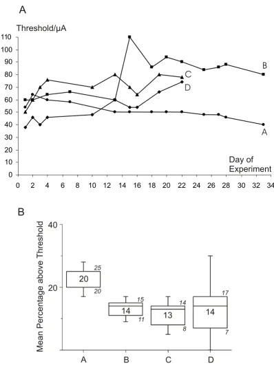
Stability of implants. Stability of stimulation threshold currents to elicit vocalizations and time course at chronic microstim-ulation probes. Electrodes were implanted at day0 of the graph A. (A) Thresholds of four chronically implanted stimmicrostim-ulation probes (A/B and C/D) are shown. Stimulation through implant A and B (animal 1) and through implant C (animal 2) elicited echolocation calls, while stimulation through implant D (animal 2) triggered communication calls. (B) "Mean percentage above threshold"-values for all four implants (A to D) as plotted in A during the actual blockade experiments. The mean stimulation current was 20% (electrode A), 14% (electrode B), 13% (electrode C) and 14% (electrode D) above threshold to ensure a vocal answer on each stimulus within the pulse train. At threshold level, not all stimuli triggered a vocalization. The median val-ues are indicated as numbers; additionally P25 and P75 valval-ues are shown in italics.
0 10 20 30 40 50 60 70 80 90 100 110
0 2 4 6 8 10 12 14 16 18 20 22 24 26 28 30 32 34
A
B
C
D
Day of
Experiment
Threshold/µA
A
Mean
Percent
age
a
bove
T
hreshold
20
40
A
B
C
D
20
14
13
14
20 25
11 15
8 14
7 17
PLA-located blockade of PAG-induced echolocation calls and communication calls
Figure 2
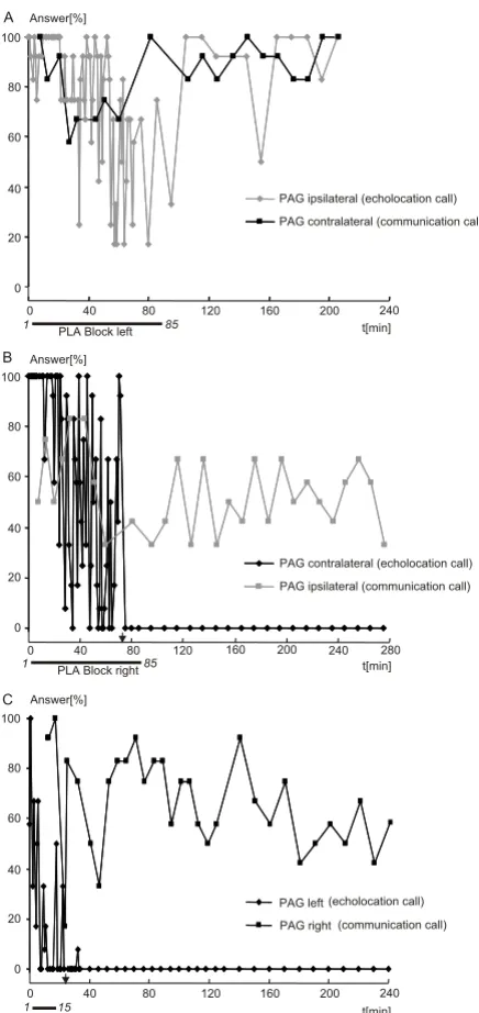
PLA-located blockade of PAG-induced echolocation calls and communication calls. The percentage of successful stimulations (y-axis) for eliciting vocalizations in the PAG is represented during and after KA induced blockade of PLA. Echolo-cation calls are graphed with diamonds, while squares are used for communiEcholo-cation calls. The kynurenic acid appliEcholo-cation is indi-cated by black horizontal bars below the x-axis. Onset and termination times of iontophoresis are given in italics. Onset and termination times of iontophoresis are indicated by numbers at both ends of the bars in italics. Stimulation success (%) was cal-culated for intervals of 2 s. Black arrows on the abscissa indicate the 25% blockade boundary. (A) Application of KA to the left PLA. The ipsilateral production of PAG-induced echolocation calls (left electrode) cannot be blocked, although a slight depres-sion can be noticed in the first half of the graph. The contralateral production of PAG-induced communication calls (right elec-trode) is only rarely influenced by the glutamate antagonist KA. (B) A blockade of the right PLA totally blocks the production of contralaterally PAG-induced echolocation calls (left electrode), while the ipsilateral production of communication calls (right electrode) again is barely influenced. Note that the curve for PAG ipsilateral (communication call) starts around 50%. (C) Bilat-eral blockade of both PLA sites again leads to a total depression of PAG-induced echolocation calls, while PAG-induced com-munication calls can be elicited across the entire experimental run. Prior to each experiment, vocal answers were stable for at least 10 minutes at a value comparable to the data shown at the start of the application of the antagonist.
PAG contralateral (communication call)
0 20 40 60 80 100
0 40 80 120 160 200 240 PAG ipsilateral (echolocation call)
t[min] Answer[%]
PLA Block left
1 85
0 20 40 60 80 100
PAG ipsilateral (communication call) PAG contralateral (echolocation call)
t[min] Answer[%]
PLA Block right 10 40 8085 120
160 200 240 280
0 20 40 60 80 100
0 40 80 120 160 200 240 PAG left
PAG right
t[min] Answer[%]
1 15 PLA Block left/right
A
B
C
and the last experimental day was -30%, +33%, +56% and +95% in experiments A, B, C and D, respectively. During experiments the stimulation current was adjusted at supra-threshold levels to provide a reliable one-to-one relationship between electrical stimuli and vocal responses. The median values of supra-threshold currents used with the chronical implants A, B, C and D were 20% (P25 = 20, P75 = 25), 14% (P25 = 11, P75 = 15), 13% (P25 = 8, P75 = 14) and 14% (P25 = 7, P75 = 17) above threshold, respectively (Fig. 1B).
Echolocation calls versus communication calls
In order to test whether the production of echolocation or communication calls could be suppressed by neurophar-macological blockade, the nonselective glutamate antago-nist kynurenic acid (KA) was iontophoretically injected into the PLA. Injections were either ipsilateral or contral-ateral to the PAG stimulation site. Twelve blockade exper-iments were conducted with consistent results in animal one (implants A and B), and seven experiments were con-ducted in animal two (implants C and D).
Blocking the PLA with KA suppressed PAG-induced echo-location calls whereas PAG-induced communication calls were less affected or not affected at all. The degree of sup-pression of echolocation calls depended on whether the PLA blockade was applied ipsilaterally or contralaterally to the PAG stimulation sites as shown in Fig. 2. An 85 min application of KA to the left PLA, i.e., the PLA ipsilateral to the PAG electrode eliciting echolocation calls and contral-ateral to the electrode triggering communication calls, did not completely suppress either echolocation calls (Fig. 2A, gray) or communication calls (Fig. 2A, black). A reduction of response probability for echolocation calls could, how-ever, be seen around 50 min after the onset of iontophore-sis. The activation probability for communication calls varied largely around 50%, and showed only a relative persistent lowering to around 40% between 50 and 90 min after the onset of KA application (Fig. 2B, gray).
A much different pattern of vocal responses appeared if the block was applied to the PLA contralateral to the PAG electrode eliciting echolocation calls. The response proba-bility for echolocation calls started to decrease about 10 min after the onset of KA application and showed great variability, until complete suppression at around minute 80 (Fig. 2B, black). The blockage persisted beyond the ter-mination of the experiment after 280 min.
When KA was applied bilaterally to both PLA sites, the suppression of PAG-induced echolocation calls occurred with a much shorter delay (about 24 min). However, PAG-induced communication calls could not be com-pletely suppressed, although the probability of eliciting communication calls showed generally greater variability
in response to KA application (Fig. 2C). A slight effect of KA could be discerned around 30 min after onset of the application; however, the effect did not persist. The slight mean decrease in elicitability, i.e., not falling below about 50%, and the high variability of elicitation probability persisted throughout the entire duration of the experi-mental run. These effects lasted beyond the 240 min, after which the experimental sessions had to be terminated.
After each blocking experiment a recovery period was inserted. Subsequently, a control experiment with electri-cal microstimulation only was started 24 hrs after the beginning of the previous experiment. In all control runs the elicitability of both types of calls had fully recovered. Calls could be elicited at standard stimulation currents, and no lesioning of PLA structures by iontophoretic cur-rents could be detected. Typically the next iontophoresis experiment started within 24 hrs after the control experiment.
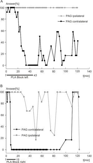
Ipsilateral vs. contralateral blockade of echolocation calls The finding that a PLA-block by KA primarily affects con-tralaterally PAG-induced echolocation calls was sup-ported by the results from following experiments, shown in Fig. 3. First, KA was applied to the left PLA for 43 min. Echolocation calls elicited in the PAG contralateral to the iontophoresis site were completely depressed about 32 min after the onset of iontophoresis and partially recov-ered about 10 min after termination of KA application. In contrast, the ipsilateral PAG-triggered echolocation calls were not affected at all. Similarly, the right PLA was blocked by KA application for 15 min. Again, the contral-ateral PAG-induced echolocation calls were blocked con-sistently about 7 min after iontophoresis onset and started to recover about 80 min after cessation of KA application. Echolocation calls elicited in the ipsilateral PAG were tem-porarily impeded, but elicitability was far from being totally blocked.
Although the depression of PAG-induced echolocation calls persisted beyond the end of the experimental session (140 min; Fig. 3A), elicitability from both PAG electrodes was fully recovered in the control experiments 24 hrs later.
Influence of sedation on animal during experiments To ensure a stable position of the animal during the ion-tophoresis experiments, Rompun® was chronically
PLA-located blockade of PAG-induced echolocation calls
Figure 3
PLA-located blockade of PAG-induced echolocation calls. At both electrodes, echolocation calls were triggered. Refer to Fig. 2 for explanations on graph. (A) Application of kynurenic acid to the left PLA does not influence the elicitability of ipsi-laterally PAG-induced echolocation calls (left electrode), whereas it lowers dramatically the efficiency of contraipsi-laterally induced echolocation calls (right electrode). The elicitability does not recover to the 75% mark within termination of the experiment at minute 120. (B) A blockade of the right PLA totally blocks the production of contralaterally PAG-induced echo-location calls (left electrode), while ipsilateral-induced echoecho-location calls (right electrode) are little affected (although some drop in elicitability can be detected). Prior to each experiment, vocal answers had to be stable for at least 10 min at a value comparable to the data shown at the application of the antagonist.
0 20 40 60 80 100
0 20 40 60 80 100 120 140
Answer[%]
t[min] PAG ipsilateral
PAG contralateral
PLA Block left 43
1
t[min] 0
20 40 60 80 100
0 20 40 60 80 100 120 140
Answer[%]
PAG contralateral
PAG ipsilateral
PLA Block right15
1
or mouth movements with communication calls, also did not change during the experiments.
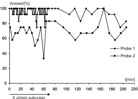
To ensure that sedation had no influence on activation of electrically induced vocalizations, a pure stimulation experiment under identical depressant conditions as applied at the blockade experiments was carried out (Fig. 4). Neither the initial dose of 0.5 ml sedative (0.04% Rompun® in 0.9% NaCl) prior to the experiment nor the
continuous application of 6 µl/min of sedative showed any influence on the ability to trigger vocalizations. The vocalizations triggered under the influence of the sedative in general showed no difference to vocalizations triggered without sedation or to vocalizations emitted spontane-ously, either in spectral or temporal patterns. Also the per-centage of vocal answers triggered by one pulse train (duration: 2s, 12 single stimuli, 12 vocal answers corre-spond to 100% vocal answer) did not decrease under the influence of the sedative, as compared to experiments without sedation (data not shown). A decline or even depression of vocal answers of the type seen in the block-ade experiments could not be detected.
Histological verification of electrode positions
Stimulation sites in the PAG and in the PLA were identi-fied by tissue lesions (Fig. 5). The lesions in both struc-tures could easily be detected in anatomical sections.
Discussion
This study has demonstrated that the neural control of echolocation calls and communication calls must have access to at least partly different neural substrates for vocalization.
Sedation of animals, stability of chronic implants and iontophoretic efficiency
The stability of the chronically implanted electrodes for electrical micro-stimulation was very satisfactory. Both echolocation calls and communication calls were elicited consistently over a long stimulation period of up to 33 days without any change in spectral composition of the calls. The slight increase of stimulation thresholds for the individual electrodes (Fig. 1) could be attributed to an accumulation of glia cells and debris caused by the pres-ence of implants. Motor reactions associated with vocali-zations also did not change during the experiments.
It is noteworthy that the onset of a contralateral PLA blockade is extraordinarily variable between 3 to almost 80 min under comparable experimental conditions (Fig. 2 and Fig. 3). Slightly different positions of the iontophore-sis probe at the PLA site may be responsible for this, as even deviations as small as 100 µm correspond to almost 15% of the mediolateral dimension (≈ 800 µm, [7]) of the PLA in P discolor. At marginal application sites, KA would have taken longer to influence the necessary number of neurons in the PLA than when injected the geometrically optimal PLA location. The onset time differences indicate that the suppressive effect of PLA inactivation depends on the of PLA neurons.
Differentially organized vocal substrates for the
production of echolocation calls and communication calls Vocalizations are complex motor patterns imbedded into differentiated behaviors of an animal. It is well estab-lished that the PAG plays an important role in vocal con-trol of communication calls [14,30], e.g., communication calls can be triggered by stimulating the PAG in several mammalian [7,31,32] and non-mammalian species [33]. In addition, echolocation calls can be elicited in bats within restricted areas of the PAG [8] that are distinct from areas in which communication calls can be triggered [7].
Outside the PAG, echolocation calls can also be elicited in a variety of brainstem areas [18], among which the PLA shows the lowest thresholds and the shortest latencies for eliciting ultrasonic vocalization. However, no communi-cation calls can be elicited from the PLA [7,18,20]. From
Sedation has no influence on electrical stimulation
Figure 4
Sedation has no influence on electrical stimulation.
Influence of sedation on vocal performances. Prior to electri-cal stimulation, an initial dose of 0.5 ml sedative (0.04% Rompun® in 0.9% NaCl) was injected subcutaneously. Start-ing 30 min later at minute 0 of the plot, a continuous dose of 6 µl/min of 0.04% Rompun® was infused subcutaneously for a total of 205 min. The elicitability of electrically triggered PAG vocalizations is shown for a time period of 205 min. Com-pared to electrically triggered vocalizations before the seda-tive was injected (data not shown), no difference can be detected. Therefore, the sedative has no influence on electri-cally elicited vocalizations. Probe 1 and 2 refers to two implanted electrodes.
0 20 40 60 80 100
0 20 40 60 80 100 120 140 160 180 200 220 Probe 1 Probe 2
0 20 40 60 80 100
0 20 40 60 80 100 120 140 160 180 200 220 Probe 1 Probe 2
t[min] Answer[%]
these findings, the hypothesis was deducted that different types of vocalizations could be modulated via at least par-tially separate and/or parallel vocal pathways in the bat.
A vocal pathway from the PAG to the nucleus retroambig-uus (NRA) for the production of communication calls has been neuroanatomically defined by several authors [1,16,32,34]. The NRA includes a group of premotor neurons which send direct projections to thoracic and upper lumbar motoneurons [17] involved in expiration, and to the nucleus ambiguus containing laryngeal and pharyngeal motoneurons [15,35]. According to current knowledge, the PLA has no direct interferences with com-ponents of this pathway. The direct descending vocal path therefore cannot account for the blockade of PAG-induced echolocation calls by inactivation of the PLA as demonstrated in this paper.
Inactivation of confined areas in the brainstem exerts sup-pressive action on vocalization also in other mammals besides bats, as demonstrated by Jürgens [36]. Here, KA injections into the ventral pons of squirrel monkeys blocked a specific type of PAG-triggered communication call with characteristic frequency modulations, whereas other call types remained unaffected.
Jürgens suggests that vocal patterns are generally control-led in different brainstem regions, and that vocalizations with frequency modulations seem to depend on an intact
periolivary region [36]. Echolocation calls of P. discolor are typically frequency-modulated calls covering a range between 45 and 100 kHz with the 3rd – 5th harmonic [37].
Our findings support the assertion by Jürgens that differ-ent vocal patterns could be controlled or modulated through activity in different brainstem regions, at least pertaining to echolocation calls and particular types of communication calls.
The effect of PLA-induced blockades on PAG-triggered vocalizations strictly depends on the side of the applica-tion, since echolocation calls can only be blocked when KA is applied contralateral to the stimulation site in the PAG. This is in contrast to what Jürgens describes for squirrel monkeys [36] where PAG-elicited vocalizations were only affected by ipsilateral, and not contralateral KA injection into the ventrolateral pons.
This difference in functional laterality may be attributed to the different brain regions involved in both studies and their specific connectivity. Since the neuroanatomical connectivity of the PLA is far from being understood, there is no straightforward explanation of this heterola-teral influence. Besides strong reciprocal connections between the PLA [18,20] of both anatomical sides, there are no major midline crossing projections known so far to our knowledge that could account for the contralateral influence. Until today no anatomical data are available to describe connections between the PAG and the PLA and
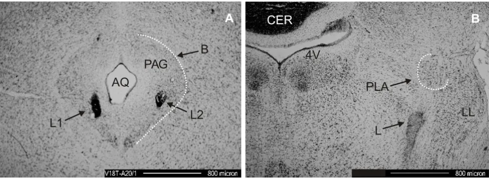
Electrical lesions
Figure 5
Electrical lesions. Histological verification of electrode locations A/B show Nissl stained 42 µm frontal sections. (A) Two lesions (L1/L2) caused by repeated electrical stimulation through chronically implanted electrodes placed into vocally active sites within the PAG. AQ, aqueduct; B, boundary between PAG and surrounding tissue. (B) Electrically induced lesion (L) 400
µm below the location of the iontophoresis probes in the PLA. Due to the 400 µm offset of the lesion below the PLA the func-tion of the PLA during further experiments was not influenced. CER, cerebellum; LL, lateral lemniscus; 4V, 4th ventricle.
AQ
PAG
B
L1
L2
4V
LL
PLA
L
from the PLA towards the region of the nucleus retroam-biguus/ambiguous complex in P. discolor. Only very few data are available from other species. In P. p. parnellii, efferents to the PAG were found when wheat germ agglutinin conjugated to horseradish peroxidase (WGA-HRP) was injected into vocally active sites of the PLA [20]. Efferents to the nucleus ambiguus from the PLA were found by Metzner using WGA-HRP [38] and connections from the lateral tegmental area (LTR) to the PAG and con-tralateral LTR were found using WGA-HRP and fluores-cent tracers [27,28]. However, these data are not sufficient to explain the effectiveness of contralateral blockades under anatomical aspects.
The enhancement of the suppression effect by bilateral KA application may have two reasons. First, reciprocal inter-connection between the two PLAs has been shown ana-tomically [20]. Second, ipsilateral blockage of the PLA also leads to reduced probability for eliciting echolocation calls, although it never reaches the level of suppression seen in most contralateral cases. The reciprocal interaction between the two PLAs is predominantly inhibitory as shown in R. rouxi (Schuller, unpublished), but the func-tional significance of this interaction is unclear. The assumption of a bilateral, but strongly unbalanced, descending control of echolocation calls via the PLA seems to fit the data more closely than does the assump-tion of a strictly unilateral organizaassump-tion. The presence of a non-functional ipsilateral PLA in addition to the silent contralateral PLA would further and more effectively decrease the probability for eliciting echolocation calls, resulting in a shorter time for onset of supression.
Based on connectional evidence (i.e., direct projections from the lateral part of the caudal PAG to the nucleus ret-roambiguus in the cat), Holstege proposed that the vocal pattern generation takes place within a final common pathway for vocalization driven by input from the PAG [15-17]. However, these findings and those of Zhang [1] that the vocal control pathway consists only of a direct connection from the PAG to the nucleus retroambiguus in the medulla oblongata are not supported by the results from the monkey [36]. Likewise, while our findings do not rule out a direct connection from the PAG to the nucleus retroambiguus (certain types of communication calls), they also demonstrate that a differentiated control for vocalizations (echolocation calls, frequency modu-lated) via parallel or at least partly separated pathways for echolocation calls could exist. This evidence of a more complicated network for vocal control at the level below the PAG in the monkey, as well as in the bat, underlines the broader significance of this concept on a mammalian level. The bat vocal control system therefore cannot be considered to be specialized, but is a general mammalian vocalization system with distinct emphasized features.
Conclusion
Communication calls and echolocation calls can be elic-ited with electrical microstimulation through chronically implanted electrodes at different sites within the PAG. Reversible blockade of the vocally active PLA in the region in which only echolocation calls can be triggered totally blocks PAG-induced echolocation calls but not communi-cation calls. Thus, the PAG-NA/NRA pathway for vocaliza-tion described in literature may not be the only pathway processing vocal activity. The PLA seems to be essential for the production of echolocation calls but not for particular types of communication calls elicited in the PAG. This suggests differential pathway organization for particular types of communication calls on one hand and echoloca-tion calls on the other hand. Whether the differentiaecholoca-tion of pathways applies to the two classes of echolocation calls and communication calls in general, or whether it is more directly dependent on specific call properties in the acoustic pattern domain, remains open to further experimentation.
Methods
Experimental design
Two male neotropical bats (P. discolor) originating from the departmental breeding colony were used for this study. During the experiments, the animals were kept under semi-natural conditions with a 12:12 hrs light cycle, at 70% relative humidity and 28°C.
Surgical preparation of the animals was done under 4% Isoflurane (CuraMED Pharma, Karlsruhe, Germany) anesthesia. After additional local application of 2% Xylo-cain® (Astia, Germany), skin and muscles overlying the
skull were cut along the midline, retracted to the sides and stabilized with sponge material (Gelastypt®, Hoechst,
Ger-many). Minor bleedings were stanched with the coagulant Orbat® (lege artis Pharma, Germany). The skull surface
was freed from tissue remains and a small holding tube was attached with a light-curing dental compound (Microglass®, Kulzer, Germany).
Experiments were conducted in an anechoic chamber, thus reducing acoustical interferences from the environ-ment and reflections of call signals. The animals were placed in a holder that prevented gross body movements and the head was immobilized by attaching the surgically affixed tube to a head holder that allowed accurate repo-sitioning (≤ 10 µm) of the animal in the stereotaxic device throughout repeated experimental procedures.
determine the skull position, and the reconstruction of the stimulation and iontophoresis sites are described elsewhere [39]. This method typically yields accuracy bet-ter than150 µm in all three dimensions.
Localization of vocally active sites within the PAG and PLA by electrical microstimulation typically started on the third postoperative day. Thirty minutes before each exper-imental session, an initial dose of 0.4 ml sedative (0.04% Rompun®/0.9% NaCl) was injected subcutaneously. The
sedated state was maintained by continuous subcutane-ous infusion of 0.04% Rompun in physiological saline with a rate between 3.5 µl to 5.5 µl/min (syringe pump: TSE-Systems, Bad Homburg, Germany). Sessions were generally limited to a maximum of 5 hrs per day.
Stimulation electrodes and iontophoresis probes were inserted through small holes of a typical diameter of 200
µm. Penetrations were made at different rostrocaudal and mediolateral inclinations in order to reach different loca-tions through the same hole. All coordinates of probe positions were referred to the reference point of the equip-ment also used to determine skull and therefore brain position, and thus could be mathematically transformed to coordinates of standard frontal sections, corresponding to a specially prepared standard working brain atlas for this bat species (T. Fenzl and A. Nixdorf, unpublished). For further verification, stimulation and iontophoresis sites within the brain were marked using electrical lesions (-4 µA DC for 5 min) through the electrodes implanted in the PAG and through the stimulation electrodes used to localize the vocally active sites in the PLA.
Electrical microstimulation
For acute microstimulation, Parylene-coated tungsten electrodes (type TM33A20, WPI Inc., Sarasota, USA) were used. Teflon-insulated silver wires (AGT0510, WPI, Sara-sota, USA, diameter: 125 µm) were implanted for chronic PAG micro-stimulation. A sharpened tungsten wire inserted into the neck musculature served as an indifferent electrode.
Electrical stimuli consisted of 15 ms long bursts with 0.1 ms long negative rectangular current pulses at 1 kHz rate, and were presented at a repetition rate of 6 bursts per s (stimulatorS48 with stimulus isolation unit PSIU6, Grass, Quincy, USA). One stimuli train lasted 2s.
During experiments, the animals were continuously mon-itored by TV under infrared light illumination (camera: Teli, Tokyo, Japan; monitor: TC-800 E4D, Osaka, Japan)) (LED array: 12 V/28 LED; Conrad Electronics, Germany).
Chronic stimulation electrodes
Sharpened Teflon®-insulated silver wires(type AGT0510,
WPI, Sarasota, USA) with a wire diameter of 125 µm and 100–200 µm bare length were secured at the skull with light curing dental compound cement at PAG positions at which appropriate calls (communication and echoloca-tion calls) could be electrically elicited. A pair of IC-socket pins was used as connectors. The electrical stimulus could be switched between electrodes using a remote controlled electric relay, interposed between the isolation unit and the stimulation electrodes.
A graphical overview of the experimental approach is pro-vided in the additional file (Additional file 1).
Iontophoresis within PLA
For reversible blockade of the PLA, the glutamate antago-nist kynurenic acid (KA; 75 mM at pH 9; Sigma-Aldrich, Steinheim, Germany) was iontophoretically applied through borosilicate glass microelectrodes with a tip diameter between 3 µm and 5 µm. Retaining current was 30 nA and ejection currents ranged between 200 nA and 250 nA, delivered by a Neurophore BH-2 system (Medical Systems Corp., Greenvale, USA).
PAG-triggered vocalizations were considered to have been blocked by iontophoresis in the PLA when 75% of electri-cal stimulations failed to elicit voelectri-calizations during a stimulation period of 10 min (coherent).
Sound recording, processing and data analysis
Vocalizations were picked up with an ultrasonic micro-phone (type 4135, Bruel & Kjaer, Darmstadt, Germany), amplified, digitally converted at 250 kHz sample rate (CIO-DAS16/M1, Computer Boards Inc., Mansfield, USA), and stored on a personal computer. The recording program was written in Agilent-VEE (version 6 pro, Agi-lent, USA). Spectral analysis of the recorded vocalizations for call identification and evaluation of frequency shifts was performed with the software "Bat Sound" (Petterson Electronic AB, Sweden). Peak amplitude of vocalizations was derived from the power spectrum of vocalizations. Call length was evaluated using the envelopes of the vocalizations.
Neuroanatomical processing
The animals were euthanized at termination of the exper-iments with Nembutal® (16 mg/100 g BW) and
(Frigocut type 2700, Reichert-Jung, Germany) into 42 µm slices and generally three adjacent series were processed. Routinely, Nissl and fiber stains [40] were applied for identification of stimulation sites. The standardized cutting procedure made it possible to refer anatomical data from individual brains to the sections of the reference brain, permitting a comparison of data from individual animals.
Animal care
Principles of laboratory animal care were followed and experiments were conducted under the regulations of the current version of German Law and Animal Protection. Reference Government of Bavaria (Az.Reg.v.Obb.211-2531-37/98).
Authors' contributions
TF developed chronic implants and designed and carried out all experiments. GS participated in the design of the study and the manuscript. Both authors read and approved the final manuscript.
Additional files
File name: additional_file_1
File format: PDF
Title of data: Graphical illustration of the experimental approach
Description of data:
The additional data (see Additional file 1) provide a graphical overview of the experimental approach by explaining the position of the implanted electrodes within the PAG together with the ipsi- and contralateral
alignments of the PLA electrodes. Additionally the remov-able connectors used on the animals are illustrated.
Additional material
Acknowledgements
The authors would like to thank Uwe Firzlaff and Andreas Nixdorf for fruit-ful discussions during the experiments, Herrmann Schweizer for his kind help on anatomical verifications of brain sections, Claudia Schulte and Horst König for their technical support, Karl-Heinz Esser (University of Hanno-ver, Germany) and Hans Erkert (University of Tübingen, Germany) for helping to establish a breeding colony of P. discolor by supplying bats and Doug Truskowsky for proofreading the manuscript. Funded by Deutsche Forschungsgemeinschaft (DFG) ref. Schu390/5-3 and Schu390/7-1.
References
1. Zhang SP, Bandler R, Davis PJ: Brain stem integration of vocali-zation: role of the nucleus retroambigualis. J Neurophysiol 1995, 74:2500-2512.
2. Lu CL, Jürgens U: Effects of chemical stimulation in the periaq-ueductal gray on vocalization in the squirrel monkey. Brain Res Bull 1993, 32:143-151.
3. Larson CR, Kistler M: Periaqueductal gray neuronal activity associated with laryngeal EMG and vocalization in the awake monkey. Neurosci Lett 1984, 46:261-266.
4. Yajima Y, Hayashi Y, Yoshii N: The midbrain central gray sub-stance as a highly sensitive neural structure for the produc-tion of ultrasonic vocalizaproduc-tion in the rat. Brain Res 1980, 198:446-452.
5. Martin JR: Motivated behaviors elicited from hypothalamus, midbrain, and pons of the guinea pig (Cavia porcellus). J Comp Physiol Psychol 1976, 90:1011-1034.
6. Apfelbach R: Electrically elicited vocalizations in the gibbon Hylobates lar (Hylobatidae), and their behavioral significance. Z Tierpsychol 1972, 30:420-430.
Table 1: Onset of KA-induced total suppression and recovery of PAG-elicited calls as shown in Figs. 2 and 3.
Call_type (PAG) KA_I/C (PLA) Tsupp [min] Trec [min] Fig.
EC I - - 2A
CC C - - 2A
EC C 75 >280 2B
CC I - - 2B
EC I/C 24 >240 2C
CC I/C - - 2C
EC I - - 3A
EC C 33 >140 3A
EC I - - 3B
EC C 8 110 3B
Call_type is either echolocation (EC) or communication (CC) call. KA application (KA_I/C) was either ipsi- (I) and/or contralateral (C) to the stimulation site in PAG. Tsupp, Trec are the times after the start of KA-application when the probability for eliciting calls was fully suppressed or fully recovered for a period of at least 10 min, respectively.
Additional File 1
Graphical illustration of the experimental approach.
Click here for file
Publish with BioMed Central and every scientist can read your work free of charge "BioMed Central will be the most significant development for disseminating the results of biomedical researc h in our lifetime."
Sir Paul Nurse, Cancer Research UK
Your research papers will be:
available free of charge to the entire biomedical community peer reviewed and published immediately upon acceptance cited in PubMed and archived on PubMed Central yours — you keep the copyright
Submit your manuscript here:
http://www.biomedcentral.com/info/publishing_adv.asp
BioMedcentral 7. Fenzl T, Schuller G: Periaqueductal gray and the region of the
paralemniscal area have different functions in the control of vocalization in the neotropical bat, Phyllostomus discolor.
Eur J Neurosci 2002, 16:1974-1986.
8. Valentine DE, Sinha SR, Moss CF: Orienting responses and vocal-izations produced by microstimulation in the superior collic-ulus of the echolocating bat, Eptesicus fuscus. J Comp Physiol A Neuroethol Sens Neural Behav Physiol 2002, 188:89-108.
9. Jürgens U: The role of the periaqueductal grey in vocal behaviour. Behav Brain Res 1994, 62:107-117.
10. Suga N, Yajima Y: Auditory-vocal integration in the midbrain of the mustached bat: Periaqueductal Gray and Reticular For-mation. In The physiological control of mammalian vocalization Edited by: Newman JD. New York: Plenum Press; 1988:87-107.
11. Suga N, Schlegel P, Shimozawa T, Simmons J: Orientation sounds evoked from echolocating bats by electrical stimulation of the brain. J Acoust Soc Am 1973, 54:793-797.
12. Jürgens U, Ploog D: Cerebral representation of vocalization in the squirrel monkey. Exp Brain Res 1970, 10:532-554.
13. Jürgens U, Pratt R: The cingular vocalization pathway in the squirrel monkey. Exp Brain Res 1979, 34:499-510.
14. Jürgens U: Neural pathways underlying vocal control. Neurosci Biobehav Rev 2002, 26:235-258.
15. Holstege G: Anatomical study of the final common pathway for vocalization in the cat. J Comp Neurol 1989, 284:242-252. 16. Vanderhorst VG, Terasawa E, Ralston HJ III, Holstege G:
Monosyn-aptic projections from the lateral periaqueductal gray to the nucleus retroambiguus in the rhesus monkey: implications for vocalization and reproductive behavior. J Comp Neurol
2000, 424:251-268.
17. Vanderhorst VG, Terasawa E, Ralston HJ III, Holstege G: Monosyn-aptic projections from the nucleus retroambiguus to motoneurons supplying the abdominal wall, axial, hindlimb, and pelvic floor muscles in the female rhesus monkey. J Comp Neurol 2000, 424:233-250.
18. Schuller G, Radtke-Schuller S: Neural control of vocalization in bats: mapping of brainstem areas with electrical microstim-ulation eliciting species-specific echolocation calls in the rufous horseshoe bat. Exp Brain Res 1990, 79:192-206.
19. Pillat J, Schuller G: Audiovocal behavior of Doppler-shift com-pensation in the horseshoe bat survives bilateral lesion of the paralemniscal tegmental area. Exp Brain Res 1998, 119:17-26. 20. Schuller G, Fischer S, Schweizer H: Significance of the
paralem-niscal tegmental area for audio-motor control in the mous-tached bat, Pteronotus p. parnellii: the afferent and efferent connections of the paralemniscal area. Eur J Neurosci 1997, 9:342-355.
21. Nixdorf A: Die prätektale Region bei der Fledermaus Phyllos-tomus discolor – eine audio-motorische Schnittstelle? Dept Biol II, Ludwig-Maximilians-Universität München 2003.
22. Metzner W: Anatomical basis for audio-vocal integration in echolocating horseshoe bats. J Comp Neurol 1996, 368:252-269. 23. Metzner W: A possible neuronal basis for Doppler shift com-pensation in echo-locating horseshoe bats. Nature 1989, 341:529-532.
24. Covey E: Response properties of single units in the dorsal nucleus of the lateral lemniscus and paralemniscal zone of an echolocating bat. J Neurophysiol 1993, 69:842-859.
25. Henkel CK, Edwards SB: The superior colliculus control of pinna movements in the cat: possible anatomical connections. J Comp Neurol 1978, 182:763-776.
26. Henkel CK: Afferent sources of a lateral midbrain tegmental zone associated with the pinnae in the cat as mapped by ret-rograde transport of horseradish peroxidase. J Comp Neurol
1981, 203:213-226.
27. Gerlach I, Thier P, Koehler W: Visual and vestibular signals in the lateral mesencephalic tegmental region of the cat. Exp Brain Res 1991, 85:641-649.
28. Gerlach I, Thier P: Brainstem afferents to the lateral mesen-cephalic tegmental region of the cat. J Comp Neurol 1995, 358:219-232.
29. Thoms G, Jürgens U: Common input of the cranial motor nuclei involved in phonation in squirrel monkey. Exp Neurol
1987, 95:85-99.
30. Jürgens U: Neuronal control of mammalian vocalization, with special reference to the squirrel monkey. Naturwissenschaften
1998, 85:376-388.
31. Kirzinger A, Jurgens U: Vocalization-correlated single-unit activity in the brain stem of the squirrel monkey. Exp Brain Res
1991, 84:545-560.
32. Zhang SP, Davis PJ, Bandler R, Carrive P: Brain stem integration of vocalization: role of the midbrain periaqueductal gray. J Neurophysiol 1994, 72:1337-1356.
33. Kennedy MC: Vocalization elicited in a lizard by electrical stimulation of the midbrain. Brain Res 1975, 91:321-325. 34. Ennis M, Xu SJ, Rizvi TA: Discrete subregions of the rat
mid-brain periaqueductal gray project to nucleus ambiguus and the periambigual region. Neuroscience 1997, 80:829-845. 35. Vanderhorst VG, Terasawa E, Ralston HJ: Monosynaptic
projec-tions from the nucleus retroambiguus region to laryngeal motoneurons in the rhesus monkey. Neuroscience 2001, 107:117-125.
36. Jürgens U: Localization of a pontine vocalization-controlling area. J Acoust Soc Am 2000, 108:1393-1396.
37. Rother G, Schmidt U: Der Einfluss visueller Information auf die Echoortung bei Phyllostomus discolor. Z Säugetierkunde 1982, 47:324-334.
38. Metzner W: Anatomical basis for audio-vocal integration in echolocating horseshoe bats. J Comp Neuro 1996, 368:252-269. 39. Schuller G, Radtke-Schuller S, Betz M: A stereotaxic method for
small animals using experimentally determined reference profiles. J Neurosci Methods 1986, 18:339-350.